Advertisement

Main menu

  • Home
  • Content
    • Current Issue
    • Accepted Manuscripts
    • Article Preview
    • Past Issue Archive
    • AJNR Case Collection
    • Case of the Week Archive
    • Classic Case Archive
    • Case of the Month Archive
  • Special Collections
    • Spinal CSF Leak Articles (Jan 2020-June 2024)
    • 2024 AJNR Journal Awards
    • Most Impactful AJNR Articles
  • Multimedia
    • AJNR Podcast
    • AJNR Scantastics
    • Video Articles
  • For Authors
    • Submit a Manuscript
    • Author Policies
    • Fast publishing of Accepted Manuscripts
    • Graphical Abstract Preparation
    • Manuscript Submission Guidelines
    • Imaging Protocol Submission
    • Submit a Case for the Case Collection
  • About Us
    • About AJNR
    • Editorial Board
  • More
    • Become a Reviewer/Academy of Reviewers
    • Subscribers
    • Permissions
    • Alerts
    • Feedback
    • Advertisers
    • ASNR Home
  • Other Publications
    • ajnr

User menu

  • Alerts
  • Log in

Search

  • Advanced search
American Journal of Neuroradiology

American Journal of Neuroradiology

ASHNR American Society of Functional Neuroradiology ASHNR American Society of Pediatric Neuroradiology ASSR
  • Alerts
  • Log in

Advanced Search

  • Home
  • Content
    • Current Issue
    • Accepted Manuscripts
    • Article Preview
    • Past Issue Archive
    • AJNR Case Collection
    • Case of the Week Archive
    • Classic Case Archive
    • Case of the Month Archive
  • Special Collections
    • Spinal CSF Leak Articles (Jan 2020-June 2024)
    • 2024 AJNR Journal Awards
    • Most Impactful AJNR Articles
  • Multimedia
    • AJNR Podcast
    • AJNR Scantastics
    • Video Articles
  • For Authors
    • Submit a Manuscript
    • Author Policies
    • Fast publishing of Accepted Manuscripts
    • Graphical Abstract Preparation
    • Manuscript Submission Guidelines
    • Imaging Protocol Submission
    • Submit a Case for the Case Collection
  • About Us
    • About AJNR
    • Editorial Board
  • More
    • Become a Reviewer/Academy of Reviewers
    • Subscribers
    • Permissions
    • Alerts
    • Feedback
    • Advertisers
    • ASNR Home
  • Follow AJNR on Twitter
  • Visit AJNR on Facebook
  • Follow AJNR on Instagram
  • Join AJNR on LinkedIn
  • RSS Feeds

Welcome to the new AJNR, Updated Hall of Fame, and more. Read the full announcements.


AJNR is seeking candidates for the position of Associate Section Editor, AJNR Case Collection. Read the full announcement.


Research ArticleBRAIN

Differential Aging of the Human Striatum: Longitudinal Evidence

Naftali Raz, Karen M. Rodrigue, Kristen M. Kennedy, Denise Head, Faith Gunning-Dixon and James D. Acker
American Journal of Neuroradiology October 2003, 24 (9) 1849-1856;
Naftali Raz
  • Find this author on Google Scholar
  • Find this author on PubMed
  • Search for this author on this site
Karen M. Rodrigue
  • Find this author on Google Scholar
  • Find this author on PubMed
  • Search for this author on this site
Kristen M. Kennedy
  • Find this author on Google Scholar
  • Find this author on PubMed
  • Search for this author on this site
Denise Head
  • Find this author on Google Scholar
  • Find this author on PubMed
  • Search for this author on this site
Faith Gunning-Dixon
  • Find this author on Google Scholar
  • Find this author on PubMed
  • Search for this author on this site
James D. Acker
  • Find this author on Google Scholar
  • Find this author on PubMed
  • Search for this author on this site
  • Article
  • Figures & Data
  • Info & Metrics
  • Responses
  • References
  • PDF
Loading

Article Figures & Data

Figures

  • Tables
  • Fig 1.
    • Download figure
    • Open in new tab
    • Download powerpoint
    Fig 1.

    An example of demarcation and tracing of three striatal regions of interest (caudate, putamen and globus pallidus) on a series of coronal images.
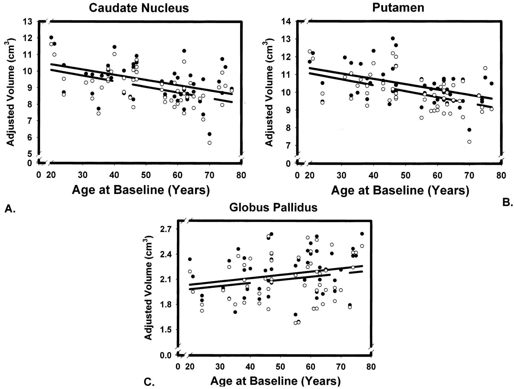
  • Fig 2.
    • Download figure
    • Open in new tab
    • Download powerpoint
    Fig 2.

    Scatter plots for two volumetric measures of three striatal regions of interest taken on 53 subjects 5 years apart. The lines indicate linear regression of adjusted volumes on age at time 1 (solid line) and time 2 (broken line). Observations at time 1 are represented by solid circles, and observations at time 2 by empty circles. Regional volumes are adjusted for the intracranial volume.

Tables

  • Figures
    • View popup
    TABLE

    Cross-Sectional Age-Related Differences in Striatal Volumes Measured Five Years Apart

    NucleusRaw VolumeRegression of ICV-Adjusted Volume on Ager12
    Time 1Time 2Time 1Time 2
    MeanSDCVMeanSDCVSlopeSErageSlopeSErage
    Caudate9.421.140.129.001.090.12−.031.010−.41−.034.009−.47.95
    Putamen10.410.980.0910.011.020.10−.030.008−.46−.033.008−.49.89
    Globus Pallidus2.160.270.122.100.270.13.004.002.22.004.002.21.93
    • Note.—CV (coefficient of variation) is a ratio of standard deviation (SD) to the mean. The regression slopes are unstandardized regression coefficients measured in cc/year. SE is standard error of slope. All coefficients for the caudate nucleus and putamen are significantly different from zero, whereas those for the globus pallidus are not. The differences in slopes between the neostriatum (caudate and putamen) and paleostriatum are statistically significant (p < .05). Correlations are Pearson product moment coefficients. All correlations r > .23 are significantly different from zero.

Back to top

In this issue

American Journal of Neuroradiology: 24 (9)
American Journal of Neuroradiology
Vol. 24, Issue 9
1 Oct 2003
  • Table of Contents
  • Index by author
Advertisement
Print
Download PDF
Email Article

Thank you for your interest in spreading the word on American Journal of Neuroradiology.

NOTE: We only request your email address so that the person you are recommending the page to knows that you wanted them to see it, and that it is not junk mail. We do not capture any email address.

Enter multiple addresses on separate lines or separate them with commas.
Differential Aging of the Human Striatum: Longitudinal Evidence
(Your Name) has sent you a message from American Journal of Neuroradiology
(Your Name) thought you would like to see the American Journal of Neuroradiology web site.
CAPTCHA
This question is for testing whether or not you are a human visitor and to prevent automated spam submissions.
Cite this article
Naftali Raz, Karen M. Rodrigue, Kristen M. Kennedy, Denise Head, Faith Gunning-Dixon, James D. Acker
Differential Aging of the Human Striatum: Longitudinal Evidence
American Journal of Neuroradiology Oct 2003, 24 (9) 1849-1856;

Citation Manager Formats

  • BibTeX
  • Bookends
  • EasyBib
  • EndNote (tagged)
  • EndNote 8 (xml)
  • Medlars
  • Mendeley
  • Papers
  • RefWorks Tagged
  • Ref Manager
  • RIS
  • Zotero
0 Responses
Respond to this article
Share
Bookmark this article
Differential Aging of the Human Striatum: Longitudinal Evidence
Naftali Raz, Karen M. Rodrigue, Kristen M. Kennedy, Denise Head, Faith Gunning-Dixon, James D. Acker
American Journal of Neuroradiology Oct 2003, 24 (9) 1849-1856;
del.icio.us logo Twitter logo Facebook logo Mendeley logo
  • Tweet Widget
  • Facebook Like
  • Google Plus One

Jump to section

  • Article
    • Abstract
    • Methods
    • Results
    • Discussion
    • Conclusion
    • Appendix
    • References
  • Figures & Data
  • Info & Metrics
  • Responses
  • References
  • PDF

Related Articles

  • No related articles found.
  • PubMed
  • Google Scholar

Cited By...

  • Effects of healthy aging on tongue-jaw kinematics during feeding behavior in rhesus macaques
  • Age-Related Brain Atrophy and the Positive Effects of Behavioral Enrichment in Middle-Aged Beagles
  • Age-related fornix decline predicts conservative response strategy-based slowing in perceptual decision-making
  • Hippocampal subfield volumes contribute to working memory interference control in aging: Evidence from longitudinal associations over 5 years
  • Hippocampal subfield volumes contribute to working memory interference control in aging: Evidence from longitudinal associations over 5 years
  • Neural asymmetry during memory encoding and its association with markers of preclinical Alzheimers Disease
  • Structural Priming Is Supported By Different Components Of Non-Declarative Memory: Evidence From Priming Across The Lifespan
  • Cardiorespiratory fitness predicts greater hippocampal volume and rate of episodic associative learning in older adults
  • Iron Level and Myelin Content in the Ventral Striatum Predict Memory Performance in the Aging Brain
  • Normal Aging in the Basal Ganglia Evaluated by Eigenvalues of Diffusion Tensor Imaging
  • Automated Optimization of Subcortical Cerebral MR Imaging-Atlas Coregistration for Improved Postoperative Electrode Localization in Deep Brain Stimulation
  • Putaminal Volume in Frontotemporal Lobar Degeneration and Alzheimer Disease: Differential Volumes in Dementia Subtypes and Controls
  • Transfer of Learning After Updating Training Mediated by the Striatum
  • Aging affects acquisition and reversal of reward-based associative learning
  • Structural changes in patients with primary generalized tonic and clonic seizures.
  • Neural Mechanisms Underlying Probabilistic Category Learning in Normal Aging
  • Differential aging of the medial temporal lobe: A study of a five-year change
  • Shrinkage of the Entorhinal Cortex over Five Years Predicts Memory Performance in Healthy Adults
  • Crossref
  • Google Scholar

This article has not yet been cited by articles in journals that are participating in Crossref Cited-by Linking.

More in this TOC Section

  • Multimodal CT Provides Improved Performance for Lacunar Infarct Detection
  • Statin Therapy Does Not Affect the Radiographic and Clinical Profile of Patients with TIA and Minor Stroke
  • Optimal MRI Sequence for Identifying Occlusion Location in Acute Stroke: Which Value of Time-Resolved Contrast-Enhanced MRA?
Show more Brain

Similar Articles

Advertisement

Indexed Content

  • Current Issue
  • Accepted Manuscripts
  • Article Preview
  • Past Issues
  • Editorials
  • Editors Choice
  • Fellow Journal Club
  • Letters to the Editor

Cases

  • Case Collection
  • Archive - Case of the Week
  • Archive - Case of the Month
  • Archive - Classic Case

Special Collections

  • Special Collections

Resources

  • News and Updates
  • Turn around Times
  • Submit a Manuscript
  • Author Policies
  • Manuscript Submission Guidelines
  • Evidence-Based Medicine Level Guide
  • Publishing Checklists
  • Graphical Abstract Preparation
  • Imaging Protocol Submission
  • Submit a Case
  • Become a Reviewer/Academy of Reviewers
  • Get Peer Review Credit from Publons

Multimedia

  • AJNR Podcast
  • AJNR SCANtastic
  • Video Articles

About Us

  • About AJNR
  • Editorial Board
  • Not an AJNR Subscriber? Join Now
  • Alerts
  • Feedback
  • Advertise with us
  • Librarian Resources
  • Permissions
  • Terms and Conditions

American Society of Neuroradiology

  • Not an ASNR Member? Join Now

© 2025 by the American Society of Neuroradiology All rights, including for text and data mining, AI training, and similar technologies, are reserved.
Print ISSN: 0195-6108 Online ISSN: 1936-959X

Powered by HighWire