
Article Figures & Data
Figures
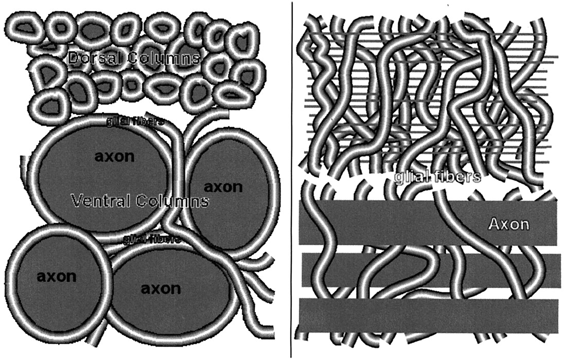
- Fig 1.
Transverse (left) and longitudinal (right) sections of lamprey spinal cord. Axonal fiber diameters range from <1 μm in the dorsal columns to 20–40 μm (Müller axons) in the ventral region. Glial fibers densely fill the space between axons.
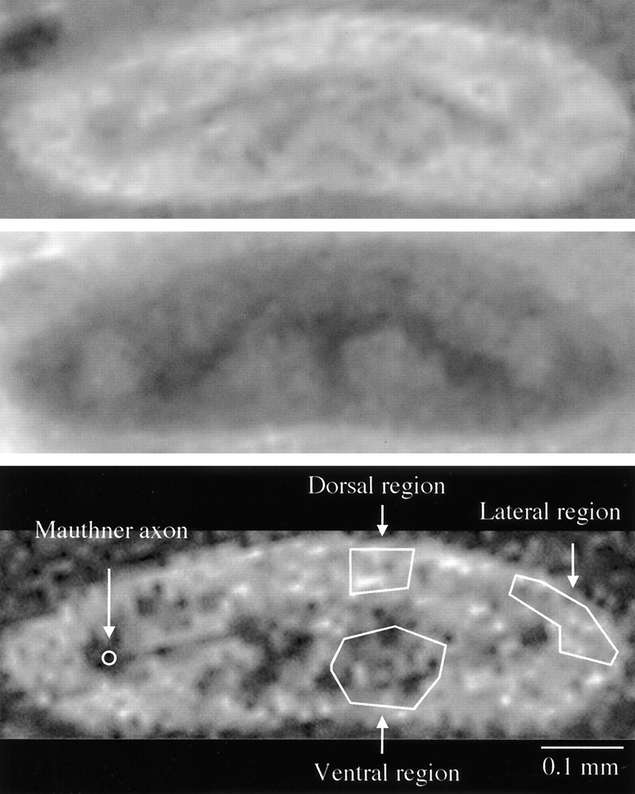
- Fig 2.
Transverse micro-MR images of normal live excised spinal cord without (top) and with (middle) MT. Bottom image is a diffusion-weighted SE image with the same geometry as that of the SE images. Overlaid ROIs (bottom) were used to calculate the MTR on the corresponding SE images.
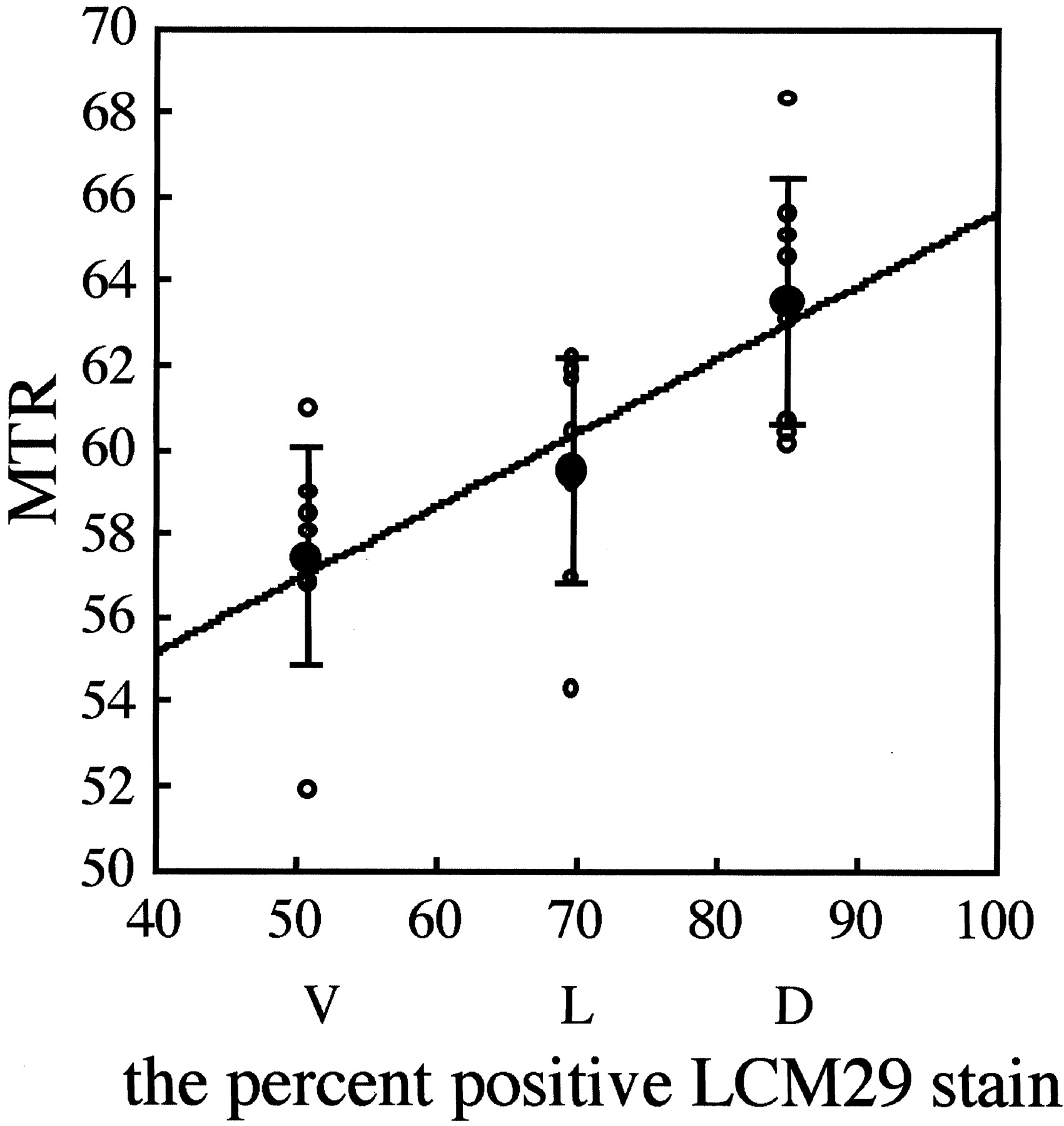
- Fig 3.
Regional MTRs in lamprey spinal cord. Values were used in the Tukey-Kramer multiple-comparison test to determine statistical significance. MTRs for each location are plotted with open circles. Closed circles are mean MTRs ± SDs.
- Fig 4.
Immunohistochemical section at a location similar to that in Figure 2 (LCM29 stain for glial keratin; original magnification, ×10). Axonal fiber diameters are 1–40 μm. Individual giant reticulospinal axons are located in the lateral region (Mauthner axon) and ventral columns (Müller axons). In the fiber tracts, spaces not occupied by axons are filled with glial cells and their processes. Threshold between negatively stained area (axoplasm) and positively stained area (glial cells and fibers) is selected manually. Image is converted to a binary image, as shown. Average of two threshold determinations is displayed in overlay regions in the areas measured. Percentage of positive staining is calculated in dorsal, lateral, and ventral columns. The Mauthner axon is not included in the lateral columns sampled for MTR measurement.
- Fig 5.
Percentage of LCM29-positive area on immunohistochemical analysis versus MTR on MR imaging. Open circles indicate MTRs; closed circles, mean MTR ± SD; D, dorsal region; L, lateral; and V, ventral. Mean MTR and percentage LCM29-positive area are significantly correlated (r2 = 0.98).
- Fig 6.
Electron photomicrograph from the lateral column of normal lamprey spinal cord. Darkly staining glial fibers (arrows) densely fill spaces between axons, which are less darkly stained. Shown are axons of varying size, including one (Axon) approximately 13 μm in diameter. Bar indicates 2 μm.


In this issue


Jump to section
Related Articles
Cited By...
- No citing articles found.