
Article Figures & Data
Figures
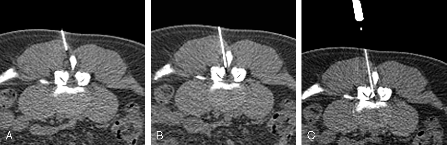
- Fig 1.
Epidural Injection Technique as demonstrated in a 38-year-old man with back pain.
A, CTF image demonstrates the needle to be appropriately angled toward the epidural space. This image was taken at the lower part of the posterior spinous process; opening used to access the epidural space, between the spinous process and medial facet, is clearly seen. The small triangle of posterior epidural fat is a useful landmark to aim for, although it is not present in some patients, particularly those with severe spinal stenosis.
B, The needle tip at the outer edge of the ligamentum flavum, corresponding to a feeling of resistance while advancing the needle, is shown. The tip can be identified by the shadowing artifact extending from it. Notice how the needle has avoided most of the erector spinae muscles during insertion, adding to the comfort level of the patient.
C, The final image in this study demonstrates contrast medium outlining the epidural space, tracking along the anterior edge of the ligamentum flavum. Although cross-filling is not seen on this image, it can be shown with a larger contrast medium injection, but this has been found to be unnecessary.
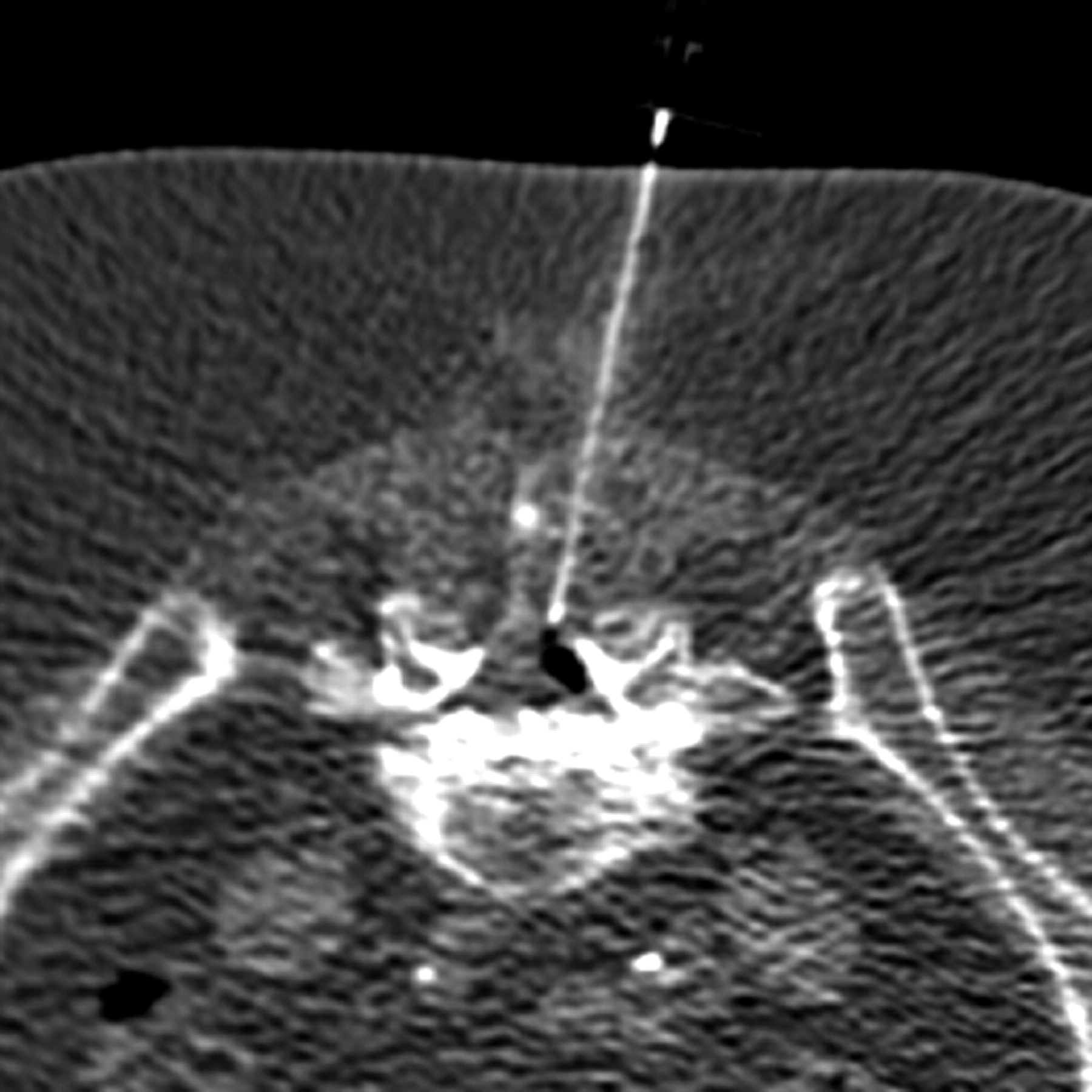
- Fig 2.
In this patient with a known allergy to iodinated contrast material, air was used as a contrast agent. The air defines the epidural space as well as contrast material, confirming appropriate needle placement.
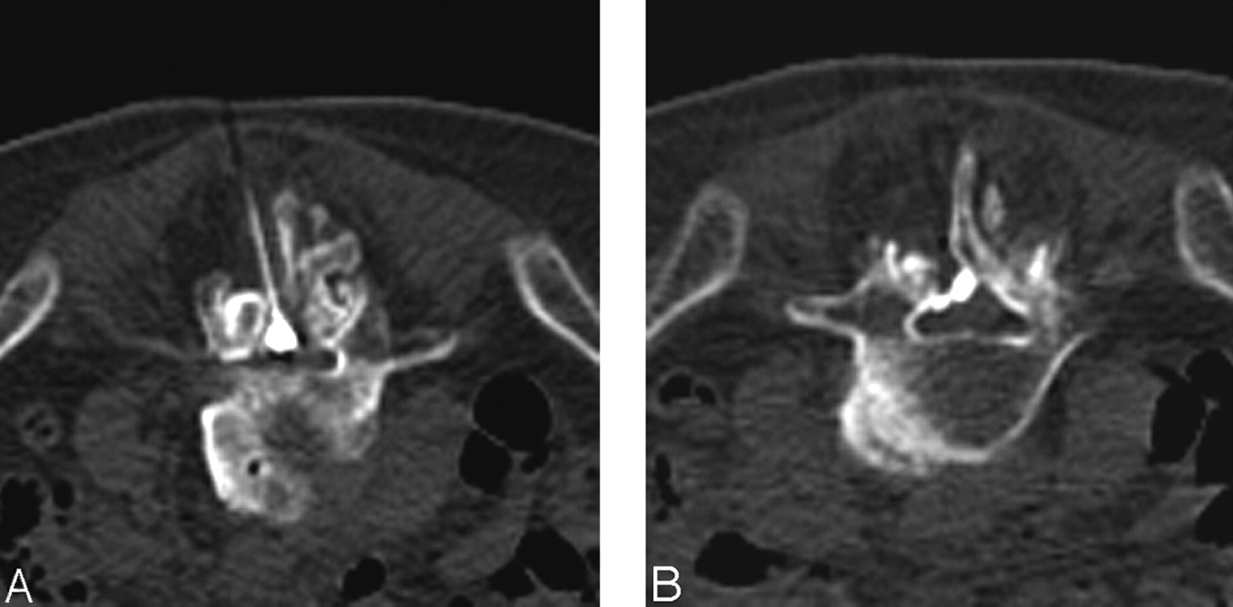
- Fig 3.
Images obtained in an 86-year-old man with scoliosis and spinal stenosis.
A, In patients such as this one, contrast medium may appear to be within the thecal sac, because it can have a globular shape at the point of injection. In reality, the thecal sac has been displaced to the right, and the patient remained asymptomatic throughout the procedure.
B, An image taken 6 mm inferior from the injection site shows that contrast material spreading within the epidural space, confirming appropriate needle placement.
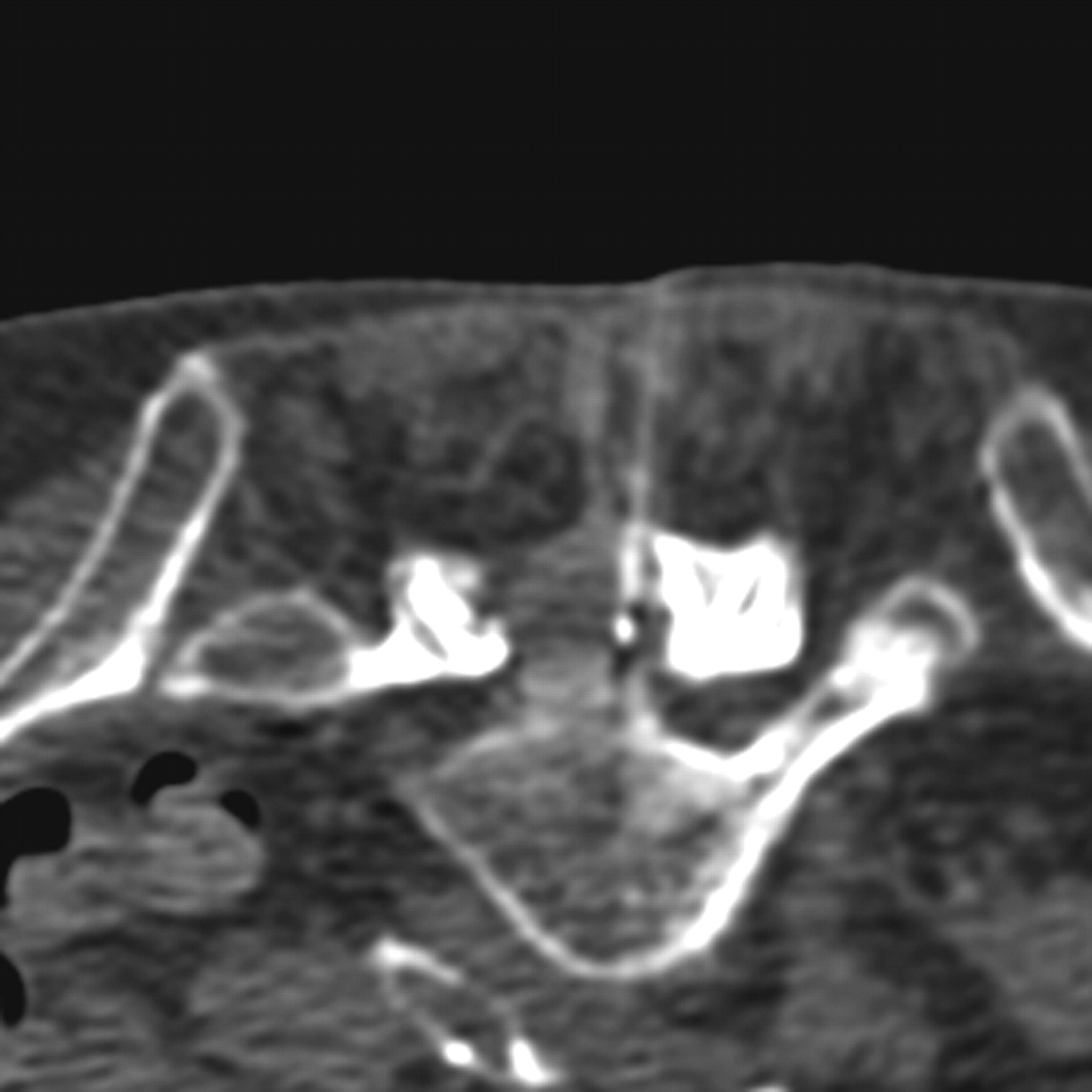
- Fig 4.
Intrathecal injection is confirmed by visualization of contrast material within the thecal sac, giving a contrast medium–fluid level, while only a small amount of epidural contrast medium is present.


In this issue


Jump to section
Related Articles
Cited By...
- Inadvertent Intrafacet Injection during Lumbar Interlaminar Epidural Steroid Injection: A Comparison of CT Fluoroscopic and Conventional Fluoroscopic Guidance
- Incidence of Inadvertent Intravascular Injection during CT Fluoroscopy-Guided Epidural Steroid Injections
- CT-Guided Lumbar Nerve Root Injections: Are We Using the Correct Radiation Dose Settings?