Advertisement

Main menu

  • Home
  • Content
    • Current Issue
    • Accepted Manuscripts
    • Article Preview
    • Past Issue Archive
    • AJNR Case Collection
    • Case of the Week Archive
    • Classic Case Archive
    • Case of the Month Archive
  • Special Collections
    • Spinal CSF Leak Articles (Jan 2020-June 2024)
    • 2024 AJNR Journal Awards
    • Most Impactful AJNR Articles
  • Multimedia
    • AJNR Podcast
    • AJNR Scantastics
    • Video Articles
  • For Authors
    • Submit a Manuscript
    • Author Policies
    • Fast publishing of Accepted Manuscripts
    • Graphical Abstract Preparation
    • Manuscript Submission Guidelines
    • Imaging Protocol Submission
    • Submit a Case for the Case Collection
  • About Us
    • About AJNR
    • Editorial Board
  • More
    • Become a Reviewer/Academy of Reviewers
    • Subscribers
    • Permissions
    • Alerts
    • Feedback
    • Advertisers
    • ASNR Home
  • Other Publications
    • ajnr

User menu

  • Alerts
  • Log in

Search

  • Advanced search
American Journal of Neuroradiology

American Journal of Neuroradiology

ASHNR American Society of Functional Neuroradiology ASHNR American Society of Pediatric Neuroradiology ASSR
  • Alerts
  • Log in

Advanced Search

  • Home
  • Content
    • Current Issue
    • Accepted Manuscripts
    • Article Preview
    • Past Issue Archive
    • AJNR Case Collection
    • Case of the Week Archive
    • Classic Case Archive
    • Case of the Month Archive
  • Special Collections
    • Spinal CSF Leak Articles (Jan 2020-June 2024)
    • 2024 AJNR Journal Awards
    • Most Impactful AJNR Articles
  • Multimedia
    • AJNR Podcast
    • AJNR Scantastics
    • Video Articles
  • For Authors
    • Submit a Manuscript
    • Author Policies
    • Fast publishing of Accepted Manuscripts
    • Graphical Abstract Preparation
    • Manuscript Submission Guidelines
    • Imaging Protocol Submission
    • Submit a Case for the Case Collection
  • About Us
    • About AJNR
    • Editorial Board
  • More
    • Become a Reviewer/Academy of Reviewers
    • Subscribers
    • Permissions
    • Alerts
    • Feedback
    • Advertisers
    • ASNR Home
  • Follow AJNR on Twitter
  • Visit AJNR on Facebook
  • Follow AJNR on Instagram
  • Join AJNR on LinkedIn
  • RSS Feeds

Welcome to the new AJNR, Updated Hall of Fame, and more. Read the full announcements.


AJNR is seeking candidates for the position of Associate Section Editor, AJNR Case Collection. Read the full announcement.


LetterLetter

Influenza-Associated Encephalitis-Encephalopathy with a Reversible Lesion in the Splenium of the Corpus Callosum: Case Report and Literature Review

Osman Kizilkilic and Sibel Karaca
American Journal of Neuroradiology November 2004, 25 (10) 1863-1864;
Osman Kizilkilic
  • Find this author on Google Scholar
  • Find this author on PubMed
  • Search for this author on this site
Sibel Karaca
  • Find this author on Google Scholar
  • Find this author on PubMed
  • Search for this author on this site
  • Article
  • Figures & Data
  • Info & Metrics
  • Responses
  • References
  • PDF
Loading

We read with interest the article by Takanashi et al (1) in the May 2004 issue of the AJNR. The authors reported two cases involving lesions of splenium of the corpus callosum in patients with influenza-associated encephalitis/encephalopathy. Although they described a reversible lesion in the splenium of the corpus callosum, they did not investigate causes other than encephalitis/encephalopathies. We would like to emphasize previously published reports regarding such lesions.

For instance, Kim et al (2) initially described transient splenial lesions, as revealed on MR imaging, in 1999. Focal nonhemorrhagic lesions of the corpus callosum are rare, but they have been described in various clinical conditions, and little is known about the underlying mechanisms. Although the pathogenesis is not understood, awareness of the fact that this is a reversible focal lesion may help prevent unnecessary invasive diagnostic and therapeutic intervention. Various pathologic conditions, such as multiple sclerosis, trauma, neoplasm, infarct, leukodystrophies (especially adrenoleukodystrophy), AIDS dementia complex, and Marchiafava-Bignami disease, may also involve the corpus callosum (3).

Kim et al (2) reported such lesions in six patients with focal epilepsy. They classified the lesion as demyelination and hypothesized that anticonvulsive drug toxicity was the cause. Similar lesions have been described in epileptic patients in the postictal period as well. These lesions have been interpreted as transient focal edema due to transient physiologic alteration in the callosal fibers caused by seizure activity. Association of such splenial lesion with acute cerebellitis due to herpes simplex, and its regression within 72 hours, has also been reported (4).

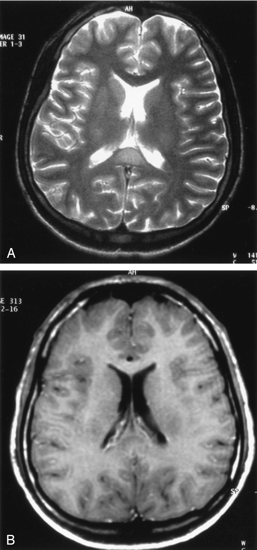
In support of these observations, we herein briefly report our experience with a 24-year-old man who presented with headache and apathy lasting for 5 days. He had no remarkable medical history, he was not receiving any medication, and he denied alcohol abuse. Scalp electroencephalograms were normal, and CSF testing showed slight elevation of protein levels. Results of blood tests were unremarkable for demyelinating or other systemic conditions, metachromatic leukodystrophy, antiphospholipid antibody syndrome, or HIV infection. Titers of serum immunoglobulins against human herpes viruses, herpes simplex virus, Epstein-Barr virus, varicella zoster virus, rubella virus, measles virus, papovavirus, enterovirus, influenza virus, and rotavirus, and Mycoplasma pneumonia were normal. Cranial MR images showed a focal nonenhancing lesion in the splenium of the corpus callosum, which totally regressed within 4 weeks without medication (Figs 1–2). We believe that this case represented an instance encephalitis/encephalopathy, but this could not be proved.

The case that Takanashi et al (1) reported was believed to have same underlying cause as ours, but to our knowledge the association of such a lesion with apathy or headache has not been reported. Polster et al (5) have suggested that such a transient splenial lesion appears to be a nonspecific endpoint of a different disease processes leading to vasogenic edema. This seems the most probable pathophysiologic explanation of such splenial lesions, as in our case.

Fig 1.
  • Download figure
  • Open in new tab
  • Download powerpoint
Fig 1.

Cranial MR images obtained in a 24-year-old man show a focal nonenhancing lesion.

A, Axial T2-weighted turbo spin echo image (TR 5000 ms, TE 99 ms; NEX 2) shows hyperintense lesion in the splenium of the corpus callosum.

B, Axial T1-weighted, contrast administered spin echo image (TR 600 ms, Te 14 ms; NEX 2) reveals hypointense character of the lesion, without contrast enhancement.

Fig 2.
  • Download figure
  • Open in new tab
  • Download powerpoint
Fig 2.

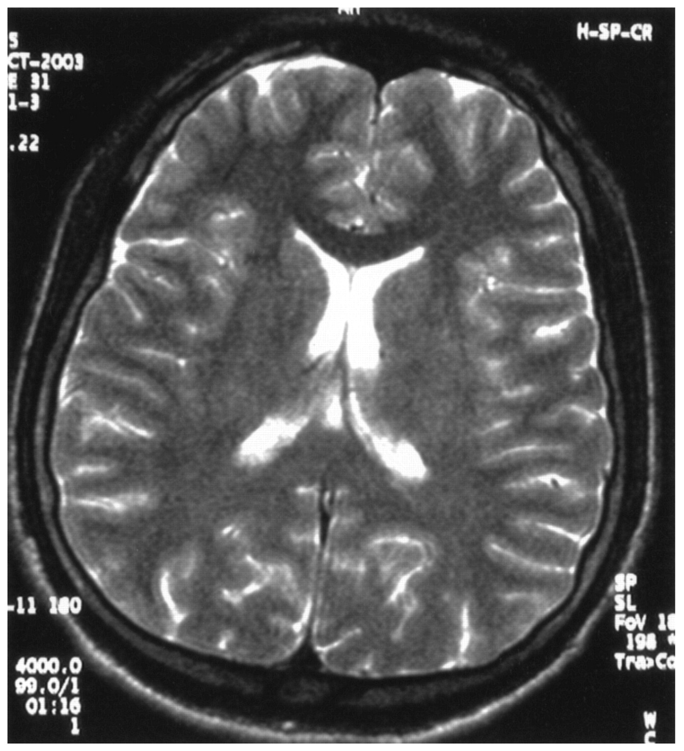
Axial T2-weighted turbo spin echo image (TR 5000 ms, TE 99 ms; NEX 2) shows resolution of the lesion in the splenium of the corpus callosum.

References

  1. ↵
    Takanashi J, Barkovich AJ, Yamaguchi K, Kohno Y. Influenza-associated encephalitis/ encephalopathy with a reversible lesion in the splenium of the corpus callosum: a case report and literature review. AJNR Am J Neuroradiol 2004;25:798–802
    Abstract/FREE Full Text
  2. ↵
    Kim SS, Chang KH, Kim ST, et al. Focal lesion in the splenium of the corpus callosum in epileptic patients: antiepileptic drug toxicity? AJNR Am J Neuroradiol 1999;20:125–129
    Abstract/FREE Full Text
  3. ↵
    Friese SA, Bitzer M, Freudenstein D, Voigt K, Kuker W. Classification of acquired lesions of the corpus callosum with MRI. Neuroradiology 2000;42:795–802
    CrossRefPubMed
  4. ↵
    Kato Z, Kozawa R, Teramoto T, Hashimoto K, Shinoda S, Kondo N. Transient lesion in the splenium of the corpus callosum in acute cerebellitis. J Child Neurol 2003;18:291–292
    Abstract/FREE Full Text
  5. ↵
    Polster T, Hoppe M, Ebner A. Transient lesion in the splenium of the corpus callosum: three further cases in epileptic patients and a pathophysiological hypothesis. J Neurol Neurosurg Psychiatry 2001;70:459–463
    Abstract/FREE Full Text
  • Copyright © American Society of Neuroradiology
PreviousNext
Back to top

In this issue

American Journal of Neuroradiology: 25 (10)
American Journal of Neuroradiology
Vol. 25, Issue 10
1 Nov 2004
  • Table of Contents
  • Index by author
Advertisement
Print
Download PDF
Email Article

Thank you for your interest in spreading the word on American Journal of Neuroradiology.

NOTE: We only request your email address so that the person you are recommending the page to knows that you wanted them to see it, and that it is not junk mail. We do not capture any email address.

Enter multiple addresses on separate lines or separate them with commas.
Influenza-Associated Encephalitis-Encephalopathy with a Reversible Lesion in the Splenium of the Corpus Callosum: Case Report and Literature Review
(Your Name) has sent you a message from American Journal of Neuroradiology
(Your Name) thought you would like to see the American Journal of Neuroradiology web site.
CAPTCHA
This question is for testing whether or not you are a human visitor and to prevent automated spam submissions.
Cite this article
Osman Kizilkilic, Sibel Karaca
Influenza-Associated Encephalitis-Encephalopathy with a Reversible Lesion in the Splenium of the Corpus Callosum: Case Report and Literature Review
American Journal of Neuroradiology Nov 2004, 25 (10) 1863-1864;

Citation Manager Formats

  • BibTeX
  • Bookends
  • EasyBib
  • EndNote (tagged)
  • EndNote 8 (xml)
  • Medlars
  • Mendeley
  • Papers
  • RefWorks Tagged
  • Ref Manager
  • RIS
  • Zotero
0 Responses
Respond to this article
Share
Bookmark this article
Influenza-Associated Encephalitis-Encephalopathy with a Reversible Lesion in the Splenium of the Corpus Callosum: Case Report and Literature Review
Osman Kizilkilic, Sibel Karaca
American Journal of Neuroradiology Nov 2004, 25 (10) 1863-1864;
del.icio.us logo Twitter logo Facebook logo Mendeley logo
  • Tweet Widget
  • Facebook Like
  • Google Plus One

Jump to section

  • Article
    • References
  • Figures & Data
  • Info & Metrics
  • Responses
  • References
  • PDF

Related Articles

  • No related articles found.
  • PubMed
  • Google Scholar

Cited By...

  • Mild encephalopathy with reversible splenial lesion in a patient with influenza A infection--first report in an adult patient in the USA
  • Crossref
  • Google Scholar

This article has not yet been cited by articles in journals that are participating in Crossref Cited-by Linking.

More in this TOC Section

  • Brain AVM’s Nidus: What if We Hadn’t Understood Anything?
  • Letter to the Editor regarding “Automated Volumetric Software in Dementia: Help or Hindrance to the Neuroradiologist?”
  • Reply:
Show more Letters

Similar Articles

Advertisement

Indexed Content

  • Current Issue
  • Accepted Manuscripts
  • Article Preview
  • Past Issues
  • Editorials
  • Editors Choice
  • Fellow Journal Club
  • Letters to the Editor

Cases

  • Case Collection
  • Archive - Case of the Week
  • Archive - Case of the Month
  • Archive - Classic Case

Special Collections

  • Special Collections

Resources

  • News and Updates
  • Turn around Times
  • Submit a Manuscript
  • Author Policies
  • Manuscript Submission Guidelines
  • Evidence-Based Medicine Level Guide
  • Publishing Checklists
  • Graphical Abstract Preparation
  • Imaging Protocol Submission
  • Submit a Case
  • Become a Reviewer/Academy of Reviewers
  • Get Peer Review Credit from Publons

Multimedia

  • AJNR Podcast
  • AJNR SCANtastic
  • Video Articles

About Us

  • About AJNR
  • Editorial Board
  • Not an AJNR Subscriber? Join Now
  • Alerts
  • Feedback
  • Advertise with us
  • Librarian Resources
  • Permissions
  • Terms and Conditions

American Society of Neuroradiology

  • Not an ASNR Member? Join Now

© 2025 by the American Society of Neuroradiology All rights, including for text and data mining, AI training, and similar technologies, are reserved.
Print ISSN: 0195-6108 Online ISSN: 1936-959X

Powered by HighWire