
Article Figures & Data
Figures
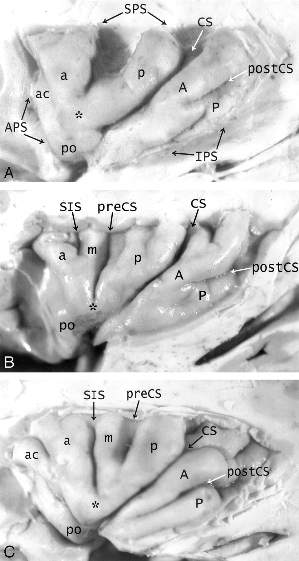
- Fig 1.
A–C, Anatomy of the convexity surface of the left insular lobe after resection of the overlying opercula, vessels, and pia-arachnoid. Gross anatomic specimens from a 50-year-old woman (A), a 63-year-old man (B), and a 71-year-old man (C). The anterior PS (APS), superior PS (SPS), and inferior PS (IPS) define the base of the insular lobe. The oblique CS of the insula subdivides the insula into larger anterior and smaller posterior lobules.
Anterior lobule: The ASG (a), MSG (m), and PSG (p) form most of the convexity surface of the anterior lobule. The short insular sulcus (SIS) separates ASG from MSG. The precentral sulcus (preCS) separates MSG from PSG. MSG shows variable size and depression below the surface of the insula. The number of gyri on the convexity surface of the anterior lobule varies substantially: two in A, three in B, and four in C. The apex (asterisk) of the insula forms by the convergence of the inferior ends of some or all of the short gyri. In A, the apex is formed only by the ASG and PSG, with no contribution from the hypoplastic MSG (most frequent type). In B and C, all three short gyri contribute to the apex. The pole (po) of the insula lies at the most anteroinferior point on the insula, near to but separate from the apex. The accessory gyrus (ac) forms a variable portion of the upper anterior face of the anterior lobule and, when large, may contribute to the convexity surface as well. In A, a prominent accessory gyrus (ac) projects anterior to the ASG, but does not reach to the convexity surface of the insula. In C, a very large accessory gyrus (ac) projects laterally to form the anteriormost gyrus on the convexity surface of the anterior lobule. The transverse gyrus forms the lower portion of the anterior face of the insula (see Fig 2).
Posterior lobule: The ALG (A) and the PLG (P) form the posterior lobule. In C, the ALG and PLG are well defined and completely separated by a complete postcentral sulcus (postCS). In A and B, the ALG and PLG are incompletely separated by shallow or incomplete postcentral sulci (postCS). In B, the dominant ALG forms the pole of the posterior lobule at the limen, while the PLG appears to branch off the posterior inferior aspect of the ALG. The superior ends of many of the insular gyri are bifid.
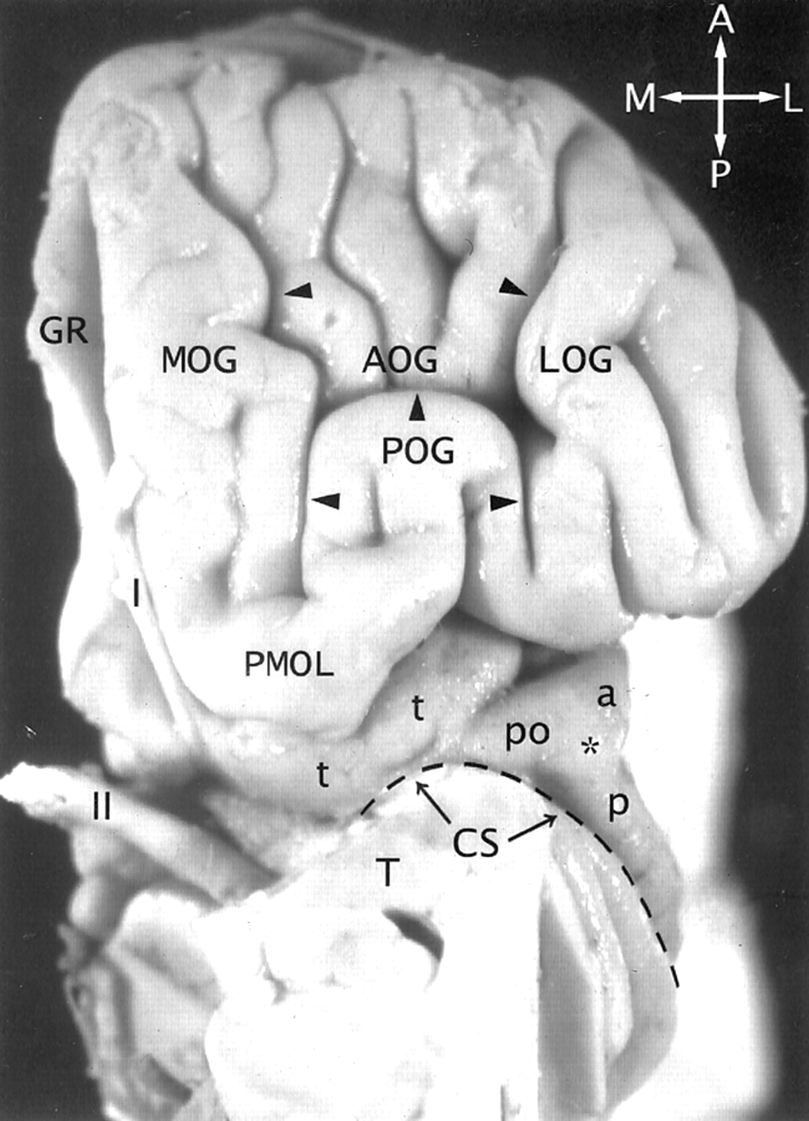
- Fig 2.
The transverse gyrus and the orbitofrontal insula relationship. Base view of the orbital surface of the left frontal lobe after resection of part of the gyrus rectus (GR) and the anterior portion of the temporal lobe (T). Medial is to the reader’s left. I indicates olfactory bulb; II, optic chiasm and tract. The H-shaped orbital sulcus (arrowheads) defines the MOG, POG, anterior orbital gyrus (AOG), and lateral orbital gyrus (LOG). At the posteromedial aspect of the orbitofrontal surface, the posterior portion of the MOG merges with the medial portion of the POG to form the prominent PMOL. PMOL gives rise to the transverse insular gyrus (t) that extends laterally to form the pole (po) of the insula just anteroinferomedial to the apex (asterisk) of the insula. The CS (dashed line) curves inferiorly immediately behind and below the apex and the pole en route to join the stem of the sylvian fissure. The ASG (a) and the PSG (p) converge to form the apex of the insula anterior to the CS. In this image, the deliberate slight rotation used to illustrate the course of the transverse gyrus from the PMOL to the pole also rotates the apex medially, so the apex does not appear to lie as far lateral in position as it would in a true base view.
- Fig 3.
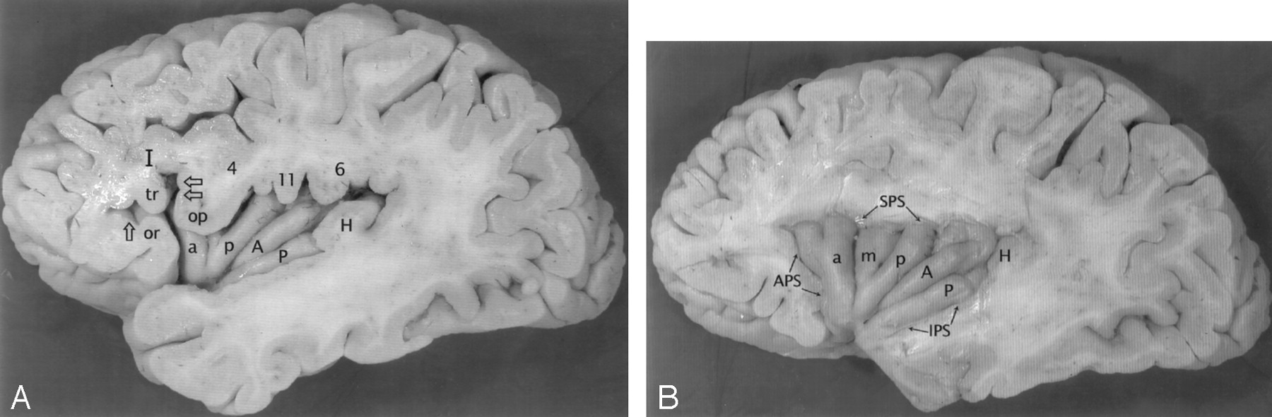
A and B, Anatomic relationship of the opercula to the insula at earlier (A) and later (B) stages of dissection of the left cerebral hemisphere of a gross anatomic specimen from a 71-year-old man. The partes orbitalis (or), triangularis (tr), and opercularis (op) of the inferior frontal gyrus (I) overlie the anterior lobule of the insula. The anterior horizontal ramus (single open arrow) of the sylvian fissure leads to the anterior PS (APS). The anterior ascending ramus (dual open arrows) of the sylvian fissure leads to the superior PS (SPS) (8). The pars opercularis (op) joins with the inferior end of the precentral gyrus (4) to form most of the frontal operculum. In this specimen, they form a “bulky” union that invaginates into the convexity surface of the anterior lobule and depresses the MSG (m) slightly below the surface of the insula. Further posteriorly, the subcentral gyrus (11) and the anterior limb of the supramarginal gyrus (6) form the rest of the frontoparietal operculum that overlies the posterior lobule. The HG (H) overlies the PLG (P), so the anteromedial surface of HG abuts onto the lateral surface of the PLG. Deeper dissection (in B) shows the origin of the HG immediately posterior to the insula, posterosuperolateral to the PLG. In this specimen, the postcentral sulcus defines separate ALG (A) and PLG (P). PLG is dominant and forms the pole of the posterior lobule at the limen.
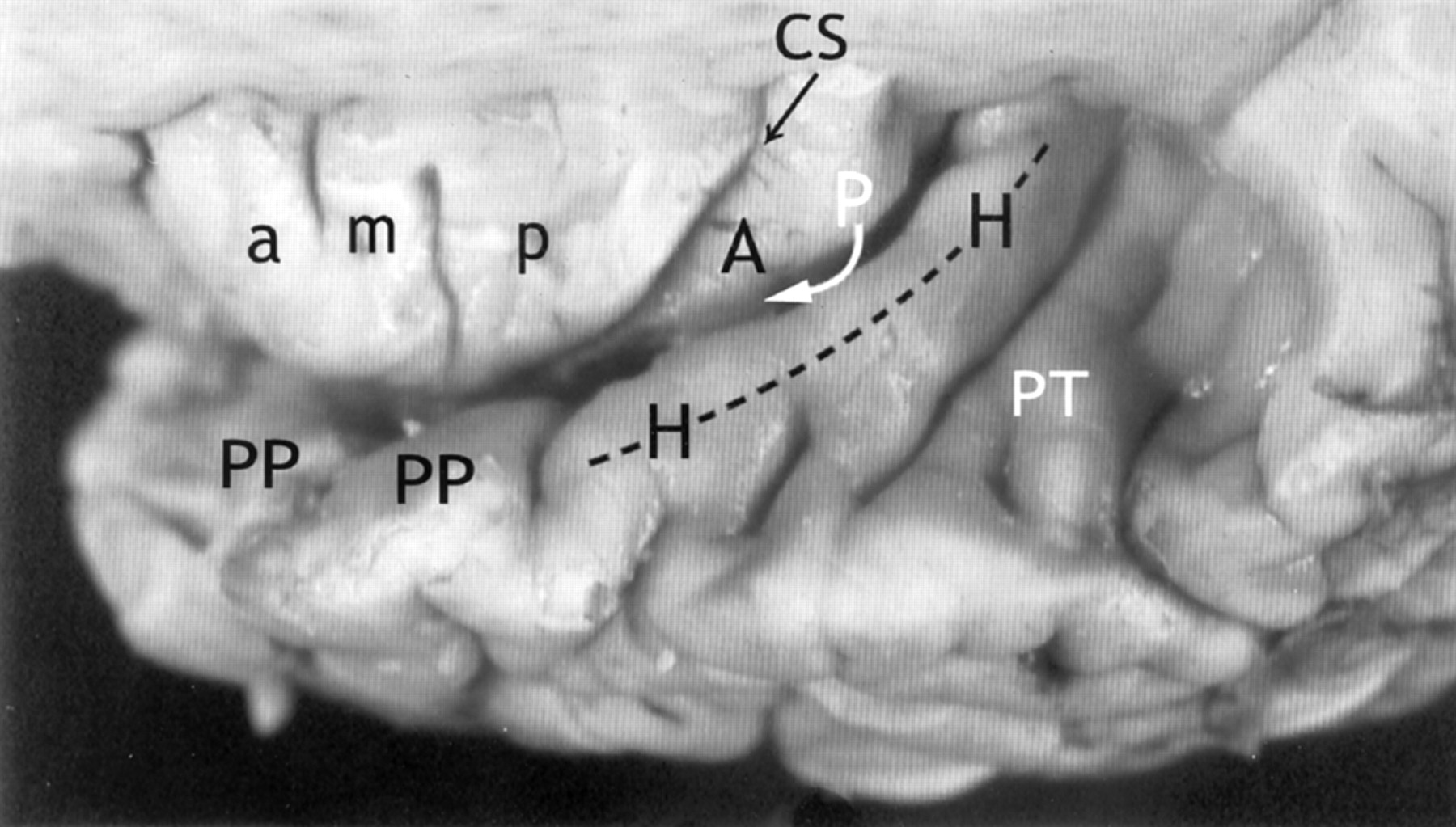
- Fig 4.
Relationship of the insula to the temporal operculum. Gross anatomic specimen from a 44-year-old woman, viewed from above and lateral after removal of the frontal and parietal opercula to expose the superior surfaces of the ASG (a), MSG (m), PSG (p), the CS, and the ALG (A). The HG (H) arises immediately posterior to the insula and curves anterolaterally around the lateral surface of the PLG (arrow), largely obscuring it in this view. The anteromedial surface of the HG abuts upon and conforms to the surface of the PLG as it crosses the superior surface of the temporal lobe. PP indicates planum polare; PT, planum temporale.
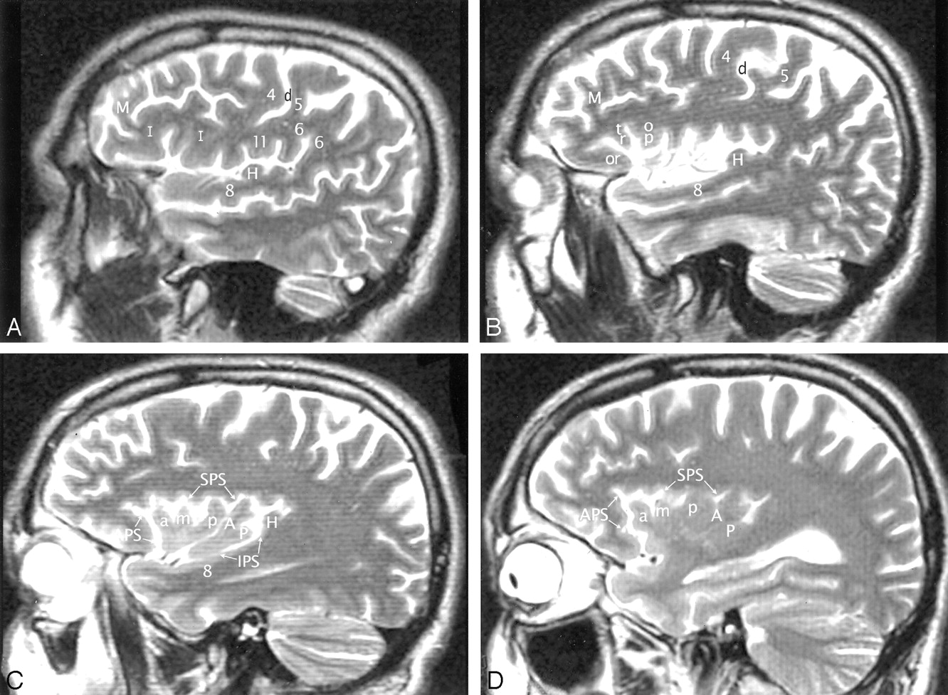
- Fig 5.
A–D, Imaging anatomy of the normal insula in a 51-year-old man. Sequential sagittal T2-weighted MR imaging sections from lateral (A) to medial (D) demonstrate the anterior (APS), superior (SPS), and inferior (IPS) PS, the ASG (a), MSG (m), and PSG (p) of the larger anterior lobule, and the ALG (A) and the branched configuration of the PLG (P) of the smaller posterior lobule. The CS (d) courses across the convexity between the precentral gyrus (4) and the postcentral gyrus (5) and then curves across the insula between the corresonding gyri of the insula (ie, between p and A). The overlying opercula interdigitate with the insular gyri with no significant distortion of insular anatomy. The HG (H) lies immediately behind the PLG and marks the posteroinferior border of the insula. M indicates middle frontal gyrus; I, inferior frontal gyrus; 6, supramarginal gyrus; 8, superior temporal gyrus; and 11, subcentral gyrus. Other abbreviations as in Figs 1–4.
- Fig 6.
A and B, Imaging anatomy of the posterior lobule on sagittal T2-weighted MR images in a 13-year-old girl (A) and a 26-year-old man (B). In A, the ALG (A) and PLG (P) are well formed and distinct, correlating with Fig 1C. In B, ALG (A) and PLG (P) are poorly separated. PLG appears to bud off the posterior aspect of the ALG, giving the posterior lobule a branched configuration, such as seen in Fig 1B. Note the convergence of the short insular gyri to form the apex (asterisk in B) as seen in Fig 1B, the course of the CS posteroinferior to the apex en route to the stem of the sylvian fissure, the relationship of the pole of the posterior lobule to the limen, and the relationship of the HG (H) to both the PLG and the entire posterior lobule.
- Fig 7.
A and B, Sagittal T2-weighted MR images of the left hemisphere in a 34-year-old woman show the opercular-insular relationship. The union of the pars opercularis (op) of the inferior frontal gyrus with the inferior end of the precentral gyrus (4) forms a very bulky “pseudomass” (Ø) that invaginates into the convexity surface of the anterior lobule, depresses the insular surface at the MSG, and splays the ASG (a) apart from the PSG (p). (See also Figs 1 and 3.)
- Fig 8.
Talairach-Tournoux relationship of the insula. Sagittal T2-weighted MR image in a 26-year-old woman. The two vertical white lines indicate the insular intersections of the coronal planes erected perpendicular to the AC-PC baseline at AC (VAC) and at PC (VPC). VAC nearly always intersects the anterior lobule, most frequently in relation to the upper portion of the precentral sulcus between the MSG (m) and the PSG (p). VPC typically defines the posterior margin of the insula.
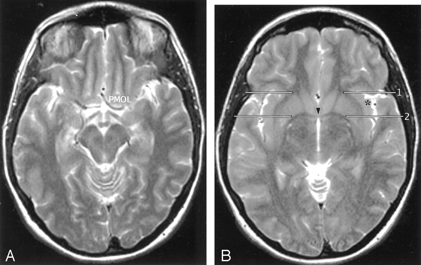
- Fig 9.
A and B, Axial T2-weighted MR images in the AC-PC plane. The MOG and POG converge to form the PMOL at the anterolateral aspect of the suprasellar cistern. The white matter of the orbitofrontal lobe continues directly around the sylvian fissure into the extreme capsule deep to the insular cortex. In axial sections obtained along the AC-PC plane, the coronal plane (1) through the junction of the anterior limb of the internal capsule with the external capsule on each side provides one landmark for the anterior border of the insula on each side. The coronal plane (2) through the midline portion (arrowhead) of the AC (VAC) intersects the anterior insula. The apex (asterisk) of the insula lies between these two planes. The coronal plane through the PC (not shown) defines the posterior margin of the insula.


Tables
Abbreviation Structure or Line PS Periinsular sulcus CS Central sulcus ASG Anterior short insular gyrus MSG Middle short insular gyrus PSG Posterior short insular gyrus ALG Anterior long insular gyrus PLG Posterior long insular gyrus HG Heschl gyrus MOG Medial orbital gyrus POG Posterior orbital gyrus PMOL Posteromedial orbital lobule AC-PC line Anterior commissure-posterior commissure baseline (ie, the Talairach-Tournoux baseline) VAC Vertical erected perpendicular to the AC-PC baseline at the AC VPC Vertical erected perpendicular to the AC-PC baseline at the PC Insular Gyri and Sulci Well Seen-Complete Well Seen-Incomplete/Partial/Hypoplastic Poorly Seen Not Seen CS 94 (15) 6 (1) - - Anterior surface of insula Transverse gyrus 100 (16) - - - Accessory gyrus 100 (16) - - - Convexity surface; anterior lobule of insula ASG 94 (15) 6 (1) - - MSG 56 (9) 31 (5) - 12 (2) PSG 94 (15) 6 (1) - - SIS 75 (12) 12 (2) 6 (1) 6 (1) PIS 75 (12) 19 (3) - 6 (1) Convexity Surface: posterior lobule of insula ALG 81 (13) 19 (3) - - PLG 56 (9) 38 (6) 6 (1) - PostCS 56 (9) 38 (6) 6 (1) - Overhanging opercula HG 100 (16) - - - Subcentral gyrus 100 (16) - - - Supramarginal gyrus 100 (16) - - - Note.—Data are percentages (number of specimen). SIS indicates short insular sulcus; PIS, precentral insular sulcus.
Insular Gyri and Sulci Left Hemisphere (n = 150) Right Hemisphere (n = 150) well Seen Poorly Seen Not Seen Bifid Well Seen Poorly Seen Not Seen Bifid ASG 99.3 (149) 0.7 (1) 0 (0) 7.3 (11) 100 (150) 0 (0) 0 (0) 13.3 (20) MSG 82 (123) 11.3 (17) 6.7 (10) 0 (0) 74 (111) 13.3 (20) 12.7 (19) 1.3 (2) PSG 97.3 (146) 2 (3) 0.7 (1) 2 (3) 99.3 (149) 0.7 (1) 0 (0) 8 (12) ALG 98.7 (148) 1.3 (2) 0 (0) 10 (15) 99.3 (149) 0.7 (1) 0 (0) 21.3 (32) PLG 59.3 (89) 20.7 (31) 20 (30) 0 (0) 57.3 (86) 18.7 (28) 24 (36) 0 (0) CS 78.7 (118)* 18 (27) 3.3 (5) NA 86.7 (130)* 10 (15) 3.3 (5) NA HG 100 (150) 0 (0) 0 (0) NA 100 (150) 0 (0) 0 (0) NA Note.—Data are percentages (number of hemispheres). NA indicates not applicable.
* Data presented are the sum of well seen-complete and well seen-partial.
- TABLE 4:
Intersection of the coronal plane VAC and VPC with the insula on sagittal MR images
Line and Intersection Left Hemisphere (n = 150) Right Hemisphere (n = 150) VAC line Anterior lobule 99.3 (149) 99.3 (149) Anterior to ASG 0 (0) 0 (0) ASG 0 (0) 0 (0) ASG/MSG 1.3 (2) 0.7 (1) MSG 23.3 (35) 24 (36) MSG/PSG 58 (87) 50.7 (76) PSG 16.7 (25) 24 (36) CS 0.7 (1) 0.7 (1) VPC line Defines posterior insula 93.3 (140) 95.3 (143) Anterior to posterior insula 5.3 (8) 4 (6) Posterior to posterior insula 1.3 (2) 0.7 (1) Note.—Data are percentages (number of hemispheres). ASG/MSG indicates short insular sulcus; MSG/PSG, precentral insular sulcus.
Feature Left Hemisphere (n = 150) Right Hemisphere (n = 150) Yes No Yes No MOG 100 (150) 0 (0) 100 (150) 0 (0) POG 100 (150) 0 (0) 100 (150) 0 (0) Posteromedial orbital lobule 98.7 (148) 1.3 (2) 99.3 (149) 0.7 (1) Continuity of white matter from the orbitofrontal to the insular lobes 98.7 (148) 1.3 (2) 98.7 (148) 1.3 (2) Apex shown as a laterally directed prominence 92 (138) 8 (12) 90.7 (136) 9.3 (14) Apex bracketed between the anterior plane through the junctions of the anterior limbs of the internal capsules with the anteriormost external capsules and the posterior plane at VAC 95.7 (132/138) 4.3 (6/138) 98.5 (134/136) 1.5 (2/136) Anterior plane defines the anterior border of the insula 86 (129) 14 (21) 88.7 (133) 11.3 (17) Note.—Data are percentages (number of hemispheres).
In this issue


Jump to section
Related Articles
Cited By...
- Overlapping Representation of Basic Tastes in the Human Gustatory Cortex
- Minimal Phrase Composition Revealed by Intracranial Recordings
- Sex differences in insular gyri responses to the cold pressor challenge
- Spontaneous Neuronal Oscillations in the Human Insula are Hierarchically Organized Traveling Waves
- Minimal phrase composition revealed by intracranial recordings
- Connection "Stripes" in the Primate Insula
- Optimizing the Detection of Subtle Insular Lesions on MRI When Insular Epilepsy Is Suspected
- Anterior insular cortex plays a critical role in interoceptive attention
- Percentage Insula Ribbon Infarction of >50% Identifies Patients Likely to Have Poor Clinical Outcome Despite Small DWI Infarct Volume
- Sex and Disease-Related Alterations of Anterior Insula Functional Connectivity in Chronic Abdominal Pain
- Admission Insular Infarction >25% Is the Strongest Predictor of Large Mismatch Loss in Proximal Middle Cerebral Artery Stroke
- Structural Abnormalities in Patients with Insular/Peri-insular Epilepsy: Spectrum, Frequency, and Pharmacoresistance
- The neuropsychological impact of insular cortex lesions
- Somatotopic Organization of Gentle Touch Processing in the Posterior Insular Cortex