
Article Figures & Data
Figures
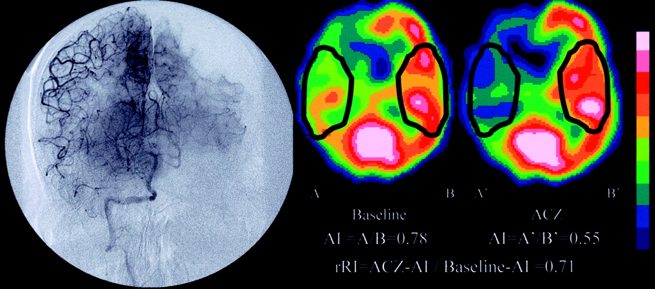
- Fig 1.
Angiogram and ACZ-challenged SPECT scans of patient 8, who had right internal carotid artery occlusion.
Left, Baseline and ACZ-challenged SPECT scans. Asymmetry index in right MCA territory of baseline scan is 0.78 (A/B) and of ACZ-challenged scan is 0.55 (A′/B′); therefore, regional reactivity index is 0.71 (0.55/0.78).
Right, Angiogram of right vertebral artery. Right MCA territory is perfused via the leptomeningeal artery from the right posterior cerebral artery (PCA). When the left PCA territory is in capillary phase, the right MCA territory is still in arteriole phase. Although the rACCT of left MCA territory is 3.9 s, that of right MCA territory is 8.8 s; therefore, the rACCT ratio is 2.3 (8.8/3.9).
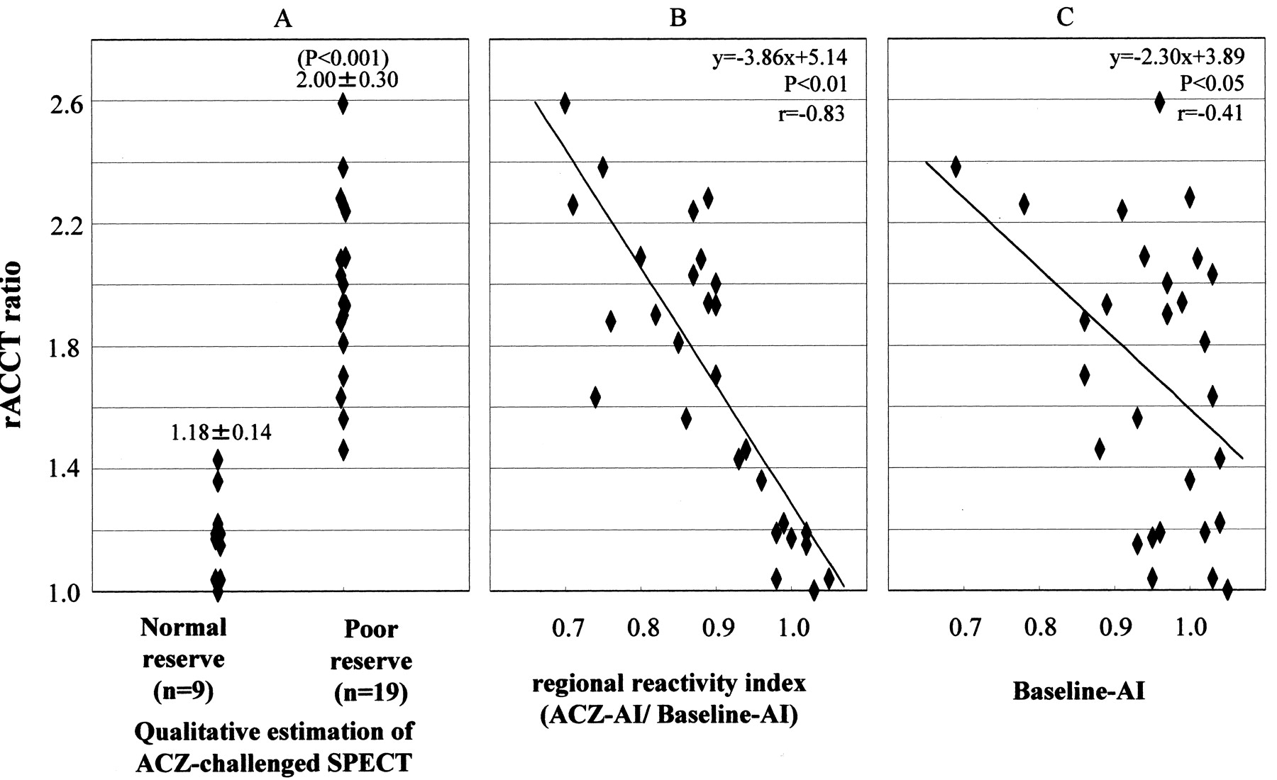
- Fig 2.
rACCT ratio of poor reserve group is larger than that of normal reserve group.
A, rACCT ratio and qualitative estimation on ACZ-challenged SPECT scan.
B, Correlation of rACCT ratio with regional reactivity index, calculated from ACZ asymmetry index/baseline asymmetry index.
C, Correlation of rACCT with asymmetry index of baseline study.


Tables
- TABLE 1:
Patient characteristics, angiographic findings, and results of acetazolamide-challenged single photon emission CT
Patient Characteristics Angiographic Findings ACZ-Challenged SPECT Occlusive Maximum rACCT (s) rACCT (s) rACCT Ratio Patient No. Sex Age (yr) Diagnosis Artery Grade (%) Occlusive Side [Flow Pattern] Normal Side Qualitative Estimation Baseline AI ACZ AI rRI 1 M 61 TIA R MCA 80 5.9 [lept(A)] 2.9 2.03 Poor 1.03 0.90 0.87 2 F 65 TIA R MCA 99 5.4 [lept(P)] 2.6 2.08 Poor 1.01 0.89 0.88 3 M 55 ATI R ICA Occlusion 3.4 [lept(P)] 2.5 1.36 Normal 1.00 0.96 0.96 4 M 68 ATI R ICA Occlusion 2.6 [Willis] 2.6 1.00 Normal 1.05 1.08 1.03 5 M 74 ATI R ICA 99 7.9 [physiol] 4.2 1.88 Poor 0.86 0.65 0.76 6 M 75 TIA R MCA Occlusion 4.9 [lept(A)] 4.7 1.04 Normal 1.03 1.01 0.98 7 M 72 TIA R ICA Occlusion 3.5 [lept(P)] 3.0 1.17 Normal 0.95 0.95 1.00 8 M 63 ATI R ICA Occlusion 8.8 [lept(P)] 3.9 2.26 Poor 0.78 0.55 0.71 9 M 70 TIA R ICA 70 3.7 [physiol] 3.1 1.19 Normal 0.96 0.98 1.02 10 M 73 Lacuna L MCA 70 3.9 [physiol] 3.4 1.15 Normal 0.93 0.95 1.02 11 M 71 ATI R ICA 99 5.7 [lept(P)] 2.4 2.38 Poor 0.69 0.52 0.75 12 F 70 ATI L ICA 70 6.8 [physiol] 3.4 2.00 Poor 0.97 0.87 0.90 13 M 71 ATI L MCA Occlusion 7.3 [lept(P)] 3.2 2.28 Poor 1.00 0.89 0.89 14 M 62 ATI R MCA 70 2.5 [physiol] 2.4 1.04 Normal 0.95 1.00 1.05 15 M 65 Lacuna L ICA 75 5.1 [physiol] 3.5 1.46 Poor 0.88 0.83 0.94 16 M 64 TIA R ICA 80 3.2 [Willis] 2.7 1.19 Normal 1.02 1.00 0.98 17 M 47 TIA L MCA Occlusion 4.7 [lept(A)] 2.1 2.24 Poor 0.91 0.79 0.87 18 M 60 TIA R ICA 99 6.3 [physiol] 3.7 1.70 Poor 0.86 0.77 0.9 19 M 53 TIA R ICA Occlusion 5.8 [lept(A)] 3.0 1.93 Poor 0.89 0.80 0.90 20 M 83 TIA L ICA 80 5.9 [lept(A)] 3.1 1.90 Poor 0.97 0.80 0.82 21 F 71 TIA R ICA 100 5.7 [lept(P)] 3.5 1.63 Poor 1.03 0.76 0.74 22 M 52 TIA R MCA 100 7.0 [lept(P)] 2.7 2.59 Poor 0.96 0.67 0.70 23 M 79 TIA R ICA 70 5.3 [lept(P)] 3.7 1.43 Normal 1.04 0.97 0.93 24 M 69 TIA R ICA 90 6.8 [lept(P)] 3.5 1.94 Poor 0.99 0.88 0.89 25 F 67 TIA R ICA 100 4.7 [lept(P)] 2.6 1.81 Poor 1.02 0.87 0.85 26 M 77 ATI R ICA 100 6.7 [EC-IC] 3.2 2.09 Poor 0.94 0.75 0.80 27 M 69 TIA L ICA 100 6.7 [Willis] 4.3 1.56 Poor 0.93 0.80 0.86 28 M 68 Lacuna R ICA 70 3.3 [physiol] 2.7 1.22 Normal 1.04 1.03 0.99 Note.—ACZ indicates acetazolamide; SPECT, single photon emission CT; rACCT, regional arteriocapillary circulation time; AI, asymmetry index; rRI, regional reactivity index; M, male; F, female; TIA, transient ischemic attack; ATI, atherothrombotic infarction; Lacuna, lacuna infarction; R, right; L, left; MCA, middle cerebral artery; ICA, internal carotid artery; lept(A), leptomeningeal collateral via anterior cerebral artery; lept(P), leptomeningeal collateral via posterior cerebral artery; Willis, circle of Willis collateral; physiol, physiological flow pattern; EC-IC, external to internal carotid collateral.
Flow Pattern n rACCT (s) mean ± SD (min-max) Non-occlusive artery 28 3.2 ± 0.6 (2.1–4.7) Physiological (70–89% stenosis) 9 4.3 ± 1.4 (2.5–6.8) Physiological (90–99% stenosis) 5 5.3 ± 2.0 (2.9–7.9) Circle of Willis collateral 8 3.3 ± 0.7 (2.5–6.7) EC-IC collateral 2 4.9 ± 2.5 (3.1–6.7) Leptomeningeal collateral 26 5.4 ± 1.3 (3.4–8.8) Note.—rACCT indicates regional arteriocapillary circulation time; min, minimum; max, maximum; EC-IC, external to internal carotid. The number of each flow pattern perfusing bilateral middle cerebral artery territories is presented. Some cases have two or three flow patterns filling the middle cerebral artery territory on the occlusive side.
In this issue


Jump to section
Related Articles
Cited By...
- Predicting clinical outcome in posterior circulation large-vessel occlusion patients with endovascular recanalisation: the GNC score
- Prediction of hyperperfusion phenomenon after carotid artery stenting and carotid angioplasty using quantitative DSA with cerebral circulation time imaging
- Prolonged cerebral circulation time is more associated with symptomatic carotid stenosis than stenosis degree or collateral circulation
- Monitoring Peri-Therapeutic Cerebral Circulation Time: A Feasibility Study Using Color-Coded Quantitative DSA in Patients with Steno-Occlusive Arterial Disease
- Quantification of Cerebrovascular Reactivity by Blood Oxygen Level-Dependent MR Imaging and Correlation with Conventional Angiography in Patients with Moyamoya Disease
- Unilateral Hemispheric Proliferation of Ivy Sign on Fluid-Attenuated Inversion Recovery Images in Moyamoya Disease Correlates Highly with Ipsilateral Hemispheric Decrease of Cerebrovascular Reserve
- Precision of Cerebrovascular Reactivity Assessment with Use of Different Quantification Methods for Hypercapnia Functional MR Imaging