Advertisement

Main menu

  • Home
  • Content
    • Current Issue
    • Accepted Manuscripts
    • Article Preview
    • Past Issue Archive
    • AJNR Case Collection
    • Case of the Week Archive
    • Classic Case Archive
    • Case of the Month Archive
  • Special Collections
    • Spinal CSF Leak Articles (Jan 2020-June 2024)
    • 2024 AJNR Journal Awards
    • Most Impactful AJNR Articles
  • Multimedia
    • AJNR Podcast
    • AJNR Scantastics
    • Video Articles
  • For Authors
    • Submit a Manuscript
    • Author Policies
    • Fast publishing of Accepted Manuscripts
    • Graphical Abstract Preparation
    • Manuscript Submission Guidelines
    • Imaging Protocol Submission
    • Submit a Case for the Case Collection
  • About Us
    • About AJNR
    • Editorial Board
  • More
    • Become a Reviewer/Academy of Reviewers
    • Subscribers
    • Permissions
    • Alerts
    • Feedback
    • Advertisers
    • ASNR Home
  • Other Publications
    • ajnr

User menu

  • Alerts
  • Log in

Search

  • Advanced search
American Journal of Neuroradiology

American Journal of Neuroradiology

ASHNR American Society of Functional Neuroradiology ASHNR American Society of Pediatric Neuroradiology ASSR
  • Alerts
  • Log in

Advanced Search

  • Home
  • Content
    • Current Issue
    • Accepted Manuscripts
    • Article Preview
    • Past Issue Archive
    • AJNR Case Collection
    • Case of the Week Archive
    • Classic Case Archive
    • Case of the Month Archive
  • Special Collections
    • Spinal CSF Leak Articles (Jan 2020-June 2024)
    • 2024 AJNR Journal Awards
    • Most Impactful AJNR Articles
  • Multimedia
    • AJNR Podcast
    • AJNR Scantastics
    • Video Articles
  • For Authors
    • Submit a Manuscript
    • Author Policies
    • Fast publishing of Accepted Manuscripts
    • Graphical Abstract Preparation
    • Manuscript Submission Guidelines
    • Imaging Protocol Submission
    • Submit a Case for the Case Collection
  • About Us
    • About AJNR
    • Editorial Board
  • More
    • Become a Reviewer/Academy of Reviewers
    • Subscribers
    • Permissions
    • Alerts
    • Feedback
    • Advertisers
    • ASNR Home
  • Follow AJNR on Twitter
  • Visit AJNR on Facebook
  • Follow AJNR on Instagram
  • Join AJNR on LinkedIn
  • RSS Feeds

Welcome to the new AJNR, Updated Hall of Fame, and more. Read the full announcements.


AJNR is seeking candidates for the position of Associate Section Editor, AJNR Case Collection. Read the full announcement.


OtherBRAIN

MR Imaging in Toxoplasmosis Encephalitis after Bone Marrow Transplantation: Paucity of Enhancement despite Fulminant Disease

Catalina Ionita, Mohammed Wasay, Lucia Balos and Rohit Bakshi
American Journal of Neuroradiology February 2004, 25 (2) 270-273;
Catalina Ionita
  • Find this author on Google Scholar
  • Find this author on PubMed
  • Search for this author on this site
Mohammed Wasay
  • Find this author on Google Scholar
  • Find this author on PubMed
  • Search for this author on this site
Lucia Balos
  • Find this author on Google Scholar
  • Find this author on PubMed
  • Search for this author on this site
Rohit Bakshi
  • Find this author on Google Scholar
  • Find this author on PubMed
  • Search for this author on this site
  • Article
  • Figures & Data
  • Info & Metrics
  • Responses
  • References
  • PDF
Loading

Article Figures & Data

Figures

  • Fig 1.
    • Download figure
    • Open in new tab
    • Download powerpoint
    Fig 1.

    Three serial MR imaging studies obtained during a period of 18 days. Images were obtained by the fluid-attenuated inversion recovery (FLAIR) sequences, with the exception of columns 4 and 6, which were obtained by the postgadolinium T1-weighted sequence. The initial study (top row) revealed a few nonspecific white matter lesions that were thought to be related to cyclosporine toxicity.

    The lesions were not seen on noncontrast T1-weighted images or diffusion-weighted images (not shown). After gadolinium administration, lesions were nonenhancing, except for the left parietal-occipital lesion, which shows faint, questionable enhancement. Because of neurologic deterioration, brain MR imaging was repeated (middle row) 6 days after the initial study, revealing worsening lesions in the subcortical white matter, cerebellum, bilateral thalamus, and basal ganglia, with no mass effect or abnormal parenchymal enhancement; there was possibly some increased leptomeningeal enhancement. During the following week, the patient became progressively obtunded and comatose. A third MR imaging study obtained 18 days after the initial study (bottom row) showed continuous progression of lesions in size and distribution, which at this time involved much of the brain. There was a remarkable lack of mass effect and lack of parenchymal enhancement of most lesions; irregular meningeal enhancement with secondary involvement (perhaps by meningeal spread) of the cortex and subcortical areas was suggested. A few lesions were hyperintense on diffusion-weighted images. Apparent diffusion coefficient maps were not obtained to distinguish restricted diffusion versus T2 shine-through (not shown). MR imaging was performed at 1.5 T with the following parameters on each day. Fast spin-echo FLAIR: 5-mm axial sequences with 1-mm section gaps, TR/TE/TI of 10,002/145/2200, matrix of 192 × 256, one signal averaged, and 22-cm field of view. T1-weighted images: 5-mm axial sequences with 1-mm section gaps, TR/TE of 500/20, matrix of 192 × 256, one signal averaged, 22-cm field of view.
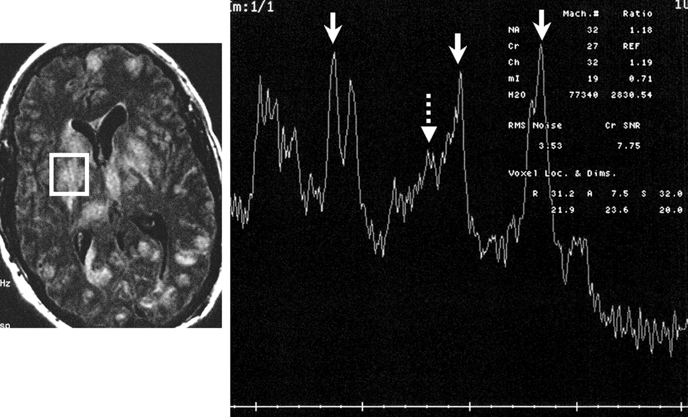
  • Fig 2.
    • Download figure
    • Open in new tab
    • Download powerpoint
    Fig 2.

    Single- voxel MR spectroscopy (right panel) by using a stimulated-echo acquisition mode with a TR of 1500 ms and TE of 30 ms. The spectral pattern is technically limited and suboptimal (note the poor baseline).

    A 2.5 × 2.5 cm voxel of the right putamen lesion was localized based on the FLAIR axial image on day 18 (left panel) at the level of the frontal horns of the lateral ventricle. The spectral pattern shows, from left to right, a moderate increase in choline (solid arrow), a moderate reduction in n-acetyl-aspartate (solid arrow), and a marked lactate peak (solid arrow). There is also a mild prominence of (amino acid) peak (dashed arrow).

  • Fig 3.
    • Download figure
    • Open in new tab
    • Download powerpoint
    Fig 3.

    Autopsy findings. Photomicrograph (magnification ×600, hematoxylin and eosin stain) of a histologic section of the cerebrum reveals multiple toxoplasma cysts (open arrow), probable neuronal cells with round, dark nuclei (arrowhead), and additional trophozoites in the extracellular space after the cysts rupture (straight arrow). The findings are consistent with those of toxoplasmosis encephalitis.

PreviousNext
Back to top

In this issue

American Journal of Neuroradiology: 25 (2)
American Journal of Neuroradiology
Vol. 25, Issue 2
1 Feb 2004
  • Table of Contents
  • Index by author
Advertisement
Print
Download PDF
Email Article

Thank you for your interest in spreading the word on American Journal of Neuroradiology.

NOTE: We only request your email address so that the person you are recommending the page to knows that you wanted them to see it, and that it is not junk mail. We do not capture any email address.

Enter multiple addresses on separate lines or separate them with commas.
MR Imaging in Toxoplasmosis Encephalitis after Bone Marrow Transplantation: Paucity of Enhancement despite Fulminant Disease
(Your Name) has sent you a message from American Journal of Neuroradiology
(Your Name) thought you would like to see the American Journal of Neuroradiology web site.
CAPTCHA
This question is for testing whether or not you are a human visitor and to prevent automated spam submissions.
Cite this article
Catalina Ionita, Mohammed Wasay, Lucia Balos, Rohit Bakshi
MR Imaging in Toxoplasmosis Encephalitis after Bone Marrow Transplantation: Paucity of Enhancement despite Fulminant Disease
American Journal of Neuroradiology Feb 2004, 25 (2) 270-273;

Citation Manager Formats

  • BibTeX
  • Bookends
  • EasyBib
  • EndNote (tagged)
  • EndNote 8 (xml)
  • Medlars
  • Mendeley
  • Papers
  • RefWorks Tagged
  • Ref Manager
  • RIS
  • Zotero
0 Responses
Respond to this article
Share
Bookmark this article
MR Imaging in Toxoplasmosis Encephalitis after Bone Marrow Transplantation: Paucity of Enhancement despite Fulminant Disease
Catalina Ionita, Mohammed Wasay, Lucia Balos, Rohit Bakshi
American Journal of Neuroradiology Feb 2004, 25 (2) 270-273;
del.icio.us logo Twitter logo Facebook logo Mendeley logo
  • Tweet Widget
  • Facebook Like
  • Google Plus One

Jump to section

  • Article
    • Abstract
    • Case Report
    • Discussion
    • Conclusion
    • References
  • Figures & Data
  • Info & Metrics
  • Responses
  • References
  • PDF

Related Articles

  • No related articles found.
  • PubMed
  • Google Scholar

Cited By...

  • Leptomeningeal Enhancement in Multiple Sclerosis and Other Neurological Diseases: A Systematic Review and Meta-Analysis
  • Spectrum and prognosis of neurologic complications after hematopoietic transplantation
  • Crossref
  • Google Scholar

This article has not yet been cited by articles in journals that are participating in Crossref Cited-by Linking.

More in this TOC Section

  • Progression of Microstructural Damage in Spinocerebellar Ataxia Type 2: A Longitudinal DTI Study
  • Usefulness of Quantitative Susceptibility Mapping for the Diagnosis of Parkinson Disease
  • Evaluating the Effects of White Matter Multiple Sclerosis Lesions on the Volume Estimation of 6 Brain Tissue Segmentation Methods
Show more Brain

Similar Articles

Advertisement

Indexed Content

  • Current Issue
  • Accepted Manuscripts
  • Article Preview
  • Past Issues
  • Editorials
  • Editors Choice
  • Fellow Journal Club
  • Letters to the Editor

Cases

  • Case Collection
  • Archive - Case of the Week
  • Archive - Case of the Month
  • Archive - Classic Case

Special Collections

  • Special Collections

Resources

  • News and Updates
  • Turn around Times
  • Submit a Manuscript
  • Author Policies
  • Manuscript Submission Guidelines
  • Evidence-Based Medicine Level Guide
  • Publishing Checklists
  • Graphical Abstract Preparation
  • Imaging Protocol Submission
  • Submit a Case
  • Become a Reviewer/Academy of Reviewers
  • Get Peer Review Credit from Publons

Multimedia

  • AJNR Podcast
  • AJNR SCANtastic
  • Video Articles

About Us

  • About AJNR
  • Editorial Board
  • Not an AJNR Subscriber? Join Now
  • Alerts
  • Feedback
  • Advertise with us
  • Librarian Resources
  • Permissions
  • Terms and Conditions

American Society of Neuroradiology

  • Not an ASNR Member? Join Now

© 2025 by the American Society of Neuroradiology All rights, including for text and data mining, AI training, and similar technologies, are reserved.
Print ISSN: 0195-6108 Online ISSN: 1936-959X

Powered by HighWire