
Article Figures & Data
Figures
- Fig 1.
FLAIR images of a healthy volunteer before (upper) and after (lower) inhalation of 100% oxygen with the use of a sealed mask. CSF hyperintensity is clearly visible in the basilar cistern (left) and sulci over the cerebral convexities (right) after oxygen inhalation.
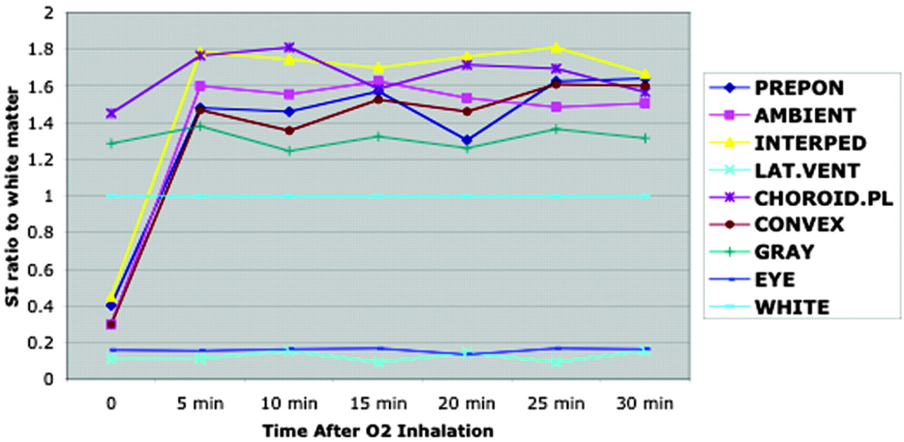
- Fig 2.
Sequential signal intensity ratio change. Signal intensity of various structures normalized to white matter as a function of time after initiation of supplemental oxygen to volunteers shows that signal intensity of prepontine, ambient, and interpeduncular cisterns and sulci over the convexity increases rapidly after oxygen inhalation. No signal intensity changes are seen in the lateral ventricles, brain parenchyma, vitreous (eye), and choroid plexus. PREPON, prepontine cistern; AMBIENT, ambient cistern; INTERPED, interpeduncular cistern; LAT.VENT, lateral ventricle; CHOROID.PL, choroid plexus; CONVEX, sulci over the convexity; GRAY, gray matter; EYE, vitreous of eye; WHITE, white matter.
- Fig 3.
Signal intensity to white matter ratio. Measurements of signal intensity ratios of various anatomic structures in healthy volunteers at 30 min with the use of two types of masks show that CSF hyperintensity is significantly higher with the sealed mask compared with the loose mask (P < .01). PREPON, prepontine cistern; AMBIENT, ambient cistern; INTERPED, interpeduncular cistern; LAT.VENT, lateral ventricle; CHOROID.PL, choroid plexus; CONVEX, sulci over the convexity; GRAY, gray matter; EYE, vitreous of eye; WHITE, white matter.
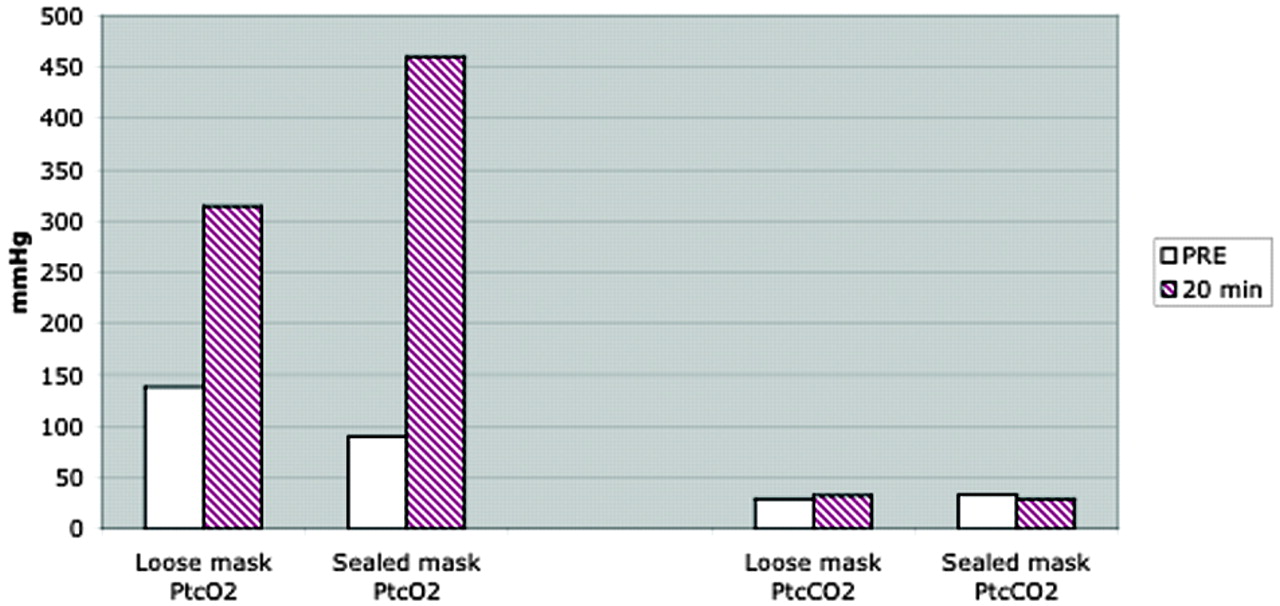
- Fig 4.
Transcutaneous oxymetry. Measurement of transcutaneous partial pressure of oxygen (PtcO2) and carbon dioxide (PtcCO2) in six healthy volunteers shows markedly high transcutaneous measurement of partial pressure of oxygen (ranging from 300–500 mmHg) after inhalation of 100% oxygen at 20 min. Transcutaneous measurement of partial pressure of oxygen was substantially higher when a sealed mask was used, compared with a loose mask. Transcutaneous measurement of partial pressure of carbon dioxide remains low, and no significant difference (P = .96) was seen between the loose mask and the sealed mask.
- Fig 5.
Lower concentrations of albumin did not significantly alter T1 relaxation time.
A, T1 relaxation time measurement of a phantom with various degrees of albumin concentration shows marked reduction of T1 relaxation time in a set of tubes with oxygen exposure. No notable changes of T1 relaxation time are seen in a set of tubes without oxygen until the albumin concentration reaches 1250 mg/dL.
B, Same data were presented as TR (R1 = 1/T1) with various degree of albumin concentration. These two graphs show nearly linear relaxivity with or without oxygen. The curves of T1 relaxation time with and without oxygen are almost parallel, suggestive of no significant synergistic effect of albumin on T1 shortening of oxygen.


In this issue


Jump to section
Related Articles
Cited By...
- Venous imaging-based biomarkers in acute ischaemic stroke
- Reduction of Oxygen-Induced CSF Hyperintensity on FLAIR MR Images in Sedated Children: Usefulness of Magnetization-Prepared FLAIR Imaging
- Elevated Cerebral Blood Volume Contributes to Increased FLAIR Signal in the Cerebral Sulci of Propofol-Sedated Children
- Blood-Brain Barrier Disruption after Cardiac Surgery
- 3D Fluid-Attenuated Inversion Recovery Imaging: Reduced CSF Artifacts and Enhanced Sensitivity and Specificity for Subarachnoid Hemorrhage