
Article Figures & Data
Figures
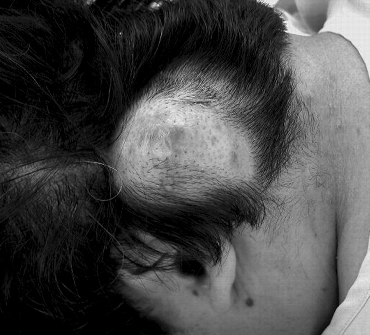
- Fig 1.
Photograph showing a large scalp tumor in the left retroauricular portion. The surface of the skin presents mild discoloration and partial alopecia.
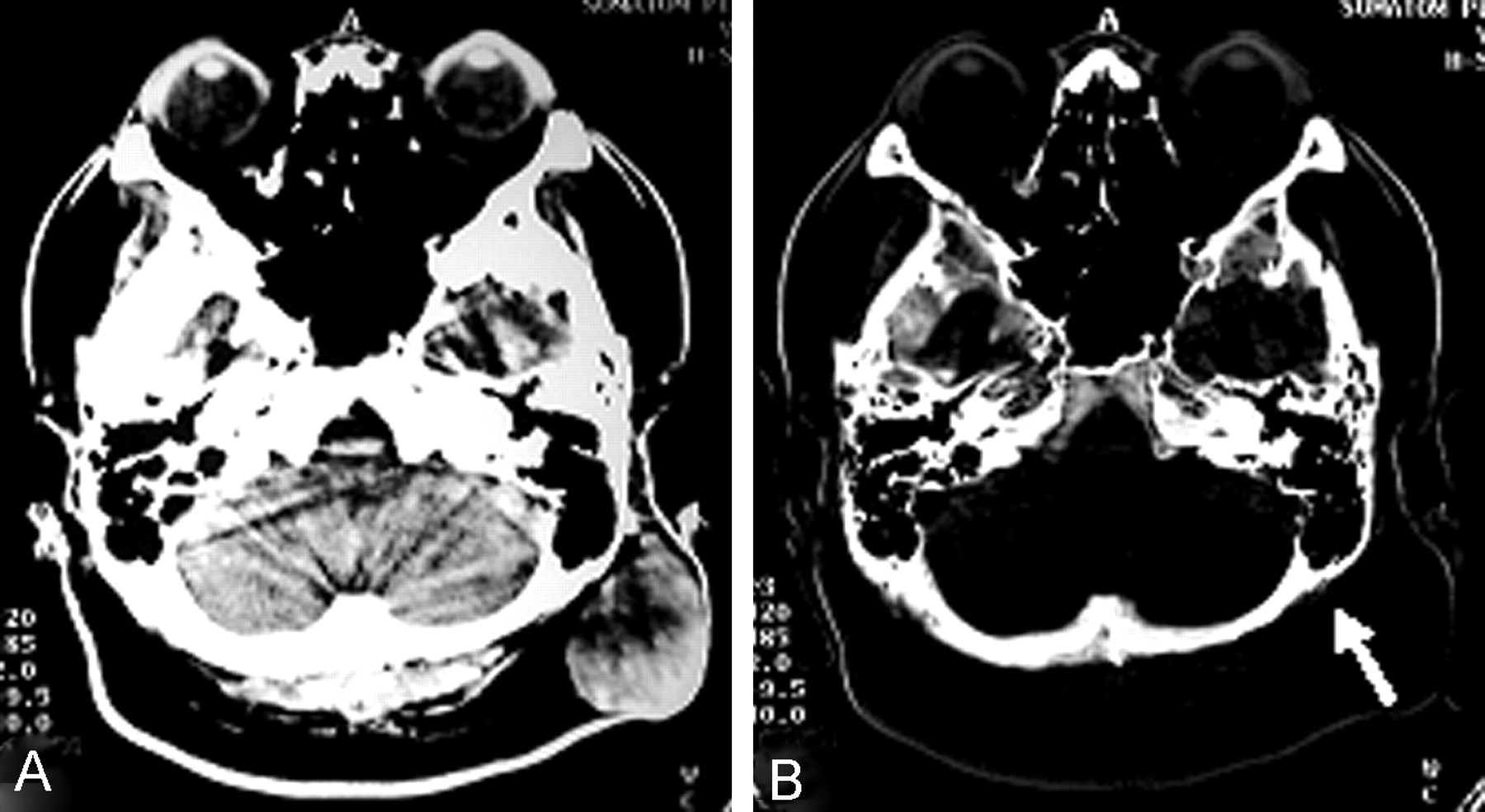
- Fig 2.
CT scan depicting a left subcutaneous mass exhibiting low isoattenuation (A) and bone image CT (B) showing erosion of the occipitomastoid bones (arrow); no evidence of tumor invasion into the skull is evident.
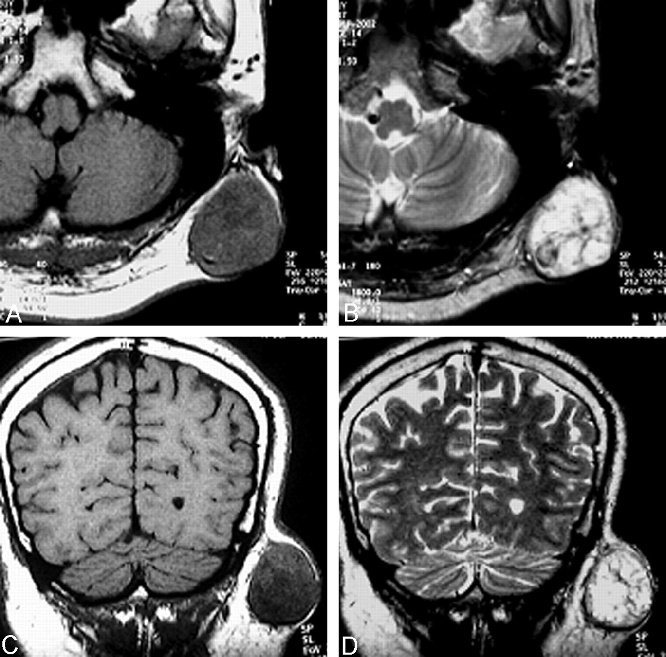
- Fig 3.
Axial (A and B) and coronal (C and D) MR images. The mass (4 × 5 cm in diameter) is seated within the subcutaneous adipose tissue. The tumor is heterogeneously hypoisointense and surrounded by a hypointense thin capsule on T1-weighted images (A and C). The multiple, hyperintense nodules are separated by a hypointense, internodular structure on T2-weighted images (B and D).
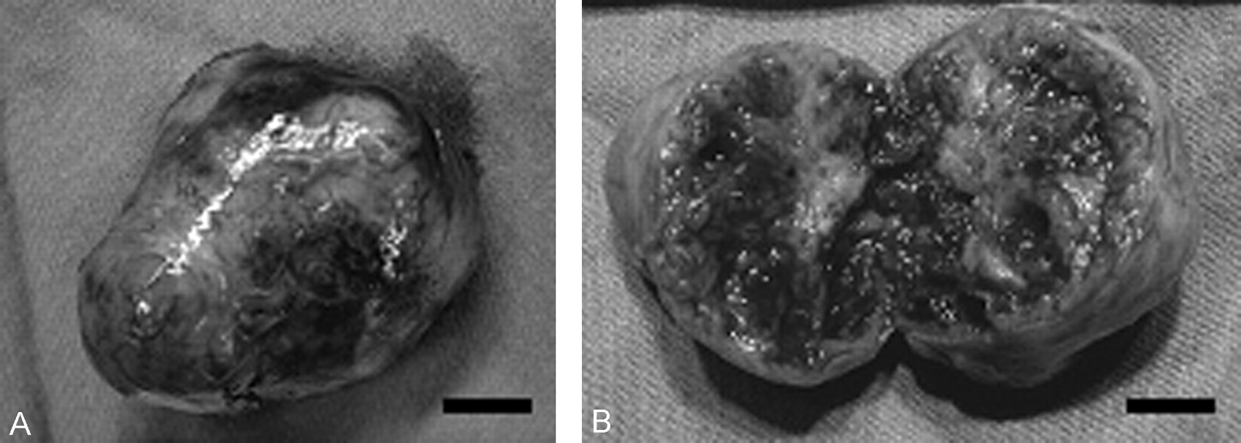
- Fig 4.
Gross specimen of the removed tumor. The tumor is covered by smooth fibrous capsule (A) and composed of well-circumscribed, soft to rubbery-firm nodules (B). The cut surface of the tumor is grayish white to yellow and there are scattered macroscopic hemorrhages (B). Scale bars: 1 cm in both A and B.
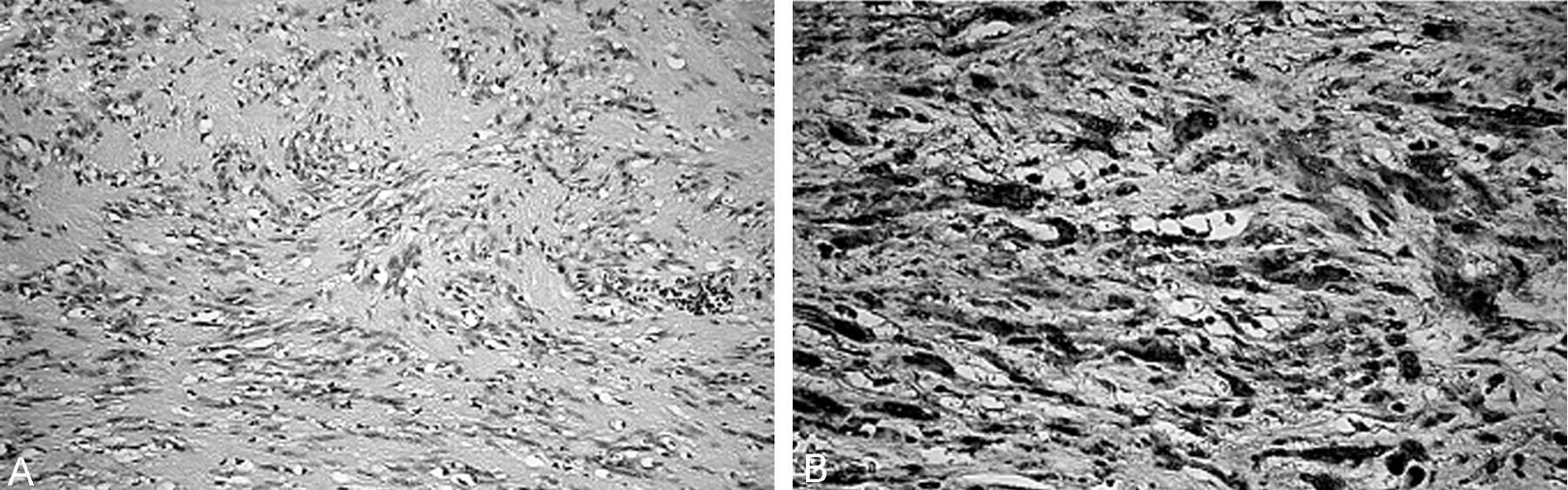
- Fig 5.
Photomicrographs of the tumor. A, The constituent cells are spindle-shaped with irregular, generally elongated nuclei and wavy, ill-defined, eosinophilic cytoplasm. (Hematoxylin-eosin, original magnification ×100) B, The tumor cells exhibit strong immunoreactivity for S-100 protein. (Original magnification ×200)


In this issue


Jump to section
Related Articles
Cited By...
- No citing articles found.