Advertisement

Main menu

  • Home
  • Content
    • Current Issue
    • Accepted Manuscripts
    • Article Preview
    • Past Issue Archive
    • Video Articles
    • AJNR Case Collection
    • Case of the Week Archive
    • Case of the Month Archive
    • Classic Case Archive
  • Special Collections
    • AJNR Awards
    • Low-Field MRI
    • Alzheimer Disease
    • ASNR Foundation Special Collection
    • Photon-Counting CT
    • View All
  • Multimedia
    • AJNR Podcasts
    • AJNR SCANtastic
    • Trainee Corner
    • MRI Safety Corner
    • Imaging Protocols
  • For Authors
    • Submit a Manuscript
    • Submit a Video Article
    • Submit an eLetter to the Editor/Response
    • Manuscript Submission Guidelines
    • Statistical Tips
    • Fast Publishing of Accepted Manuscripts
    • Graphical Abstract Preparation
    • Imaging Protocol Submission
    • Author Policies
  • About Us
    • About AJNR
    • Editorial Board
    • Editorial Board Alumni
  • More
    • Become a Reviewer/Academy of Reviewers
    • Subscribers
    • Permissions
    • Alerts
    • Feedback
    • Advertisers
    • ASNR Home

User menu

  • Alerts
  • Log in

Search

  • Advanced search
American Journal of Neuroradiology

American Journal of Neuroradiology

ASHNR American Society of Functional Neuroradiology ASHNR American Society of Pediatric Neuroradiology ASSR
  • Alerts
  • Log in

Advanced Search

  • Home
  • Content
    • Current Issue
    • Accepted Manuscripts
    • Article Preview
    • Past Issue Archive
    • Video Articles
    • AJNR Case Collection
    • Case of the Week Archive
    • Case of the Month Archive
    • Classic Case Archive
  • Special Collections
    • AJNR Awards
    • Low-Field MRI
    • Alzheimer Disease
    • ASNR Foundation Special Collection
    • Photon-Counting CT
    • View All
  • Multimedia
    • AJNR Podcasts
    • AJNR SCANtastic
    • Trainee Corner
    • MRI Safety Corner
    • Imaging Protocols
  • For Authors
    • Submit a Manuscript
    • Submit a Video Article
    • Submit an eLetter to the Editor/Response
    • Manuscript Submission Guidelines
    • Statistical Tips
    • Fast Publishing of Accepted Manuscripts
    • Graphical Abstract Preparation
    • Imaging Protocol Submission
    • Author Policies
  • About Us
    • About AJNR
    • Editorial Board
    • Editorial Board Alumni
  • More
    • Become a Reviewer/Academy of Reviewers
    • Subscribers
    • Permissions
    • Alerts
    • Feedback
    • Advertisers
    • ASNR Home
  • Follow AJNR on Twitter
  • Visit AJNR on Facebook
  • Follow AJNR on Instagram
  • Join AJNR on LinkedIn
  • RSS Feeds

AJNR Awards, New Junior Editors, and more. Read the latest AJNR updates

OtherBrain

Noninvasive MR Cisternography with Fluid-Attenuated Inversion Recovery and 100% Supplemental O2 in the Evaluation of Neurocysticercosis

Flávio Braga, Antônio J. Rocha, Hélio R. Gomes, Guinel Hernandez Filho, Carlos J. Silva and Ricardo B. Fonseca
American Journal of Neuroradiology February 2004, 25 (2) 295-297;
Flávio Braga
  • Find this author on Google Scholar
  • Find this author on PubMed
  • Search for this author on this site
Antônio J. Rocha
  • Find this author on Google Scholar
  • Find this author on PubMed
  • Search for this author on this site
Hélio R. Gomes
  • Find this author on Google Scholar
  • Find this author on PubMed
  • Search for this author on this site
Guinel Hernandez Filho
  • Find this author on Google Scholar
  • Find this author on PubMed
  • Search for this author on this site
Carlos J. Silva
  • Find this author on Google Scholar
  • Find this author on PubMed
  • Search for this author on this site
Ricardo B. Fonseca
  • Find this author on Google Scholar
  • Find this author on PubMed
  • Search for this author on this site
  • Article
  • Figures & Data
  • Supplemental
  • Info & Metrics
  • Responses
  • References
  • PDF
Loading

Article Figures & Data

Figures

  • Fig 1.
    • Download figure
    • Open in new tab
    • Download powerpoint
    Fig 1.

    A 40-year-old man with parenchymal neurocysticercosis and suspected subarachnoid involvement.

    A, MR FLAIR image (1.0 T; TR, 11,000 ms; TE, 140 ms; TI, 2600 ms) shows no extraaxial lesions.

    B, FLAIR image after 100% O2 for 5 minutes shows two small cysts in the Sylvian fissures confirming racemous neurocysticercosis.
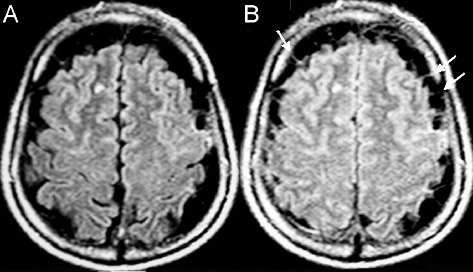
  • Fig 2.
    • Download figure
    • Open in new tab
    • Download powerpoint
    Fig 2.

    A 57-year-old man with multiple vesicular lesions in the brain parenchyma.

    A, FLAIR image shows prominent CSF space along the cerebral convexities.

    B, FLAIR image after 100% O2 for 5 minutes does not show the expected increased signal intensity of the CSF due to the presence of multiple clustered extraaxial cysts. The walls of the cysts (arrows) are better visualized after 100% O2.

  • Fig 3.
    • Download figure
    • Open in new tab
    • Download powerpoint
    Fig 3.

    A 34-year-old male patient with known neurocysticercosis.

    A, B, and C, FLAIR images show multiple vesicular lesions in the parenchyma, mainly along the cortical surfaces. The perimesencephalic cisterns are prominent and contain hyperintense foci that could represent the expected flow artifact or cystic lesions with scoleces (B).

    D, E, and F, FLAIR after 100% O2 shows increased signal intensity in the sulci and basal cisterns, allowing the visualization of a greater number of cortical lesions in the frontal, temporal, and occipital lobes. Also, the increased signal intensity of the CSF confirms that there is no cystic lesion in the perimesencephalic cisterns (E).

PreviousNext
Back to top

In this issue

American Journal of Neuroradiology: 25 (2)
American Journal of Neuroradiology
Vol. 25, Issue 2
1 Feb 2004
  • Table of Contents
  • Index by author
Advertisement
Print
Download PDF
Email Article

Thank you for your interest in spreading the word on American Journal of Neuroradiology.

NOTE: We only request your email address so that the person you are recommending the page to knows that you wanted them to see it, and that it is not junk mail. We do not capture any email address.

Enter multiple addresses on separate lines or separate them with commas.
Noninvasive MR Cisternography with Fluid-Attenuated Inversion Recovery and 100% Supplemental O2 in the Evaluation of Neurocysticercosis
(Your Name) has sent you a message from American Journal of Neuroradiology
(Your Name) thought you would like to see the American Journal of Neuroradiology web site.
CAPTCHA
This question is for testing whether or not you are a human visitor and to prevent automated spam submissions.
Cite this article
Flávio Braga, Antônio J. Rocha, Hélio R. Gomes, Guinel Hernandez Filho, Carlos J. Silva, Ricardo B. Fonseca
Noninvasive MR Cisternography with Fluid-Attenuated Inversion Recovery and 100% Supplemental O2 in the Evaluation of Neurocysticercosis
American Journal of Neuroradiology Feb 2004, 25 (2) 295-297;

Citation Manager Formats

  • BibTeX
  • Bookends
  • EasyBib
  • EndNote (tagged)
  • EndNote 8 (xml)
  • Medlars
  • Mendeley
  • Papers
  • RefWorks Tagged
  • Ref Manager
  • RIS
  • Zotero
0 Responses
Respond to this article
Share
Bookmark this article
Noninvasive MR Cisternography with Fluid-Attenuated Inversion Recovery and 100% Supplemental O2 in the Evaluation of Neurocysticercosis
Flávio Braga, Antônio J. Rocha, Hélio R. Gomes, Guinel Hernandez Filho, Carlos J. Silva, Ricardo B. Fonseca
American Journal of Neuroradiology Feb 2004, 25 (2) 295-297;
del.icio.us logo Twitter logo Facebook logo Mendeley logo
  • Tweet Widget
  • Facebook Like
  • Google Plus One

Jump to section

  • Article
    • Abstract
    • Discussion
    • Conclusion
    • References
  • Figures & Data
  • Supplemental
  • Info & Metrics
  • Responses
  • References
  • PDF

Related Articles

  • PubMed
  • Google Scholar

Cited By...

  • Neurocysticercosis: some of the essentials
  • NEW CONCEPTS IN THE DIAGNOSIS AND MANAGEMENT OF NEUROCYSTICERCOSIS (TAENIA SOLIUM)
  • Crossref
  • Google Scholar

This article has not yet been cited by articles in journals that are participating in Crossref Cited-by Linking.

More in this TOC Section

  • Predictors of Reperfusion in Patients with Acute Ischemic Stroke
  • Enhanced Axonal Metabolism during Early Natalizumab Treatment in Relapsing-Remitting Multiple Sclerosis
  • Progression of Microstructural Damage in Spinocerebellar Ataxia Type 2: A Longitudinal DTI Study
Show more Brain

Similar Articles

Advertisement

Indexed Content

  • Current Issue
  • Accepted Manuscripts
  • Article Preview
  • Past Issues
  • Editorials
  • Editor's Choice
  • Fellows' Journal Club
  • Letters to the Editor
  • Video Articles

Cases

  • Case Collection
  • Archive - Case of the Week
  • Archive - Case of the Month
  • Archive - Classic Case

More from AJNR

  • Trainee Corner
  • Imaging Protocols
  • MRI Safety Corner
  • Book Reviews

Multimedia

  • AJNR Podcasts
  • AJNR Scantastics

Resources

  • Turnaround Time
  • Submit a Manuscript
  • Submit a Video Article
  • Submit an eLetter to the Editor/Response
  • Manuscript Submission Guidelines
  • Statistical Tips
  • Fast Publishing of Accepted Manuscripts
  • Graphical Abstract Preparation
  • Imaging Protocol Submission
  • Evidence-Based Medicine Level Guide
  • Publishing Checklists
  • Author Policies
  • Become a Reviewer/Academy of Reviewers
  • News and Updates

About Us

  • About AJNR
  • Editorial Board
  • Editorial Board Alumni
  • Alerts
  • Permissions
  • Not an AJNR Subscriber? Join Now
  • Advertise with Us
  • Librarian Resources
  • Feedback
  • Terms and Conditions
  • AJNR Editorial Board Alumni

American Society of Neuroradiology

  • Not an ASNR Member? Join Now

© 2025 by the American Society of Neuroradiology All rights, including for text and data mining, AI training, and similar technologies, are reserved.
Print ISSN: 0195-6108 Online ISSN: 1936-959X

Powered by HighWire