Advertisement

Main menu

  • Home
  • Content
    • Current Issue
    • Accepted Manuscripts
    • Article Preview
    • Past Issue Archive
    • Video Articles
    • AJNR Case Collection
    • Case of the Week Archive
    • Case of the Month Archive
    • Classic Case Archive
  • Special Collections
    • AJNR Awards
    • Low-Field MRI
    • Alzheimer Disease
    • ASNR Foundation Special Collection
    • Photon-Counting CT
    • View All
  • Multimedia
    • AJNR Podcasts
    • AJNR SCANtastic
    • Trainee Corner
    • MRI Safety Corner
    • Imaging Protocols
  • For Authors
    • Submit a Manuscript
    • Submit a Video Article
    • Submit an eLetter to the Editor/Response
    • Manuscript Submission Guidelines
    • Statistical Tips
    • Fast Publishing of Accepted Manuscripts
    • Graphical Abstract Preparation
    • Imaging Protocol Submission
    • Author Policies
  • About Us
    • About AJNR
    • Editorial Board
    • Editorial Board Alumni
  • More
    • Become a Reviewer/Academy of Reviewers
    • Subscribers
    • Permissions
    • Alerts
    • Feedback
    • Advertisers
    • ASNR Home

User menu

  • Alerts
  • Log in

Search

  • Advanced search
American Journal of Neuroradiology

American Journal of Neuroradiology

ASHNR American Society of Functional Neuroradiology ASHNR American Society of Pediatric Neuroradiology ASSR
  • Alerts
  • Log in

Advanced Search

  • Home
  • Content
    • Current Issue
    • Accepted Manuscripts
    • Article Preview
    • Past Issue Archive
    • Video Articles
    • AJNR Case Collection
    • Case of the Week Archive
    • Case of the Month Archive
    • Classic Case Archive
  • Special Collections
    • AJNR Awards
    • Low-Field MRI
    • Alzheimer Disease
    • ASNR Foundation Special Collection
    • Photon-Counting CT
    • View All
  • Multimedia
    • AJNR Podcasts
    • AJNR SCANtastic
    • Trainee Corner
    • MRI Safety Corner
    • Imaging Protocols
  • For Authors
    • Submit a Manuscript
    • Submit a Video Article
    • Submit an eLetter to the Editor/Response
    • Manuscript Submission Guidelines
    • Statistical Tips
    • Fast Publishing of Accepted Manuscripts
    • Graphical Abstract Preparation
    • Imaging Protocol Submission
    • Author Policies
  • About Us
    • About AJNR
    • Editorial Board
    • Editorial Board Alumni
  • More
    • Become a Reviewer/Academy of Reviewers
    • Subscribers
    • Permissions
    • Alerts
    • Feedback
    • Advertisers
    • ASNR Home
  • Follow AJNR on Twitter
  • Visit AJNR on Facebook
  • Follow AJNR on Instagram
  • Join AJNR on LinkedIn
  • RSS Feeds

AJNR Awards, New Junior Editors, and more. Read the latest AJNR updates

Getting new auth cookie, if you see this message a lot, tell someone!
Research ArticleBrain

Prediction of Neurologic Deterioration in Patients with Lacunar Infarction in the Territory of the Lenticulostriate Artery Using Perfusion CT

Mikito Yamada, Shinichi Yoshimura, Yasuhiko Kaku, Toru Iwama, Hirotaka Watarai, Takashi Andoh and Noboru Sakai
American Journal of Neuroradiology March 2004, 25 (3) 402-408;
Mikito Yamada
  • Find this author on Google Scholar
  • Find this author on PubMed
  • Search for this author on this site
Shinichi Yoshimura
  • Find this author on Google Scholar
  • Find this author on PubMed
  • Search for this author on this site
Yasuhiko Kaku
  • Find this author on Google Scholar
  • Find this author on PubMed
  • Search for this author on this site
Toru Iwama
  • Find this author on Google Scholar
  • Find this author on PubMed
  • Search for this author on this site
Hirotaka Watarai
  • Find this author on Google Scholar
  • Find this author on PubMed
  • Search for this author on this site
Takashi Andoh
  • Find this author on Google Scholar
  • Find this author on PubMed
  • Search for this author on this site
Noboru Sakai
  • Find this author on Google Scholar
  • Find this author on PubMed
  • Search for this author on this site
  • Article
  • Figures & Data
  • Info & Metrics
  • Responses
  • References
  • PDF
Loading

Article Figures & Data

Figures

  • Tables
  • Fig 1.
    • Download figure
    • Open in new tab
    • Download powerpoint
    Fig 1.

    Setting of regions of interest and representative images of CBF, CBV, and MTT. On the CBF map on perfusion CT scans, a round small region of interest was set at the region with decreased CBF in the territory of the lenticulostriate artery (region of interest 1 [ROI-1]). Another region of interest was set in the mirror position to region of interest 1 in the contralateral hemisphere (region of interest 2 [ROI-2]). Using these two regions of interest, CBF, CBV, and MTT were measured. MRI, MR image.

  • Fig 2.
    • Download figure
    • Open in new tab
    • Download powerpoint
    Fig 2.

    Changes of infarction size on MR images (MRI). Infarction sizes were measured on initial FLAIR images and follow-up T2-weighted MR images. Significant difference was shown between the two groups in infarction size on follow-up MR images (P = .037) but not on initial MR images (P = .814). n.s., not significant.

  • Fig 3.
    • Download figure
    • Open in new tab
    • Download powerpoint
    Fig 3.

    Comparison of CBF, CBV, and MTT in control and progress groups. Significant difference was shown in MTT value between the two groups (P < .001) but not in CBF value (P = .052) or CBV value (P = .349) in region of interest 1. Significant differences were shown between the two groups in CBF ratio (P = .016) and MTT ratio (P < .001) but not in CBV ratio (P = .695). n.s., not significant.
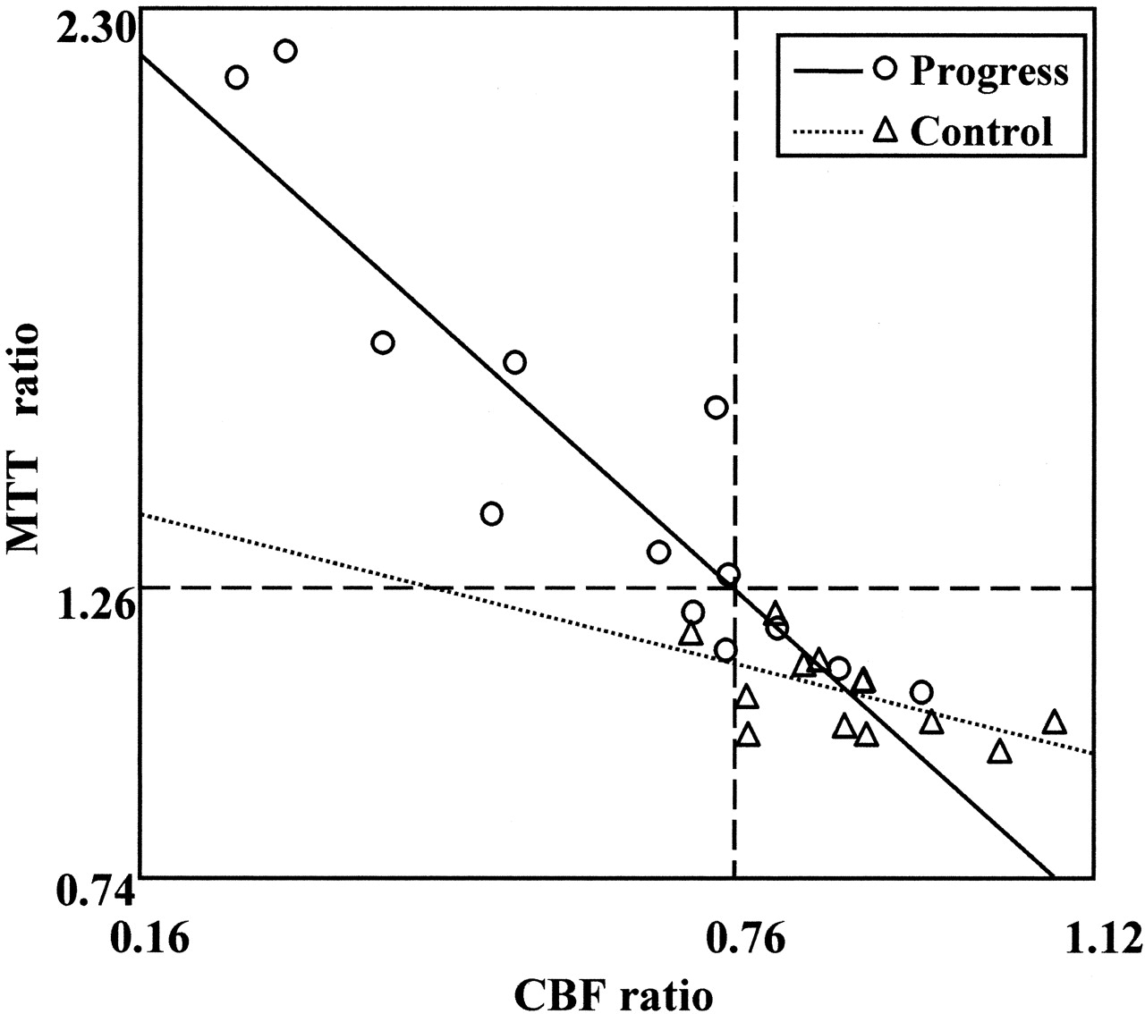
  • Fig 4.
    • Download figure
    • Open in new tab
    • Download powerpoint
    Fig 4.

    Relationships between CBF and MTT ratio. With cutoff lines at <0.76 in the CBF ratio and at >1.26 in the MTT ratio, sensitivity was 76.3%, specificity was 92.3%, positive predictive value was 90.9%, and negative predictive value was 80.8%.

Tables

  • Figures
    • View popup
    TABLE 1:

    Characteristics of patients at admission

    Control (n = 13)Progress (n = 13)P value*
    Age (yr)69.5 ± 7.870.1 ± 7.40.830
    Sex n (%)M, 9 (69.2); F, 4 (30.8)M, 7 (53.8); F, 6 (46.2)0.419
    Cardiac disease n (%)1 (7.7)2 (15.4)0.538
    Diabetes n (%)3 (23.1)2 (15.4)0.618
    Hypertension n (%)8 (61.5)9 (69.2)0.680
    Hypercholesterolemia n (%)5 (38.5)2 (15.4)0.179
    Antiplatelet n (%)3 (23.1)4 (30.8)0.658
    Smoking n (%)5 (38.5)3 (23.1)0.394
    Previous cerebral disease n (%)4 (30.8)4 (30.8)
    Admission NIHSS (points)2.2 ± 0.82.0 ± 0.80.615
    Time to perfusion CT (hr)9.8 ± 8.410.2 ± 6.90.837
    • Note.—M indicates male; F, female; NIHSS, National Institute of Health Stroke Scale. All values are expressed as number of patients (percentage) or average ± SD.

    • * Logistic analysis was conducted for the control and progress groups.

    • View popup
    TABLE 2:

    Summary of infarction size, values, and ratios of cerebral blood flow, cerebral blood volume, and mean transit time

    Control (n = 13)Progress (n = 13)P value*
    Infarction size on MR image
     Initial FLAIR image (mm2)50.8 ± 21.748.7 ± 21.00.814
     Follow-up T2WI (mm2)53.5 ± 21.478.3 ± 34.30.037
    Value and ratio on perfusion CT scan
     CBF value (ml/100 g/min)26.1 ± 7.019.6 ± 9.00.052
     CBV value (ml/100 g)4.3 ± 0.94.8 ± 1.90.349
     MTT value (second)10.0 ± 1.314.5 ± 3.5<0.001
     CBF ratio0.9 ± 0.10.6 ± 0.20.016
     CBV ratio1.0 ± 0.21.0 ± 0.30.695
     MTT ratio1.1 ± 0.11.5 ± 0.4<0.001
    • Note.—FLAIR indicates fluid-attenuated inversion recovery; T2WI, T2-weighted image; CBF, cerebral blood flow; CBV, cerebral blood volume; MTT, mean transit time. All values are expressed as average ± SD.

    • * The differences in value and ratio of cerebral blood flow, cerebral blood volume, and mean transit time were analyzed statistically between the two groups by using the unpaired Student’s t test.

PreviousNext
Back to top

In this issue

American Journal of Neuroradiology: 25 (3)
American Journal of Neuroradiology
Vol. 25, Issue 3
1 Mar 2004
  • Table of Contents
  • Index by author
Advertisement
Print
Download PDF
Email Article

Thank you for your interest in spreading the word on American Journal of Neuroradiology.

NOTE: We only request your email address so that the person you are recommending the page to knows that you wanted them to see it, and that it is not junk mail. We do not capture any email address.

Enter multiple addresses on separate lines or separate them with commas.
Prediction of Neurologic Deterioration in Patients with Lacunar Infarction in the Territory of the Lenticulostriate Artery Using Perfusion CT
(Your Name) has sent you a message from American Journal of Neuroradiology
(Your Name) thought you would like to see the American Journal of Neuroradiology web site.
CAPTCHA
This question is for testing whether or not you are a human visitor and to prevent automated spam submissions.
Cite this article
Mikito Yamada, Shinichi Yoshimura, Yasuhiko Kaku, Toru Iwama, Hirotaka Watarai, Takashi Andoh, Noboru Sakai
Prediction of Neurologic Deterioration in Patients with Lacunar Infarction in the Territory of the Lenticulostriate Artery Using Perfusion CT
American Journal of Neuroradiology Mar 2004, 25 (3) 402-408;

Citation Manager Formats

  • BibTeX
  • Bookends
  • EasyBib
  • EndNote (tagged)
  • EndNote 8 (xml)
  • Medlars
  • Mendeley
  • Papers
  • RefWorks Tagged
  • Ref Manager
  • RIS
  • Zotero
0 Responses
Respond to this article
Share
Bookmark this article
Prediction of Neurologic Deterioration in Patients with Lacunar Infarction in the Territory of the Lenticulostriate Artery Using Perfusion CT
Mikito Yamada, Shinichi Yoshimura, Yasuhiko Kaku, Toru Iwama, Hirotaka Watarai, Takashi Andoh, Noboru Sakai
American Journal of Neuroradiology Mar 2004, 25 (3) 402-408;
del.icio.us logo Twitter logo Facebook logo Mendeley logo
  • Tweet Widget
  • Facebook Like
  • Google Plus One

Jump to section

  • Article
    • Abstract
    • Methods
    • Results
    • Discussion
    • Conclusion
    • References
  • Figures & Data
  • Info & Metrics
  • Responses
  • References
  • PDF

Related Articles

  • No related articles found.
  • PubMed
  • Google Scholar

Cited By...

  • Early Neurologic Deterioration in Lacunar Stroke: Clinical and Imaging Predictors and Association With Long-term Outcome
  • Lacunar stroke: mechanisms and therapeutic implications
  • Perfusion Deficits and Mismatch in Patients with Acute Lacunar Infarcts Studied with Whole-Brain CT Perfusion
  • Multimodal CT Provides Improved Performance for Lacunar Infarct Detection
  • Neuroimaging Markers for Early Neurologic Deterioration in Single Small Subcortical Infarction
  • CT perfusion improves diagnostic accuracy and confidence in acute ischaemic stroke
  • Crossref
  • Google Scholar

This article has not yet been cited by articles in journals that are participating in Crossref Cited-by Linking.

More in this TOC Section

  • Fast Contrast-Enhanced 4D MRA and 4D Flow MRI Using Constrained Reconstruction (HYPRFlow): Potential Applications for Brain Arteriovenous Malformations
  • Multimodal CT Provides Improved Performance for Lacunar Infarct Detection
  • Optimal MRI Sequence for Identifying Occlusion Location in Acute Stroke: Which Value of Time-Resolved Contrast-Enhanced MRA?
Show more Brain

Similar Articles

Advertisement

Indexed Content

  • Current Issue
  • Accepted Manuscripts
  • Article Preview
  • Past Issues
  • Editorials
  • Editor's Choice
  • Fellows' Journal Club
  • Letters to the Editor
  • Video Articles

Cases

  • Case Collection
  • Archive - Case of the Week
  • Archive - Case of the Month
  • Archive - Classic Case

More from AJNR

  • Trainee Corner
  • Imaging Protocols
  • MRI Safety Corner
  • Book Reviews

Multimedia

  • AJNR Podcasts
  • AJNR Scantastics

Resources

  • Turnaround Time
  • Submit a Manuscript
  • Submit a Video Article
  • Submit an eLetter to the Editor/Response
  • Manuscript Submission Guidelines
  • Statistical Tips
  • Fast Publishing of Accepted Manuscripts
  • Graphical Abstract Preparation
  • Imaging Protocol Submission
  • Evidence-Based Medicine Level Guide
  • Publishing Checklists
  • Author Policies
  • Become a Reviewer/Academy of Reviewers
  • News and Updates

About Us

  • About AJNR
  • Editorial Board
  • Editorial Board Alumni
  • Alerts
  • Permissions
  • Not an AJNR Subscriber? Join Now
  • Advertise with Us
  • Librarian Resources
  • Feedback
  • Terms and Conditions
  • AJNR Editorial Board Alumni

American Society of Neuroradiology

  • Not an ASNR Member? Join Now

© 2025 by the American Society of Neuroradiology All rights, including for text and data mining, AI training, and similar technologies, are reserved.
Print ISSN: 0195-6108 Online ISSN: 1936-959X

Powered by HighWire