
Article Figures & Data
Figures
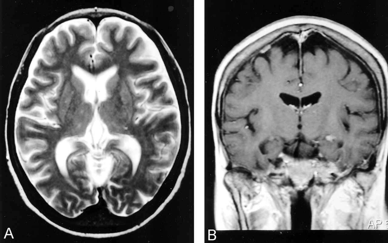
- Fig 1.
Images were obtained at the initial admission before chemotherapy began. Axial T2-weighted (3500/90) MR images (A) reveal minimum high-signal-intensity lesions in the both thalamus and putamen bilaterally. Coronal T1-weighted (458/14) MR image (B) shows that relatively thick meninges and bone marrow of the skull are enhanced after contrast material administration.
- Fig 2.
rCBF images at three different sections calculated by the I-123 IMP ARG method before the first course of chemotherapy (A), after the first course of chemotherapy (B), 2 hours after the recovery from the left hemiparesis (C), and 2 months before death (D) are shown. The same color scale is used to display quantitative rCBF images through four sequential I-123 IMP SPECT studies.
- Fig 3.
Images obtained on February 6, 2001, 10 hours after the onset of right hemiparesis. Postcontrast head CT shows that the vascular structures in the left cerebrum are enhanced as compared with those in the other side through the level of the third ventricle (A) and the body of the lateral ventricles (B).
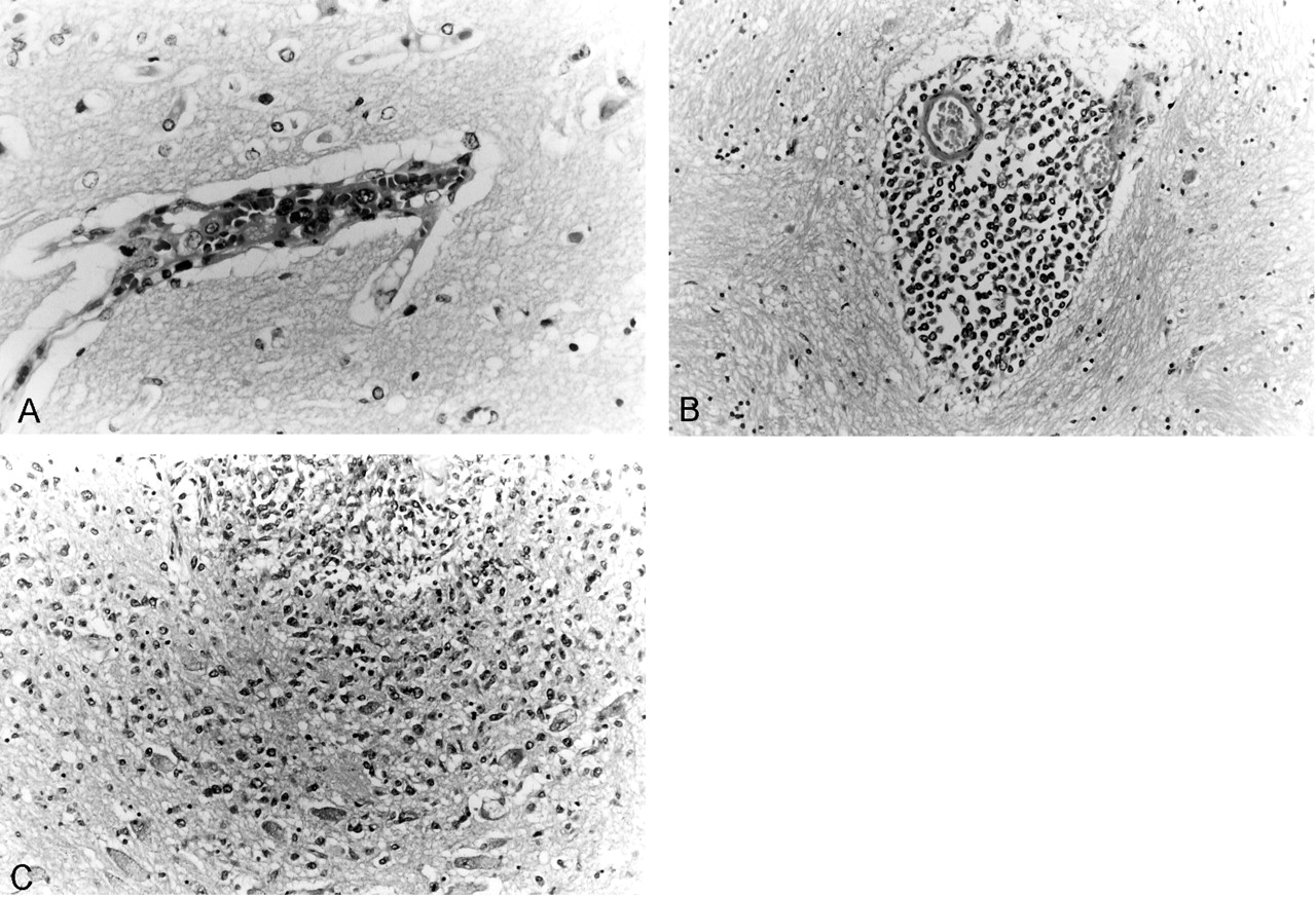
- Fig 4.
Histologic specimens (Hematoxylin-eosin stain) reveal infiltration of neoplastic lymphoid cells within the lumen of a small vessel of the brain parenchyma (A, ×400), especially concentrated in the Virchow-Robin perivascular space (B, ×200), and in the brain parenchyma (C, ×200).


Tables
- TABLE 1:
Summary of the values of rCBF calculated by the I-123 IMP ARG method in sequential studies
Examination Date ACA MCA PCA WM BG Cellrb Right Left Right Left Right Left Right Left Right Left Right Left November 30, 1999 54–60 57–65 58–62 59–63 54–67 48–60 40 45 79 87 64 66 December 20, 1999 30–36 33–37 33–36 33–37 35–40 32–38 22 21 42 48 42 40 February 9, 2001 35–45 48–60 36–43 58–70 36–46 49–61 29 42 45 54 56 51 February 20, 2001 45–60 43–56 47–59 46–68 50–59 46–56 36 27 56 67 64 65 Note.—ACA, cerebral cortex under anterior cerebral artery; BG, basal ganglia; Cellrb, cerebellum; MCA, cerebral cortex under middle cerebral artery; PCA, cerebral cortex under posterior cerebral artery; WM, semioval center.
Date Chemotherapy Examination Findings November 26, 1999 MRI (Fig 1) Minimum high intensity in the basal ganglia and thalamus on T2-weighted, dural/arachnoid enhancement with Gd-DTPA November 30, 1999 CHOP I-123 IMP (Fig 2A) Very high rCBF throughout the brain before the first half-dose CHOP December 20, 1999 I-123 IMP (Fig 2B) rCBF decreased to a level within normal range after the two half-dose CHOP February 5, 2001 THE-COP February 6, 2001 CT (Fig 3) Increased vascular densities in the left cerebral hemisphere, 6 hours after the onset of right hemiparesis February 9, 2001 I-123 IMP (Fig 2C) High rCBF in the left cerebral hemisphere and the right cerebellum, 2 hours after the recovery from right hemiparesis February 19–22, 2001 EPOCH February 20, 2001 I-123 IMP (Fig 2D) Relatively high rCBF at similar degree in both the right and the left cerebral hemispheres.
In this issue


Jump to section
Related Articles
Cited By...
- No citing articles found.