Advertisement

Main menu

  • Home
  • Content
    • Current Issue
    • Accepted Manuscripts
    • Article Preview
    • Past Issue Archive
    • AJNR Case Collection
    • Case of the Week Archive
    • Classic Case Archive
    • Case of the Month Archive
  • Special Collections
    • Spinal CSF Leak Articles (Jan 2020-June 2024)
    • 2024 AJNR Journal Awards
    • Most Impactful AJNR Articles
  • Multimedia
    • AJNR Podcast
    • AJNR Scantastics
    • Video Articles
  • For Authors
    • Submit a Manuscript
    • Author Policies
    • Fast publishing of Accepted Manuscripts
    • Graphical Abstract Preparation
    • Manuscript Submission Guidelines
    • Imaging Protocol Submission
    • Submit a Case for the Case Collection
  • About Us
    • About AJNR
    • Editorial Board
  • More
    • Become a Reviewer/Academy of Reviewers
    • Subscribers
    • Permissions
    • Alerts
    • Feedback
    • Advertisers
    • ASNR Home
  • Other Publications
    • ajnr

User menu

  • Alerts
  • Log in

Search

  • Advanced search
American Journal of Neuroradiology

American Journal of Neuroradiology

ASHNR American Society of Functional Neuroradiology ASHNR American Society of Pediatric Neuroradiology ASSR
  • Alerts
  • Log in

Advanced Search

  • Home
  • Content
    • Current Issue
    • Accepted Manuscripts
    • Article Preview
    • Past Issue Archive
    • AJNR Case Collection
    • Case of the Week Archive
    • Classic Case Archive
    • Case of the Month Archive
  • Special Collections
    • Spinal CSF Leak Articles (Jan 2020-June 2024)
    • 2024 AJNR Journal Awards
    • Most Impactful AJNR Articles
  • Multimedia
    • AJNR Podcast
    • AJNR Scantastics
    • Video Articles
  • For Authors
    • Submit a Manuscript
    • Author Policies
    • Fast publishing of Accepted Manuscripts
    • Graphical Abstract Preparation
    • Manuscript Submission Guidelines
    • Imaging Protocol Submission
    • Submit a Case for the Case Collection
  • About Us
    • About AJNR
    • Editorial Board
  • More
    • Become a Reviewer/Academy of Reviewers
    • Subscribers
    • Permissions
    • Alerts
    • Feedback
    • Advertisers
    • ASNR Home
  • Follow AJNR on Twitter
  • Visit AJNR on Facebook
  • Follow AJNR on Instagram
  • Join AJNR on LinkedIn
  • RSS Feeds

Welcome to the new AJNR, Updated Hall of Fame, and more. Read the full announcements.


AJNR is seeking candidates for the position of Associate Section Editor, AJNR Case Collection. Read the full announcement.


OtherHEAD AND NECK

Amyloidosis Concurrently Involving the Sinonasal Cavities and Larynx

Shy-Chyi Chin, Girish Fatterpeckar, Chuan-Hsiang Kao, Cheng-Yu Chen and Peter M. Som
American Journal of Neuroradiology April 2004, 25 (4) 636-638;
Shy-Chyi Chin
  • Find this author on Google Scholar
  • Find this author on PubMed
  • Search for this author on this site
Girish Fatterpeckar
  • Find this author on Google Scholar
  • Find this author on PubMed
  • Search for this author on this site
Chuan-Hsiang Kao
  • Find this author on Google Scholar
  • Find this author on PubMed
  • Search for this author on this site
Cheng-Yu Chen
  • Find this author on Google Scholar
  • Find this author on PubMed
  • Search for this author on this site
Peter M. Som
  • Find this author on Google Scholar
  • Find this author on PubMed
  • Search for this author on this site
  • Article
  • Figures & Data
  • Info & Metrics
  • Responses
  • References
  • PDF
Loading

Abstract

Summary: Localized amyloidosis is an uncommon benign disorder. The purpose of this report is to present the case of a 21-year-old man who had localized amyloidosis simultaneously involving the sinonasal cavities and the larynx. The rarer sinonasal lesion demonstrated CT findings of adjacent “fluffy” bone changes, possibly representing a new finding suggestive of this disorder. At MR imaging, the amyloid had signal intensity similar to that of skeletal muscle on T1- and T2-weighted images. After contrast material administration, the amyloid enhanced at most minimally, but peripheral enhancement about the mass was present. The importance of this case lies in the multifocal presentation of this uncommon disorder, and the imaging findings herein may provide a new sign of this paranasal sinus disease.

Amyloidosis is characterized by extracellular deposits of amyloid (1). Although uncommon, about 150 cases of amyloidosis have been documented since 1935 in the otolaryngology literature (2–5). There has, however, been a paucity of descriptions of its imaging findings in the head and neck (6). The purpose of this report is to present the CT and MR findings of this rare disorder concurrently involving the sinonasal cavities and the larynx.

Case Report

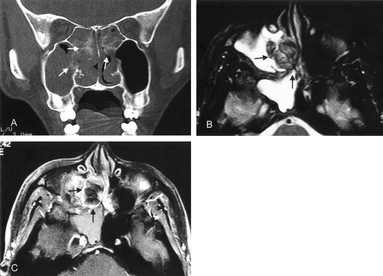
A 21-year-old man presented to the ear, nose, and throat clinic with a 1-year history of slowly progressive nasal stuffiness and dysphonia. Clinical examination demonstrated whitish, ulcerative, and swollen nasal conchae with serous secretions present in the adjacent osteomeatal infundibulum, ethmoid, and maxillary sinuses. Laryngoscopy disclosed a submucosal firm mass involving the left false fold and effacing the ipsilateral laryngeal ventricle. The patient denied any history of familial or hereditary disease or any notable allergies. CT and MR imaging studies were performed. The paranasal sinus CT showed that the affected bony conchae and sinus walls had a “fluffy” and somewhat hyperplastic reaction adjacent to the soft tissue mass that filled both nasal fossae (right greater than left) and the ethmoid sinuses (Fig 1A). At MR imaging, the lesion had low to intermediate signal intensity on both T1- and T2-weighted images and demonstrated peripheral enhancement on the contrast-enhanced study. The remaining soft tissues within the paranasal sinuses had high T2-weighted signal intensity, which is typical of obstructive secretions (Fig 1B and C). The MR study demonstrated a submucosal lesion in the larynx involving the left false cord and obliteration of the left laryngeal ventricle. The lesion also had low T1- and T2-weighted signal intensities and enhanced primarily along its periphery (Fig 2 A and B). The patient underwent surgical excision of both lesions and had an uneventful recovery. Histopathologic analysis with Congo Red stain confirmed the diagnosis of amyloidosis. Immunohistochemical examination revealed a positive staining pattern for six and eight light chain immunoglobulins. A thorough systemic workup for additional amyloid deposits revealed no evidence of other disease.

Fig 1.
  • Download figure
  • Open in new tab
  • Download powerpoint
Fig 1.

A, Coronal CT viewed at wide window settings shows fluffy calcifications (arrows) of the turbinates and sinus walls adjacent to a nasal mass. Soft tissue windowing shows entrapped secretions and inflammatory mucosal thickening in the right maxillary and sphenoid sinuses.

B, Axial T2-weighted MR image (TR/TE, 3000/96 ms) through the paranasal sinuses, demonstrates a predominantly hypointense lesion (arrows) involving the right sinonasal cavity. In contrast, entrapped secretions in the right maxillary and sphenoid sinuses have high T2-weighted signal intensity.

C, Contrast-enhanced, axial fat-suppressed T1-weighted image (TR/TE, 540/12 ms) demonstrates peripheral enhancement of the lesion (arrows).

Fig 2.
  • Download figure
  • Open in new tab
  • Download powerpoint
Fig 2.

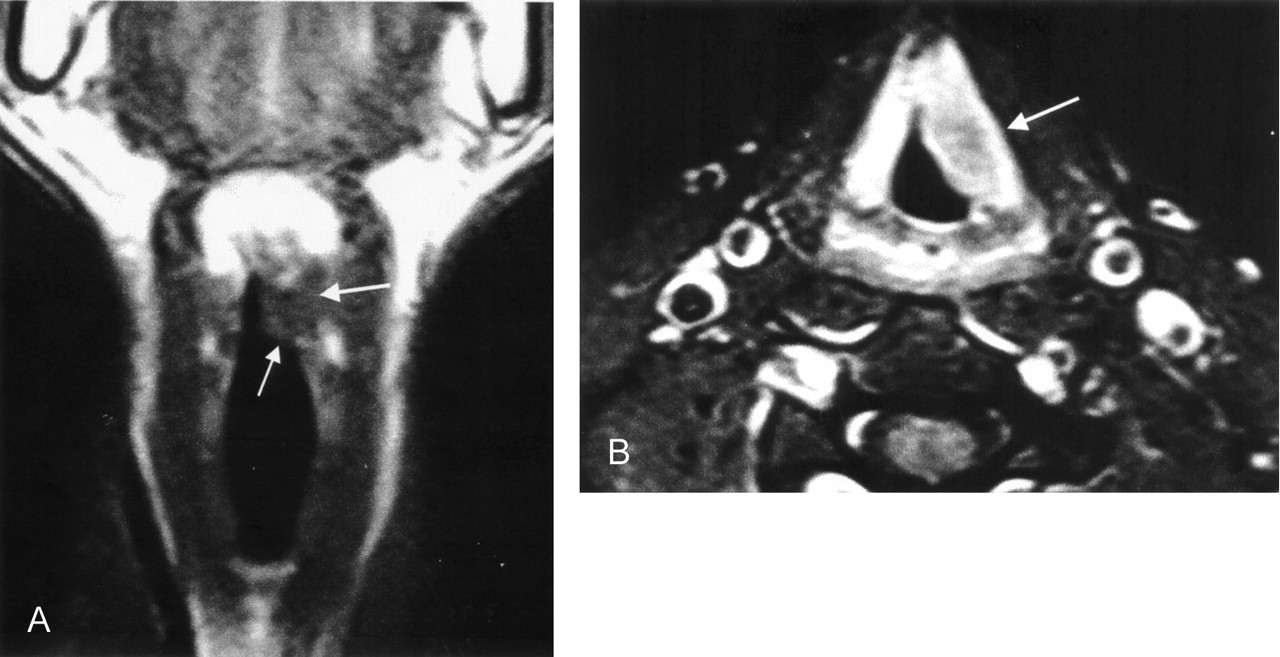
A, Coronal T1-weighted image through the larynx demonstrates a mass (arrows) involving the left false vocal cord with hypointensity similar to adjacent muscle. The lesion also has low signal intensity on T2-weighted images (not shown).

B, Contrast-enhanced axial T1-weighted, fat-suppressed image demonstrates a minimally enhanced submucosal lesion (arrow) surrounded by intense mucosal enhancement.

Discussion

Amyloidosis refers to a heterogeneous group of disorders that share the idiopathic extracellular accumulation of amyloid in tissues. If the deposition is extensive, it may interfere with organ or tissue function and even lead to death (7). Amyloid is a linear, nonbranching, fibrillar proteinaceous material that is arranged in a highly organized fashion (1). Amyloidosis is now classified as primary amyloidosis, myeloma-associatedamyloid, localized amyloid (laryngeal), secondary amyloidosis, familial amyloidosis, senile amyloidosis, and dialysis-associated amyloidosis (8, 9). In a large review, 70% of the cases were primary, 19% were localized, 4% were familial, and 3% were secondary (7).

About 19% of the cases involved the head and neck, and the larynx is the most commonly affected area, although amyloid has been reported in virtually all head and neck sites (10, 11). To our knowledge, only two case reports have described the simultaneous involvement of the larynx and the sinonasal cavities (12, 13).

Although the otolaryngology literature has described amyloidosis in detail, little has been mentioned regarding its imaging appearance in the radiology literature. Calcification has been mentioned as a nonspecific CT finding (6). In the paranasal sinuses, however, this case had a “fluffy” appearance in the sinonasal cavity bones adjacent to the amyloid deposits. It is possible that the deposition of the proteinaceous amyloid fibrils in the submucosal layers of the sinonasal cavities incited an osteoblastic reaction that resulted in the “fluffy” bone changes noted. Although sinonasal calcifications can also be seen in inspissated secretions, fungal mycetomas, cartilaginous tumors, and olfactory neuroblastomas, the “fluffy” bone changes have not been seen with these diseases.

The MR findings of a prolonged T2 relaxation time and signal intensity characteristics similar to those of skeletal muscle on T1-weighted and T2-weighted images have been described in the radiology literature (6). This is not surprising in light of the fact that that the highly organized ∃-pleated sheet ultrastructure of amyloid is similar to the multilayered, myofibrillar ultrastructure of skeletal muscle (14).

Although the amyloid itself does not enhance with contrast material administration, peripheral enhancement in the region of the amyloid deposits has been noted. This may be caused by the foreign-body giant-cell reaction that is evoked about the amyloid deposits (15). The lack of enhancement of the amyloid deposits helps to distinguish them from cellular tumors, all of which enhance to varying degrees. As mentioned above, CT findings of desiccated secretions and fungal mycetomas can be seen as hyperattenuated foci, and MR may disclose low T2-weighted signal intensity or signal intensity voids. However, these inflammatory diseases do not cause the “fluffy” bone changes in the adjacent sinonasal walls noted in the case of amyloidosis.

In the larynx, the amyloid deposits are submucosal and homogeneous and are not associated with the cartilage changes. They are firm lesions that tend to occur in the supraglottic larynx, although all laryngeal sites may be affected (7). The differential diagnosis includes other submucosal diseases such as laryngeal sarcoidosis, lymphoma, and pseudotumor. Submucosal lesions such as paragangliomas and hemangiomas are more localized on CT scans and MR images, and the entire lesion enhances.

Although the final diagnosis of a mass requires that the pathologist be the final arbiter, in this patient the “fluffy” bone appearance in the paranasal sinus walls occurring concurrently in a patient with a submucosal supraglottic mass may suggest the diagnosis of this rare disorder.

References

  1. ↵
    Cohen AS, Calkens E. Electron microscopic observations on a fibrous component in amyloid of diverse origins. Nature 1959;183:1202–1203
    CrossRefPubMed
  2. ↵
    Simpson GT, Skinner M, Strong MS, Coheb AS. Localized amyloidosis of the head and neck and upper aerodigestive and lower respiratory tracts. Ann Otol Rhinol Laryngol 1984;93:374–379
    PubMed
  3. Kramer R, Som ML. Local tumor-like deposits of amyloid. Arch Otolaryngol 1935;21:324–334
    CrossRef
  4. Johner CH, Widen AH, Sahgal S. Amyloidosis of the head and neck. Trans Am Acad Ophthalmol Otolaryngol 1972;76:1354–1355
    PubMed
  5. ↵
    Tsikoudas A, Martin-Hirsch DP, Woodhead CJ. Primary sinonasal amyloidosis. J Laryngol Otol 2001;115:55–56
    PubMed
  6. ↵
    Gean-Marton AD, Kirsch CF, Vezina LG, et al. Focal amyloidosis of the head and neck: evaluation with CT and MR imaging. Radiology 1991;181:521–525
    PubMed
  7. ↵
    Barnes, L. Miscellaneous disorders of the head and neck. In: Barnes L, ed. Surgical Pathology of the Head and Neck. Vol 3, 2nd ed. New York: Marcel Dekker;2001 :2191–2193
  8. ↵
    Cohen AS, Jones LA. Amyloid and amyloidosis. Curr Opin Rheumatol 1992;4:94–105
    PubMed
  9. ↵
    Husby G, Araki S, Benditt EP, et al. The 1990 guidelines for nomenclature and classification of amyloid and amyloidosis. In: Natvig JB, ed. Amyloid and Amyloidosis. Dordecht: Kluwer Academic;1990 :7–11
  10. ↵
    Lewis JE, Olsen KD, Kurtin PJ, Kyle RA. Laryngeal amyloidosis: a clinicopathologic and immunohistochemical review. Otolaryngol Head Neck Surg 1992;106:372–377
    PubMed
  11. ↵
    Thompson LDR. Pathology of the larynx, hypopharynx, and trachea. In: Fu YS, Wenig BM, Abemayor E, eds. Head and Neck Pathology with Clinical Correlations. 1st ed. Philadelphia: Churchill Livingstone;2001 :379–380
  12. ↵
    Birchall D, Fields JM, Poon CL. Case report: focal amyloidosis of the maxillary antrum: plain film, CT and MR appearances. Clin Radiol 1997;52:392–394
    PubMed
  13. ↵
    Sataloff RT, Abaza M, Abaza NA. Amyloidosis of the larynx. Ear Nose Throat J 2001;80:369–370
    PubMed
  14. ↵
    Banker BQ, Girvin JP. The ultrastructure features of the mammalian muscle spindle. J Neuropathol Exp Neurol 1971;30:155–195
    PubMed
  15. ↵
    Barnes EL Jr, Zafar T. Laryngeal amyloidosis: clinicopathologic study of seven cases. Ann Otol Rhinol Laryngol 1977;86:856–863
    PubMed
  • Received April 10, 2003.
  • Accepted after revision June 30, 2003.
  • Copyright © American Society of Neuroradiology
View Abstract
PreviousNext
Back to top

In this issue

American Journal of Neuroradiology: 25 (4)
American Journal of Neuroradiology
Vol. 25, Issue 4
1 Apr 2004
  • Table of Contents
  • Index by author
Advertisement
Print
Download PDF
Email Article

Thank you for your interest in spreading the word on American Journal of Neuroradiology.

NOTE: We only request your email address so that the person you are recommending the page to knows that you wanted them to see it, and that it is not junk mail. We do not capture any email address.

Enter multiple addresses on separate lines or separate them with commas.
Amyloidosis Concurrently Involving the Sinonasal Cavities and Larynx
(Your Name) has sent you a message from American Journal of Neuroradiology
(Your Name) thought you would like to see the American Journal of Neuroradiology web site.
CAPTCHA
This question is for testing whether or not you are a human visitor and to prevent automated spam submissions.
Cite this article
Shy-Chyi Chin, Girish Fatterpeckar, Chuan-Hsiang Kao, Cheng-Yu Chen, Peter M. Som
Amyloidosis Concurrently Involving the Sinonasal Cavities and Larynx
American Journal of Neuroradiology Apr 2004, 25 (4) 636-638;

Citation Manager Formats

  • BibTeX
  • Bookends
  • EasyBib
  • EndNote (tagged)
  • EndNote 8 (xml)
  • Medlars
  • Mendeley
  • Papers
  • RefWorks Tagged
  • Ref Manager
  • RIS
  • Zotero
0 Responses
Respond to this article
Share
Bookmark this article
Amyloidosis Concurrently Involving the Sinonasal Cavities and Larynx
Shy-Chyi Chin, Girish Fatterpeckar, Chuan-Hsiang Kao, Cheng-Yu Chen, Peter M. Som
American Journal of Neuroradiology Apr 2004, 25 (4) 636-638;
del.icio.us logo Twitter logo Facebook logo Mendeley logo
  • Tweet Widget
  • Facebook Like
  • Google Plus One

Jump to section

  • Article
    • Abstract
    • Case Report
    • Discussion
    • References
  • Figures & Data
  • Info & Metrics
  • Responses
  • References
  • PDF

Related Articles

  • No related articles found.
  • PubMed
  • Google Scholar

Cited By...

  • No citing articles found.
  • Crossref
  • Google Scholar

This article has not yet been cited by articles in journals that are participating in Crossref Cited-by Linking.

More in this TOC Section

  • Correlation of Apparent Diffusion Coefficient at 3T with Prognostic Parameters of Retinoblastoma
  • Parathyroid Lesions: Characterization with Dual-Phase Arterial and Venous Enhanced CT of the Neck
  • Efficacy of Diffusion-Weighted Imaging for the Differentiation between Lymphomas and Carcinomas of the Nasopharynx and Oropharynx: Correlations of Apparent Diffusion Coefficients and Histologic Features
Show more Head and Neck

Similar Articles

Advertisement

Indexed Content

  • Current Issue
  • Accepted Manuscripts
  • Article Preview
  • Past Issues
  • Editorials
  • Editors Choice
  • Fellow Journal Club
  • Letters to the Editor

Cases

  • Case Collection
  • Archive - Case of the Week
  • Archive - Case of the Month
  • Archive - Classic Case

Special Collections

  • Special Collections

Resources

  • News and Updates
  • Turn around Times
  • Submit a Manuscript
  • Author Policies
  • Manuscript Submission Guidelines
  • Evidence-Based Medicine Level Guide
  • Publishing Checklists
  • Graphical Abstract Preparation
  • Imaging Protocol Submission
  • Submit a Case
  • Become a Reviewer/Academy of Reviewers
  • Get Peer Review Credit from Publons

Multimedia

  • AJNR Podcast
  • AJNR SCANtastic
  • Video Articles

About Us

  • About AJNR
  • Editorial Board
  • Not an AJNR Subscriber? Join Now
  • Alerts
  • Feedback
  • Advertise with us
  • Librarian Resources
  • Permissions
  • Terms and Conditions

American Society of Neuroradiology

  • Not an ASNR Member? Join Now

© 2025 by the American Society of Neuroradiology All rights, including for text and data mining, AI training, and similar technologies, are reserved.
Print ISSN: 0195-6108 Online ISSN: 1936-959X

Powered by HighWire