Advertisement

Main menu

  • Home
  • Content
    • Current Issue
    • Accepted Manuscripts
    • Article Preview
    • Past Issue Archive
    • AJNR Case Collection
    • Case of the Week Archive
    • Classic Case Archive
    • Case of the Month Archive
  • Special Collections
    • Spinal CSF Leak Articles (Jan 2020-June 2024)
    • 2024 AJNR Journal Awards
    • Most Impactful AJNR Articles
  • Multimedia
    • AJNR Podcast
    • AJNR Scantastics
    • Video Articles
  • For Authors
    • Submit a Manuscript
    • Author Policies
    • Fast publishing of Accepted Manuscripts
    • Graphical Abstract Preparation
    • Manuscript Submission Guidelines
    • Imaging Protocol Submission
    • Submit a Case for the Case Collection
  • About Us
    • About AJNR
    • Editorial Board
  • More
    • Become a Reviewer/Academy of Reviewers
    • Subscribers
    • Permissions
    • Alerts
    • Feedback
    • Advertisers
    • ASNR Home
  • Other Publications
    • ajnr

User menu

  • Alerts
  • Log in

Search

  • Advanced search
American Journal of Neuroradiology

American Journal of Neuroradiology

ASHNR American Society of Functional Neuroradiology ASHNR American Society of Pediatric Neuroradiology ASSR
  • Alerts
  • Log in

Advanced Search

  • Home
  • Content
    • Current Issue
    • Accepted Manuscripts
    • Article Preview
    • Past Issue Archive
    • AJNR Case Collection
    • Case of the Week Archive
    • Classic Case Archive
    • Case of the Month Archive
  • Special Collections
    • Spinal CSF Leak Articles (Jan 2020-June 2024)
    • 2024 AJNR Journal Awards
    • Most Impactful AJNR Articles
  • Multimedia
    • AJNR Podcast
    • AJNR Scantastics
    • Video Articles
  • For Authors
    • Submit a Manuscript
    • Author Policies
    • Fast publishing of Accepted Manuscripts
    • Graphical Abstract Preparation
    • Manuscript Submission Guidelines
    • Imaging Protocol Submission
    • Submit a Case for the Case Collection
  • About Us
    • About AJNR
    • Editorial Board
  • More
    • Become a Reviewer/Academy of Reviewers
    • Subscribers
    • Permissions
    • Alerts
    • Feedback
    • Advertisers
    • ASNR Home
  • Follow AJNR on Twitter
  • Visit AJNR on Facebook
  • Follow AJNR on Instagram
  • Join AJNR on LinkedIn
  • RSS Feeds

Welcome to the new AJNR, Updated Hall of Fame, and more. Read the full announcements.


AJNR is seeking candidates for the position of Associate Section Editor, AJNR Case Collection. Read the full announcement.


OtherSPINE

MR Imaging Findings of Spinal Posterior Column Involvement in a Case of Miller Fisher Syndrome

Nobuhiro Inoue, Homare Ichimura, Satoshi Goto, Yoichiro Hashimoto and Yukitaka Ushio
American Journal of Neuroradiology April 2004, 25 (4) 645-648;
Nobuhiro Inoue
  • Find this author on Google Scholar
  • Find this author on PubMed
  • Search for this author on this site
Homare Ichimura
  • Find this author on Google Scholar
  • Find this author on PubMed
  • Search for this author on this site
Satoshi Goto
  • Find this author on Google Scholar
  • Find this author on PubMed
  • Search for this author on this site
Yoichiro Hashimoto
  • Find this author on Google Scholar
  • Find this author on PubMed
  • Search for this author on this site
Yukitaka Ushio
  • Find this author on Google Scholar
  • Find this author on PubMed
  • Search for this author on this site
  • Article
  • Figures & Data
  • Supplemental
  • Info & Metrics
  • Responses
  • References
  • PDF
Loading

This article has a correction. Please see:

  • Erratum - June 01, 2004

Abstract

Summary: The site of lesions causing ataxia in Miller Fisher syndrome (MFS) remains in dispute. A 43-year-old man manifested rapidly progressive left-sided ptosis, bilateral abducens palsy, areflexia, and severe ataxia. Initial MR imaging showed confined lesions of the cauda equina with gadolinium enhancement. A diagnosis of MFS was made, and the patient underwent immunotherapy. His ophthalmoplegia disappeared, but other symptoms remained. Five months after onset, MR imaging disclosed lesions confined to the spinal posterior column, which were considered to result from involvement of posterior nerve roots of the cauda equina and to be responsible for his remaining severe ataxia.

Miller Fisher syndrome (MFS) is characterized by the clinical triad of ophthalmoplegia, ataxia, and areflexia (1), whereas its site of lesions, particularly of ataxia, is a matter of controversy (2–5) As a variant form of Guillain-Barré syndrome (GBS), MFS is generally thought to result from peripheral neuropathy (3, 6–8). Several MR imaging studies also have shown CNS abnormalities in MFS (9–13), which suggests that central lesions also are responsible for some clinical aspects of MFS. Here we report a patient with persistent severe ataxia in features of MFS whose MR imaging at the chronic stage showed lesions confined to the posterior column of the spinal cord.

Case Reports

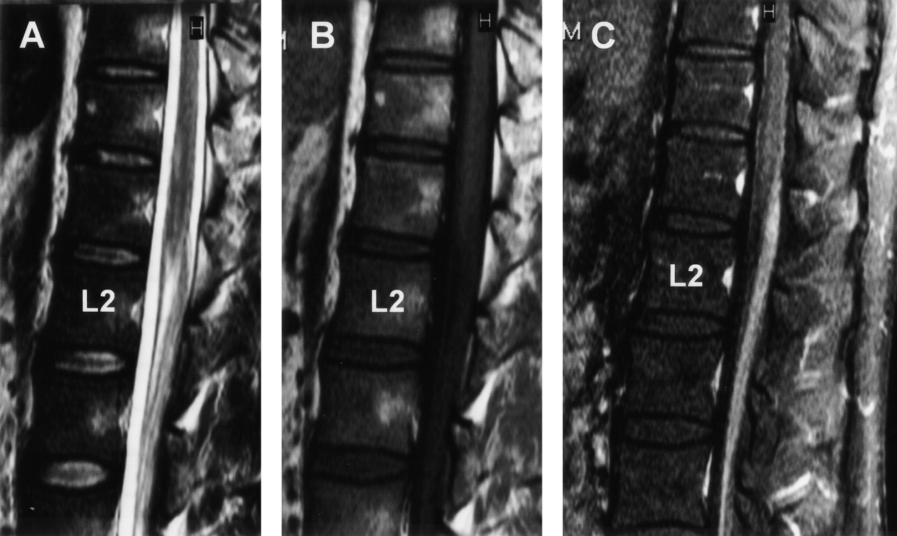
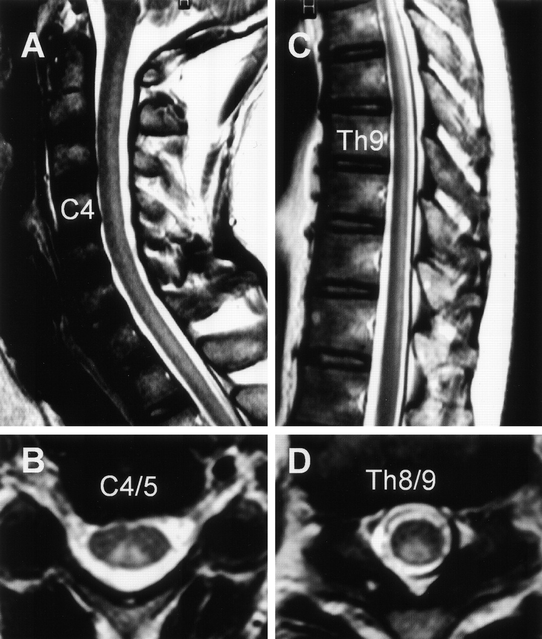
One month after suffering an upper respiratory tract infection with flulike symptoms, a 43-year-old man experienced dysesthesia and hypesthesia in all extremities followed by the development of diplopia and ataxic gait. On admission, he was alert and oriented. Speech was not dysarthric. Left-sided ptosis and bilateral abducens palsy were noted. Because of severe truncal and limb ataxia, he could not maintain an upright position without support and was unable to walk, even with assistance. Deep tendon reflexes were negative. In all extremities, there was a marked deterioration of position and vibratory sensation with modest dysesthesia and hypesthesia. Mild weakness of muscle strength was also noted. On CSF examination, the protein content was 134 mg/dL; the cell count was six cells/μL. Laboratory data, including CPK, were all within normal limits. Antibodies to gangliosides (GQ1b, GM1, GM1b, GM2, GD1a, GaNac-GD1a, GD1b, GD2, GT1a, and GT1b) and oligoclonal band were not detectable. The serum treponema pallidum hemagglutination test was negative. MR imaging performed on admission disclosed no apparent abnormalities in the brain, including the brain stem and cerebellum (Fig 1). At spinal MR imaging, however, gadolinium enhancement of the cauda equina was evident (Fig 2). A diagnosis of MFS was made, and the patient received 40 mg/kg/day of Gamma-globulin by intravenous administration for 5 days. Thereafter, his ophthalmoplegia disappeared, although severe ataxia, positional deficit, and lack of vibratory sensation persisted. He was seriously disabled, mainly because of truncal and limb ataxia. Somatosensory evoked-potential examination revealed no response to electric stimulation. Five months after onset, a repeat MR imaging study of his brain and spinal cord was performed and demonstrated on T2-weighed images hyperintense lesions confined to the spinal posterior column extending from the level of C1–T12 (Fig 3). No abnormalities were present on T1-weighed images or on the gadolinium-enhanced study.

Fig 1.
  • Download figure
  • Open in new tab
  • Download powerpoint
Fig 1.

MR imaging findings of the posterior fossa on admission. A, Axial view at the level of medulla and cerebellum. B, Axial view at the level of pons and cerebellum. C, Axial view at the level of midbrain. T2-weighted image, T1-weighted image, and gadolinium-enhanced images of each section indicated as 1, 2, and 3, respectively.

Fig 2.
  • Download figure
  • Open in new tab
  • Download powerpoint
Fig 2.

MR imaging findings of the sagittal spinal cord on admission at the lumbosacral level. A, T2-weighted image. B, T1-weighted image. C, Gadolinium-enhanced image. There is strong enhancement of the cauda equinae.

Fig 3.
  • Download figure
  • Open in new tab
  • Download powerpoint
Fig 3.

MR imaging findings on the spinal cord at 5 months after onset. Sagittal (A and C) and axial (B and D) views of T2-weighed images showing the hyperintense lesions confined to the spinal posterior column extending from the level of C1–Th12. A, Sagittal image of the cervical spine. B, Axial image at the level of C4/ C5. C, Sagittal image in the thoracic spine. D, Axial image at the level of T8/T9.

Discussion

Almost 50 years ago, Miller Fisher described a rare clinical syndrome with rapid evolving severe ataxia, ophthalmoplegia, and areflexia (MFS). Currently, MFS is generally considered a variant of GBS, but pathophysiology continues to be debated. The prognosis of MFS is usually benign (14,) but disabling or even fatal outcomes are occasionally found (15). Our patient presented the clinical triad of MFS with prior infection. CSF analysis showed a highly elevated protein content and a low cell count. Laboratory data including serologic tests and the initial MR imaging findings were not suggestive for other diseases such as neurosyphilis or multiple sclerosis, and we made a diagnosis of MFS. Indeed, immunotherapy abolished his cranial nerve symptoms; however, severe ataxia due to positional deficit and lack of vibratory sensation persisted, rendering him severely disabled and unable to lead an independent life.

In a recent study of 50 consecutive patients with MFS (14), the incidence of impaired vibratory or deep sensation was less than 20%, and all patients were minimally or not at all disabled 6 months after onset. With respect to the characteristic clinical features and prognosis of MFS, our patient was unusual because he continued to manifest a severe, unresolved deficit of deep sensations.

Ataxia is the most controversial symptom in MFS. Fisher interpreted it as the manifestation of an unusual peripheral neuron lesion, although he also raised the possibility that it might be of cerebellar origin (1). Other authors have supported his concept (3, 16, 17), and Kuwabara et al (18) recently suggested that MFS patients have dysfunction of the proprioceptive afferent system and their special sensory ataxia may be caused by the selective involvement of muscle spindle afferents. On the contrary, several authors proposed that ataxia in MFS is caused by the lesion of the cerebellar efferent or afferent pathway in the brain stem (4, 10, 11, 13, 19–21). On repeat MR imaging studies in our patient, although lesions of the cauda equina were initially found on the admission, lesions confined to the spinal posterior column were noted at 5 months after onset. On the basis of these MR imaging findings with anatomic considerations, we suggest that in our patient the anterograde degeneration of the posterior tracts in the spinal cord may occur secondarily to the initial involvement of posterior nerve roots of the cauda equina, resulting in his unresolved severe ataxia. In an autopsy analysis of ataxic forms of GBS, Richter reported degeneration of sensory roots, root entry zones, and posterior tracts (22). In some patients with GBS, MR imaging revealed involvement of posterior nerve roots, but not the posterior column, of the spinal cord (23, 24). With respect to lesion localization, debate continues on whether pure radicular involvement explains all the symptoms in MFS or whether ataxia is attributable to central and long-tract involvement. The imaging findings in our patient, who manifested involvement of the spinal posterior column, represent an important addition to the current understanding of pathophysiology of MFS.

Acknowledgments

We thank Dr. Nobuhiro Yuki of the Department of Neurology, Dokkyo University School of Medicine, Tochigi, Japan, for reviewing an earlier draft of the manuscript and providing critical suggestions.

References

  1. ↵
    Fisher M. An unusual variant of acute idiopathic polyneuritis (syndrome of ophthalmoplegia, ataxia and areflexia). N Engl J Med 1956;255:57–65
  2. ↵
    Ropper AH. The CNS in Gullain-Barré syndrome. Arch Neurol 1983;40:397–398
    CrossRefPubMed
  3. ↵
    Ropper AH, Shahani B. Proposed mechanism of ataxia in Fisher’s syndrome. Arch Neurol 1983;40:537–538
    CrossRefPubMed
  4. ↵
    Meienberg OR. Lesion site in Fisher’s syndrome. Arch Neurol 1984;41:250–251
    CrossRefPubMed
  5. ↵
    Al-Din ASN. The nosological position of the ophthalmoplegia, ataxia and areflexia syndrome: “the spectrum hypothesis.” Acta Neurol Scand 1987;75:287–294
    PubMed
  6. ↵
    Fross RD, Daube JR. Neuropathy in the Miller Fisher syndrome: clinical and electrophysiological findings. Neurology 1987;37:1493–1498
    Abstract/FREE Full Text
  7. Jamal GA, Ballantyne JP. The localization of the lesion in patients with acute ophthalmoplegia, ataxia, and areflexia (Miller Fisher syndrome): a serial multimodal neurophysiological study. Brain 1988;111:95–114
    Abstract/FREE Full Text
  8. ↵
    Sauron B, Bouche P, Cathala EP, et al. Miller Fisher syndrome: clinical evidence of peripheral origin in 10 cases. Neurology 1984;34:953–956
    Abstract/FREE Full Text
  9. ↵
    Giroud M, Mousson C, Chalopin JM, et al. Miller Fisher syndrome and pontine abnormalities on MRI: a case report. J Neurol 1990;237:489–490
    CrossRefPubMed
  10. ↵
    Petty RKH, Duncan R, Jamal GA, et al. Brainstem encephalitis and the Miller Fisher syndrome. J Neurol Neurosurg Psychiatry 1993;56:201–203
    Abstract/FREE Full Text
  11. ↵
    Urushitani M, Udaka Fukashi, Kameyama M. Miller Fisher–Guillain-Barré overlap syndrome with enhancing lesions in the spinocerebellar tracts. J Neurol Neurosurg Psychiatry 1995;58:241–243
    Abstract/FREE Full Text
  12. Echaniz-Laguna A, Battaglia F, Heymann R, et al. The Miller Fisher syndrome: neurophysiologic and MRI evidence of both peripheral and central origin in one case. J Neurol 2000;247:980–982
    CrossRefPubMed
  13. ↵
    Tezer FI, Gurer G, Karatas H, et al. Involvement of the central nervous system in Miller Fisher syndrome: a case report. Clin Neurol Neurosurg 2002;104:377–379
    CrossRefPubMed
  14. ↵
    Mori M, Kuwabara S, Fukutake T, et al. Clinical features and prognosis of Miller Fisher syndrome. Neurology 2001;56:1104–1106
    Abstract/FREE Full Text
  15. ↵
    Blau I, Casson I, Lieberman A, Weiss E. The not-so-benign Miller Fisher syndrome. Arch Neurol 1980;37:384–385
    CrossRefPubMed
  16. ↵
    Landaú WM, Glenn C, Dust G. MRI in Miller Fisher variant of Guillain-Barré syndrome. Neurology 1987;37:1431
  17. ↵
    Ropper AH. Three patients with Fisher’s syndrome. Neurology 1988;38:1630–1631
    Abstract/FREE Full Text
  18. ↵
    Kuwabara S, Asahina M, Nakajima M, et al. Special sensory ataxia in Miller Fisher syndrome detected by postural body sway analysis. Ann Neurol 1999;45:533–536
    CrossRefPubMed
  19. ↵
    Al-Din AN, Anderson M, Edwin R, Bickerstaff IH. Brainstem encephalitis and the syndrome of Miller Fisher. Brain 1982;105:481–495
    Abstract/FREE Full Text
  20. Barontini F, Sita D. The nosological position of Fisher’s syndrome (ophthalmoplegia, ataxia, and areflexia). J Neurol 1983;229:33–44
    CrossRefPubMed
  21. ↵
    Taphoom MJB, Lanting P, Hazenberg GJ, et al. Observations on the lesion site in the Miller Fisher syndrome: presentation of a case including CT and MRI. Acta Neurol Scand 1989;90:167–169
  22. ↵
    Richter RB. The ataxic form of polyradiculoneuritis (Landry-Guillain-Barré syndrome). J Neuropathol Exp Neurol 1962;21:171–184
  23. ↵
    Crino PB, Zimmerman R, Laskowitz D, et al. Magnetic resonance imaging of the cauda equina in Guillain-Barré syndrome. Neurology 1994;44:1334–1336
    Abstract/FREE Full Text
  24. ↵
    Byun WM, Park WK, Park BH, et al. Guillain-Barré syndrome: MR imaging findings of the spine in eight patients. Radiology 1988;208:137–141
  • Received May 20, 2003.
  • Accepted after revision June 24, 2003.
  • Copyright © American Society of Neuroradiology
View Abstract
PreviousNext
Back to top

In this issue

American Journal of Neuroradiology: 25 (4)
American Journal of Neuroradiology
Vol. 25, Issue 4
1 Apr 2004
  • Table of Contents
  • Index by author
Advertisement
Print
Download PDF
Email Article

Thank you for your interest in spreading the word on American Journal of Neuroradiology.

NOTE: We only request your email address so that the person you are recommending the page to knows that you wanted them to see it, and that it is not junk mail. We do not capture any email address.

Enter multiple addresses on separate lines or separate them with commas.
MR Imaging Findings of Spinal Posterior Column Involvement in a Case of Miller Fisher Syndrome
(Your Name) has sent you a message from American Journal of Neuroradiology
(Your Name) thought you would like to see the American Journal of Neuroradiology web site.
CAPTCHA
This question is for testing whether or not you are a human visitor and to prevent automated spam submissions.
Cite this article
Nobuhiro Inoue, Homare Ichimura, Satoshi Goto, Yoichiro Hashimoto, Yukitaka Ushio
MR Imaging Findings of Spinal Posterior Column Involvement in a Case of Miller Fisher Syndrome
American Journal of Neuroradiology Apr 2004, 25 (4) 645-648;

Citation Manager Formats

  • BibTeX
  • Bookends
  • EasyBib
  • EndNote (tagged)
  • EndNote 8 (xml)
  • Medlars
  • Mendeley
  • Papers
  • RefWorks Tagged
  • Ref Manager
  • RIS
  • Zotero
0 Responses
Respond to this article
Share
Bookmark this article
MR Imaging Findings of Spinal Posterior Column Involvement in a Case of Miller Fisher Syndrome
Nobuhiro Inoue, Homare Ichimura, Satoshi Goto, Yoichiro Hashimoto, Yukitaka Ushio
American Journal of Neuroradiology Apr 2004, 25 (4) 645-648;
del.icio.us logo Twitter logo Facebook logo Mendeley logo
  • Tweet Widget
  • Facebook Like
  • Google Plus One

Jump to section

  • Article
    • Abstract
    • Case Reports
    • Discussion
    • Acknowledgments
    • References
  • Figures & Data
  • Supplemental
  • Info & Metrics
  • Responses
  • References
  • PDF

Related Articles

  • Erratum
  • PubMed
  • Google Scholar

Cited By...

  • Immunolocalization of GQ1b and Related Gangliosides in Human Extraocular Neuromuscular Junctions and Muscle Spindles
  • Crossref
  • Google Scholar

This article has not yet been cited by articles in journals that are participating in Crossref Cited-by Linking.

More in this TOC Section

  • Bern Score Validity for SIH
  • MP2RAGE 7T in MS Lesions of the Cervical Spine
  • Deep Learning for STIR Spine MRI Quality
Show more Spine

Similar Articles

Advertisement

Indexed Content

  • Current Issue
  • Accepted Manuscripts
  • Article Preview
  • Past Issues
  • Editorials
  • Editors Choice
  • Fellow Journal Club
  • Letters to the Editor

Cases

  • Case Collection
  • Archive - Case of the Week
  • Archive - Case of the Month
  • Archive - Classic Case

Special Collections

  • Special Collections

Resources

  • News and Updates
  • Turn around Times
  • Submit a Manuscript
  • Author Policies
  • Manuscript Submission Guidelines
  • Evidence-Based Medicine Level Guide
  • Publishing Checklists
  • Graphical Abstract Preparation
  • Imaging Protocol Submission
  • Submit a Case
  • Become a Reviewer/Academy of Reviewers
  • Get Peer Review Credit from Publons

Multimedia

  • AJNR Podcast
  • AJNR SCANtastic
  • Video Articles

About Us

  • About AJNR
  • Editorial Board
  • Not an AJNR Subscriber? Join Now
  • Alerts
  • Feedback
  • Advertise with us
  • Librarian Resources
  • Permissions
  • Terms and Conditions

American Society of Neuroradiology

  • Not an ASNR Member? Join Now

© 2025 by the American Society of Neuroradiology All rights, including for text and data mining, AI training, and similar technologies, are reserved.
Print ISSN: 0195-6108 Online ISSN: 1936-959X

Powered by HighWire