Advertisement

Main menu

  • Home
  • Content
    • Current Issue
    • Accepted Manuscripts
    • Article Preview
    • Past Issue Archive
    • AJNR Case Collection
    • Case of the Week Archive
    • Classic Case Archive
    • Case of the Month Archive
  • Special Collections
    • Spinal CSF Leak Articles (Jan 2020-June 2024)
    • 2024 AJNR Journal Awards
    • Most Impactful AJNR Articles
  • Multimedia
    • AJNR Podcast
    • AJNR Scantastics
    • Video Articles
  • For Authors
    • Submit a Manuscript
    • Author Policies
    • Fast publishing of Accepted Manuscripts
    • Graphical Abstract Preparation
    • Manuscript Submission Guidelines
    • Imaging Protocol Submission
    • Submit a Case for the Case Collection
  • About Us
    • About AJNR
    • Editorial Board
  • More
    • Become a Reviewer/Academy of Reviewers
    • Subscribers
    • Permissions
    • Alerts
    • Feedback
    • Advertisers
    • ASNR Home
  • Other Publications
    • ajnr

User menu

  • Alerts
  • Log in

Search

  • Advanced search
American Journal of Neuroradiology

American Journal of Neuroradiology

ASHNR American Society of Functional Neuroradiology ASHNR American Society of Pediatric Neuroradiology ASSR
  • Alerts
  • Log in

Advanced Search

  • Home
  • Content
    • Current Issue
    • Accepted Manuscripts
    • Article Preview
    • Past Issue Archive
    • AJNR Case Collection
    • Case of the Week Archive
    • Classic Case Archive
    • Case of the Month Archive
  • Special Collections
    • Spinal CSF Leak Articles (Jan 2020-June 2024)
    • 2024 AJNR Journal Awards
    • Most Impactful AJNR Articles
  • Multimedia
    • AJNR Podcast
    • AJNR Scantastics
    • Video Articles
  • For Authors
    • Submit a Manuscript
    • Author Policies
    • Fast publishing of Accepted Manuscripts
    • Graphical Abstract Preparation
    • Manuscript Submission Guidelines
    • Imaging Protocol Submission
    • Submit a Case for the Case Collection
  • About Us
    • About AJNR
    • Editorial Board
  • More
    • Become a Reviewer/Academy of Reviewers
    • Subscribers
    • Permissions
    • Alerts
    • Feedback
    • Advertisers
    • ASNR Home
  • Follow AJNR on Twitter
  • Visit AJNR on Facebook
  • Follow AJNR on Instagram
  • Join AJNR on LinkedIn
  • RSS Feeds

Welcome to the new AJNR, Updated Hall of Fame, and more. Read the full announcements.


AJNR is seeking candidates for the position of Associate Section Editor, AJNR Case Collection. Read the full announcement.


LetterLetter

Mid-Anterior Surface of the Callosal Splenium: Subependymal or Subpial?

Akira Yamamoto, Yukio Miki, Yasutaka Fushimi, Tsutomu Okada and Hidekazu Tomimoto
American Journal of Neuroradiology April 2004, 25 (4) 664-665;
Akira Yamamoto
  • Find this author on Google Scholar
  • Find this author on PubMed
  • Search for this author on this site
Yukio Miki
  • Find this author on Google Scholar
  • Find this author on PubMed
  • Search for this author on this site
Yasutaka Fushimi
  • Find this author on Google Scholar
  • Find this author on PubMed
  • Search for this author on this site
Tsutomu Okada
  • Find this author on Google Scholar
  • Find this author on PubMed
  • Search for this author on this site
Hidekazu Tomimoto
  • Find this author on Google Scholar
  • Find this author on PubMed
  • Search for this author on this site
  • Article
  • Figures & Data
  • Info & Metrics
  • Responses
  • References
  • PDF
Loading

We read with interest the recent article by Pekala et al in the AJNR (1). They described that focal high signal intensity on FLAIR images may be seen in the anterior “subependymal” region of the splenium after radiation therapy or with aging. We would like to point out the following anatomic information that is related to some of the statements made in the article.

The corpus callosum is the largest transverse commissure connecting the cerebral hemispheres. Posteriorly, the corpus callosum is attached with the fornix and hippocampal commissure. On each side, its inferior surface roofs the lateral ventricle, covered by the ependyma (2).

The velum interpositum is located in the roof of the third ventricle below the body of the fornix. The upper and lower walls of the velum interpositum are formed by two membranous layers of tela choroidea in the roof of the third ventricle. The layer that is attached to the lower surface of the fornix and hippocampal commissure forms the upper wall. The lower wall is attached to the superior surface of the pineal body and tectum posteriorly (3).

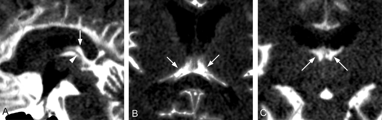
The double layers of pia mater, the tela choroidea, cover the ependymal roof of the third ventricle. The anterior aspect of the tela choroidea is closed at the foramen of Monro, where the pia mater folds on itself. The posterior ends remain open, and between the two ends is cistern of velum interpositum (also known as cistern of the transverse fissure or cistern of the roof of the third ventricle), which abuts on the midanterior surface of the splenium and communicates with the quadrigeminal cistern (4) (Fig 1).

In conclusion, the midanterior surface of the callosal splenium abuts on the subarachnoid cistern (cistern of velum interpositum), but not on the ventricles. The correct nomenclature is the anterior “subpial” region of the callosal splenium.

Fig 1.
  • Download figure
  • Open in new tab
  • Download powerpoint
Fig 1.

Sagittal (A), axial (B), and coronal (C) MDCT cisternograms showing filling of contrast material in the subarachnoid cisterns and fourth ventricle but not yet in the third and lateral ventricles. Cistern of velum interpositum (arrows) communicates with the quadrigeminal cistern around the internal cerebral veins (arrowhead). The midanterior surface of the callosal splenium abuts on the cistern of velum interpositum, not on the ventricle.

References

  1. ↵
    Pekala JS, Mamourian AC, Wishart HA, et al. Focal lesion in the splenium of the corpus callosum on FLAIR MR images: a common finding with aging and after brain radiation therapy. AJNR Am J Neuroradiol 2003;24:855–861
    Abstract/FREE Full Text
  2. ↵
    Pearson C. Cerebral cortex. In: Williams PL, Bannister LH, Berry MM, et al, eds. Gray’s Anatomy. 38th ed. London: Churchill Livingstone;1995 :1141–1186
  3. ↵
    Rhoton AL Jr. Microsurgical anatomy of the lateral ventricles. In: Wilkins RH, Rengachary SS, eds. Neurosurgery. 2nd ed. New York: McGraw-Hill;1996 :1419–1434
  4. ↵
    Spiegel SM, Nixon B, TerBrugge K, et al. Arachnoid cyst of the velum interpositum. AJNR Am J Neuroradiol 1988;9:981–983
    FREE Full Text
  • Copyright © American Society of Neuroradiology
PreviousNext
Back to top

In this issue

American Journal of Neuroradiology: 25 (4)
American Journal of Neuroradiology
Vol. 25, Issue 4
1 Apr 2004
  • Table of Contents
  • Index by author
Advertisement
Print
Download PDF
Email Article

Thank you for your interest in spreading the word on American Journal of Neuroradiology.

NOTE: We only request your email address so that the person you are recommending the page to knows that you wanted them to see it, and that it is not junk mail. We do not capture any email address.

Enter multiple addresses on separate lines or separate them with commas.
Mid-Anterior Surface of the Callosal Splenium: Subependymal or Subpial?
(Your Name) has sent you a message from American Journal of Neuroradiology
(Your Name) thought you would like to see the American Journal of Neuroradiology web site.
CAPTCHA
This question is for testing whether or not you are a human visitor and to prevent automated spam submissions.
Cite this article
Akira Yamamoto, Yukio Miki, Yasutaka Fushimi, Tsutomu Okada, Hidekazu Tomimoto
Mid-Anterior Surface of the Callosal Splenium: Subependymal or Subpial?
American Journal of Neuroradiology Apr 2004, 25 (4) 664-665;

Citation Manager Formats

  • BibTeX
  • Bookends
  • EasyBib
  • EndNote (tagged)
  • EndNote 8 (xml)
  • Medlars
  • Mendeley
  • Papers
  • RefWorks Tagged
  • Ref Manager
  • RIS
  • Zotero
0 Responses
Respond to this article
Share
Bookmark this article
Mid-Anterior Surface of the Callosal Splenium: Subependymal or Subpial?
Akira Yamamoto, Yukio Miki, Yasutaka Fushimi, Tsutomu Okada, Hidekazu Tomimoto
American Journal of Neuroradiology Apr 2004, 25 (4) 664-665;
del.icio.us logo Twitter logo Facebook logo Mendeley logo
  • Tweet Widget
  • Facebook Like
  • Google Plus One

Jump to section

  • Article
    • References
  • Figures & Data
  • Info & Metrics
  • Responses
  • References
  • PDF

Related Articles

  • No related articles found.
  • PubMed
  • Google Scholar

Cited By...

  • No citing articles found.
  • Crossref
  • Google Scholar

This article has not yet been cited by articles in journals that are participating in Crossref Cited-by Linking.

More in this TOC Section

  • Brain AVM’s Nidus: What if We Hadn’t Understood Anything?
  • Letter to the Editor regarding “Automated Volumetric Software in Dementia: Help or Hindrance to the Neuroradiologist?”
  • Reply:
Show more Letters

Similar Articles

Advertisement

Indexed Content

  • Current Issue
  • Accepted Manuscripts
  • Article Preview
  • Past Issues
  • Editorials
  • Editors Choice
  • Fellow Journal Club
  • Letters to the Editor

Cases

  • Case Collection
  • Archive - Case of the Week
  • Archive - Case of the Month
  • Archive - Classic Case

Special Collections

  • Special Collections

Resources

  • News and Updates
  • Turn around Times
  • Submit a Manuscript
  • Author Policies
  • Manuscript Submission Guidelines
  • Evidence-Based Medicine Level Guide
  • Publishing Checklists
  • Graphical Abstract Preparation
  • Imaging Protocol Submission
  • Submit a Case
  • Become a Reviewer/Academy of Reviewers
  • Get Peer Review Credit from Publons

Multimedia

  • AJNR Podcast
  • AJNR SCANtastic
  • Video Articles

About Us

  • About AJNR
  • Editorial Board
  • Not an AJNR Subscriber? Join Now
  • Alerts
  • Feedback
  • Advertise with us
  • Librarian Resources
  • Permissions
  • Terms and Conditions

American Society of Neuroradiology

  • Not an ASNR Member? Join Now

© 2025 by the American Society of Neuroradiology All rights, including for text and data mining, AI training, and similar technologies, are reserved.
Print ISSN: 0195-6108 Online ISSN: 1936-959X

Powered by HighWire