Advertisement

Main menu

  • Home
  • Content
    • Current Issue
    • Accepted Manuscripts
    • Article Preview
    • Past Issue Archive
    • AJNR Case Collection
    • Case of the Week Archive
    • Classic Case Archive
    • Case of the Month Archive
  • Special Collections
    • Spinal CSF Leak Articles (Jan 2020-June 2024)
    • 2024 AJNR Journal Awards
    • Most Impactful AJNR Articles
  • Multimedia
    • AJNR Podcast
    • AJNR Scantastics
    • Video Articles
  • For Authors
    • Submit a Manuscript
    • Author Policies
    • Fast publishing of Accepted Manuscripts
    • Graphical Abstract Preparation
    • Manuscript Submission Guidelines
    • Imaging Protocol Submission
    • Submit a Case for the Case Collection
  • About Us
    • About AJNR
    • Editorial Board
  • More
    • Become a Reviewer/Academy of Reviewers
    • Subscribers
    • Permissions
    • Alerts
    • Feedback
    • Advertisers
    • ASNR Home
  • Other Publications
    • ajnr

User menu

  • Alerts
  • Log in

Search

  • Advanced search
American Journal of Neuroradiology

American Journal of Neuroradiology

ASHNR American Society of Functional Neuroradiology ASHNR American Society of Pediatric Neuroradiology ASSR
  • Alerts
  • Log in

Advanced Search

  • Home
  • Content
    • Current Issue
    • Accepted Manuscripts
    • Article Preview
    • Past Issue Archive
    • AJNR Case Collection
    • Case of the Week Archive
    • Classic Case Archive
    • Case of the Month Archive
  • Special Collections
    • Spinal CSF Leak Articles (Jan 2020-June 2024)
    • 2024 AJNR Journal Awards
    • Most Impactful AJNR Articles
  • Multimedia
    • AJNR Podcast
    • AJNR Scantastics
    • Video Articles
  • For Authors
    • Submit a Manuscript
    • Author Policies
    • Fast publishing of Accepted Manuscripts
    • Graphical Abstract Preparation
    • Manuscript Submission Guidelines
    • Imaging Protocol Submission
    • Submit a Case for the Case Collection
  • About Us
    • About AJNR
    • Editorial Board
  • More
    • Become a Reviewer/Academy of Reviewers
    • Subscribers
    • Permissions
    • Alerts
    • Feedback
    • Advertisers
    • ASNR Home
  • Follow AJNR on Twitter
  • Visit AJNR on Facebook
  • Follow AJNR on Instagram
  • Join AJNR on LinkedIn
  • RSS Feeds

Welcome to the new AJNR, Updated Hall of Fame, and more. Read the full announcements.


AJNR is seeking candidates for the position of Associate Section Editor, AJNR Case Collection. Read the full announcement.


Research ArticleBRAIN

Anatomic Dissection Tractography: A New Method for Precise MR Localization of White Matter Tracts

E. Leon Kier, Lawrence H. Staib, Lawrence M. Davis and Richard A. Bronen
American Journal of Neuroradiology May 2004, 25 (5) 670-676;
E. Leon Kier
  • Find this author on Google Scholar
  • Find this author on PubMed
  • Search for this author on this site
Lawrence H. Staib
  • Find this author on Google Scholar
  • Find this author on PubMed
  • Search for this author on this site
Lawrence M. Davis
  • Find this author on Google Scholar
  • Find this author on PubMed
  • Search for this author on this site
Richard A. Bronen
  • Find this author on Google Scholar
  • Find this author on PubMed
  • Search for this author on this site
  • Article
  • Figures & Data
  • Supplemental
  • Info & Metrics
  • Responses
  • References
  • PDF
Loading

Article Figures & Data

Figures

  • Fig 1.
    • Download figure
    • Open in new tab
    • Download powerpoint
    Fig 1.

    Flowchart summarizes the stages of dissection tractography, which involves two methods: MR-assisted dissection and depiction of dissected tracts on MR images. In the first stage, MR imaging during the initial dissection helps prevent damage to the tracts under scrutiny. In the second stage, coregistration of 3D MR images of the specimen before and after the completion of dissection allows for the coregistration of a traced, dissected tract and the intact specimen. This in turn allows the creation of cross-sectional MR images with the traced white matter tract superimposed (Fig 4).

  • Fig 2.
    • Download figure
    • Open in new tab
    • Download powerpoint
    Fig 2.

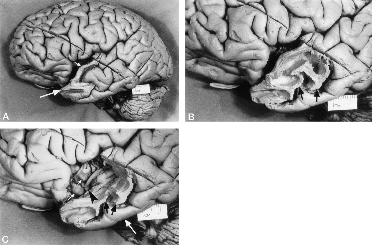
    Photographs of the lateral surface of a cerebral hemisphere show stages in the dissection of the temporal lobe.

    A, Initially, the white matter of the anterior part of superior temporal gyrus (small arrow) and temporal lobe pole (large arrow) was isolated by progressive removal of the gray matter.

    B, With further dissection, the white matter of the anterior segment of the middle temporal gyrus (arrows) was isolated by progressive removal of the gray matter.

    C, To visualize the insular gyri (small white arrows) covered by the middle cerebral artery (arrowhead) and its insular branches, the white matter of the anterior segment of the superior temporal gyrus was completely removed by dissection. A portion of the white matter of the middle temporal gyrus (black arrows) was also removed by dissection. The inferior temporal gyrus (large white arrow) was left intact.

  • Fig 3.
    • Download figure
    • Open in new tab
    • Download powerpoint
    Fig 3.

    Method used to identify the location of the dissected surface of the uncinate fasciculus on reformatted cross-sectional MR images.

    A, Photograph of the lateral surface of the dissected brain specimen. Branches of the middle cerebral artery have been dissected away, leaving the horizontal segment (M1) of the middle cerebral artery as a landmark (arrowhead). The lower part of the cortex of the insular gyri (white arrows) and the posterior orbital gyrus (black arrow) have also been removed by dissection. A segment of the surface of the uncinate fasciculus (transparent red) is visible and courses from the temporal lobe into the white matter of the insula. It passes anteriorly around the middle cerebral artery and into the posterior orbital gyrus. To demonstrate the underlying dissected uncinate fasciculus, we used a 10% transparent red that results in a pink appearance.

    B, 3D MR rendering of the dissected specimen in A demonstrates the accurate surface anatomy that can be depicted in this way. A segment of the surface of the uncinate fasciculus is color coded transparent red. The middle cerebral artery (arrowhead), the cortex of the insular gyri (white arrows), and the posterior orbital gyrus (black arrow) can be identified.

    C and D, Coronal (C) and axial (D) reformatted cross-sectional MR images show the location of the dissected segment of the surface of the uncinate fasciculus (arrows). These cross-sectional images were generated at a level indicated by a cursor (X) placed on the surface of the uncinate fasciculus on the 3D MR rendering in B. The position of the cursor on the cross-sectional images indicates the surface of the uncinate fasciculus. Coregistration of the cursor permitted comparison of localization on the dissected surface with corresponding coronal and axial MR images.

  • Fig 4.
    • Download figure
    • Open in new tab
    • Download powerpoint
    Fig 4.

    Method for MR localization of the dissected surface of the uncinate fasciculus on a 3D MR rendering and on multiplanar reformatted MR images of the specimen before dissection.

    A, 3D MR rendering shows the undissected lateral surface of the brain.

    B, 3D MR rendering shows the dissected lateral surface of the brain.

    C, 3D MR rendering with the surface of the dissected segment of the uncinate fasciculus traced and color-coded red.

    D, Coronal reformatted cross-sectional MR image of the dissected specimen in C shows the location of the dissected segment of the surface of the uncinate fasciculus in red.

    E, Coronal reformatted cross-sectional MR image of the coregistered undissected specimen shows the color-coded tracing from D. This resulted in the identification of the precise MR location of the tract on the multiplanar reformatted images of the undissected specimen.

PreviousNext
Back to top

In this issue

American Journal of Neuroradiology: 25 (5)
American Journal of Neuroradiology
Vol. 25, Issue 5
1 May 2004
  • Table of Contents
  • Index by author
Advertisement
Print
Download PDF
Email Article

Thank you for your interest in spreading the word on American Journal of Neuroradiology.

NOTE: We only request your email address so that the person you are recommending the page to knows that you wanted them to see it, and that it is not junk mail. We do not capture any email address.

Enter multiple addresses on separate lines or separate them with commas.
Anatomic Dissection Tractography: A New Method for Precise MR Localization of White Matter Tracts
(Your Name) has sent you a message from American Journal of Neuroradiology
(Your Name) thought you would like to see the American Journal of Neuroradiology web site.
CAPTCHA
This question is for testing whether or not you are a human visitor and to prevent automated spam submissions.
Cite this article
E. Leon Kier, Lawrence H. Staib, Lawrence M. Davis, Richard A. Bronen
Anatomic Dissection Tractography: A New Method for Precise MR Localization of White Matter Tracts
American Journal of Neuroradiology May 2004, 25 (5) 670-676;

Citation Manager Formats

  • BibTeX
  • Bookends
  • EasyBib
  • EndNote (tagged)
  • EndNote 8 (xml)
  • Medlars
  • Mendeley
  • Papers
  • RefWorks Tagged
  • Ref Manager
  • RIS
  • Zotero
0 Responses
Respond to this article
Share
Bookmark this article
Anatomic Dissection Tractography: A New Method for Precise MR Localization of White Matter Tracts
E. Leon Kier, Lawrence H. Staib, Lawrence M. Davis, Richard A. Bronen
American Journal of Neuroradiology May 2004, 25 (5) 670-676;
del.icio.us logo Twitter logo Facebook logo Mendeley logo
  • Tweet Widget
  • Facebook Like
  • Google Plus One

Jump to section

  • Article
    • Abstract
    • Methods
    • Results
    • Discussion
    • Conclusions
    • References
  • Figures & Data
  • Supplemental
  • Info & Metrics
  • Responses
  • References
  • PDF

Related Articles

  • Erratum
  • PubMed
  • Google Scholar

Cited By...

  • Principles and Limitations of Computational Algorithms in Clinical Diffusion Tensor MR Tractography
  • Crossref
  • Google Scholar

This article has not yet been cited by articles in journals that are participating in Crossref Cited-by Linking.

More in this TOC Section

  • Optimal MRI Sequence for Identifying Occlusion Location in Acute Stroke: Which Value of Time-Resolved Contrast-Enhanced MRA?
  • SWI or T2*: Which MRI Sequence to Use in the Detection of Cerebral Microbleeds? The Karolinska Imaging Dementia Study
  • Progression of Microstructural Damage in Spinocerebellar Ataxia Type 2: A Longitudinal DTI Study
Show more Brain

Similar Articles

Advertisement

Indexed Content

  • Current Issue
  • Accepted Manuscripts
  • Article Preview
  • Past Issues
  • Editorials
  • Editors Choice
  • Fellow Journal Club
  • Letters to the Editor

Cases

  • Case Collection
  • Archive - Case of the Week
  • Archive - Case of the Month
  • Archive - Classic Case

Special Collections

  • Special Collections

Resources

  • News and Updates
  • Turn around Times
  • Submit a Manuscript
  • Author Policies
  • Manuscript Submission Guidelines
  • Evidence-Based Medicine Level Guide
  • Publishing Checklists
  • Graphical Abstract Preparation
  • Imaging Protocol Submission
  • Submit a Case
  • Become a Reviewer/Academy of Reviewers
  • Get Peer Review Credit from Publons

Multimedia

  • AJNR Podcast
  • AJNR SCANtastic
  • Video Articles

About Us

  • About AJNR
  • Editorial Board
  • Not an AJNR Subscriber? Join Now
  • Alerts
  • Feedback
  • Advertise with us
  • Librarian Resources
  • Permissions
  • Terms and Conditions

American Society of Neuroradiology

  • Not an ASNR Member? Join Now

© 2025 by the American Society of Neuroradiology All rights, including for text and data mining, AI training, and similar technologies, are reserved.
Print ISSN: 0195-6108 Online ISSN: 1936-959X

Powered by HighWire