
Article Figures & Data
Figures
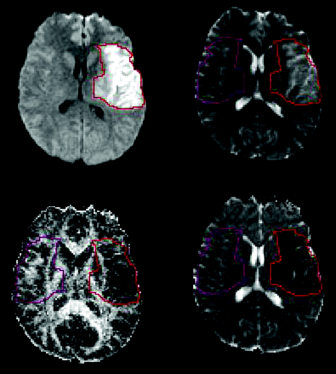
- Fig 1.
Tracing method for the VOIs. The infarct was outlined on isotropic DWI, section by section, by using a free-hand method. On each section, the ROI was flipped about the vertical axis and placed in the same location on contralateral normal side to obtain a mirror-image ROI. All section ROIs were combined to provide one stoke and one contralateral normal VOI for each patient. All VOIs were projected onto inherently coregistered echo-planar T2WIs, FA maps, and ADC maps. Top left, Isotropic DWI. Top right, Echo-planar T2WI. Bottom left, FA map. Bottom right, ADC map.
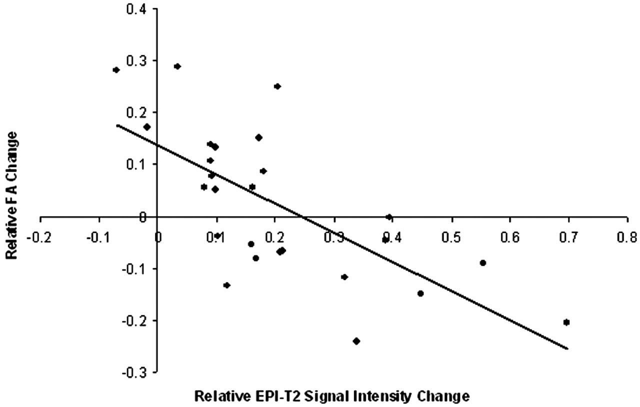
- Fig 2.
Graph demonstrates a highly significant inverse linear correlation between FA and T2 signal intensity (r = −0.691, P = .00009). Note that all patients with a relative increase in T2 signal intensity of < 0.10 also have increased relative FA values.
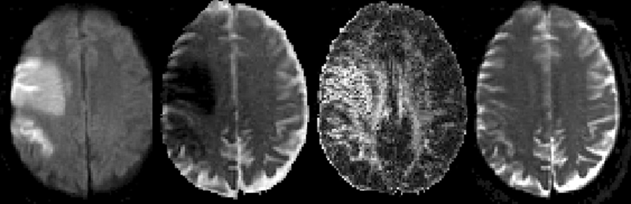
- Fig 3.
Images in a 59-year-old man who underwent an MR examination 8 hours after the onset of symptoms. An acute infarction in the right middle cerebral artery distribution was observed; this was due to thromboembolism. The FA map demonstrates increased signal intensity, whereas the echo-planar T2WI shows no hyperintensity. This is an example of how increased FA was associated with minimal T2 change. Far left, Isotropic DWI. Middle left, ADC map. Middle right, FA map. Far right, echo-planar T2WI.
- Fig 4.
Images in a 71-year-old woman who presented with slurred speech and left-sided weakness and who was imaged 11 hours after the onset of these symptoms. DWI demonstrates an acute infarction involving the right MCA territory. Decreased FA (far left) and increased T2 signal intensity (far right) was observed in the right corona radiata. This is an example of how decreased FA was associated with increased T2 signal intensity. Far left, Isotropic DWI. Middle left, ADC map. Middle right, FA map Far right, Echo-planar T2WI.


Tables
Results in 26 patients with acute stroke
Territory Volume (mm3) Interval from Onset to Imaging (hours) Relative Change FA ADC T2 R MCA 84 7 0.288 −0.27 0.036 L MCA 1969 4 0.281 −0.449 −0.069 L MCA 1254 6.5 0.25 −0.438 0.206 R ACA/MCA 10436 3.5 0.173 −0.251 −0.017 R MCA 2694 5 0.152 −0.365 0.174 R MCA 5682 8 0.139 −0.514 0.092 L ACA 1191 3.5 0.133 −0.323 0.099 R MCA 2217 3 0.107 −0.144 0.092 L MCA 3992 6 0.086 −0.453 0.181 L PCA 846 4.5 0.079 −0.322 0.094 R MCA 4831 1.3 0.057 −0.175 0.162 R MCA 195 6 0.056 −0.248 0.08 L MCA 6043 12 0.052 −0.55 0.099 L MCA 102 5 −0.001 −0.344 0.396 R ACA/MCA 233 9 −0.038 −0.17 0.103 R MCA 4567 9 −0.045 −0.38 0.388 L ACA/MCA 9046 3 −0.054 −0.228 0.16 R MCA 585 7 −0.066 −0.116 0.213 R MCA 1699 3.5 −0.068 −0.255 0.209 L MCA 6240 6 −0.081 −0.316 0.169 R MCA 152 3 −0.09 −0.331 0.555 L MCA 435 12 −0.116 −0.3 0.32 L MCA 3838 6 −0.134 −0.187 0.12 R MCA 883 11 −0.149 −0.423 0.449 R MCA 452 12 −0.204 −0.226 0.697 L MCA 718 11 −0.241 −0.307 0.341 Note.—FA, ADC, and T2 values were obtained from initial MR examinations and the relative values were used to express changes in FA, ADC and T2 signal. ACA indicates anterior cerebral artery; MCA, middle cerebral artery; PCA, posterior cerebral artery.
In this issue


Jump to section
Related Articles
Cited By...
- Structural integrity following focused ultrasound thalamotomy and its correlation with tremor relief
- White Matter Ischemic Changes in Hyperacute Ischemic Stroke: Voxel-Based Analysis Using Diffusion Tensor Imaging and MR Perfusion
- Increased Corticospinal Tract Fractional Anisotropy Can Discriminate Stroke Onset Within the First 4.5 Hours