
Article Figures & Data
Figures
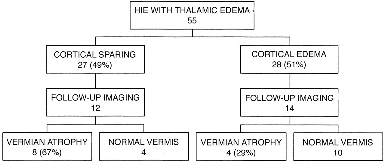
- Fig 1.
Schematic diagram shows frequency of vermian atrophy in children after neonatal hypoxic-ischemic encephalopathy with thalamic edema.
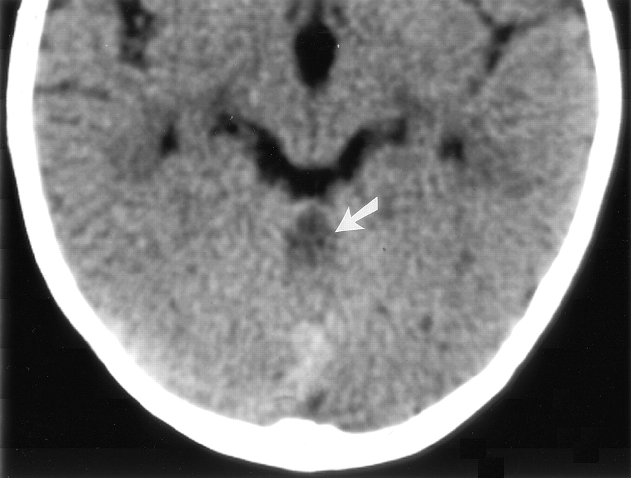
- Fig 2.
Axial view CT scan obtained through the posterior fossa in a 10-month-old female infant (patient 3), who was born before arrival at the hospital, shows focal hypoattenuation in the superior vermis (arrow). The thalami were small but with normal attenuation (not shown). This patient had thalamic edema with normal cortex shown by neonatal CT.
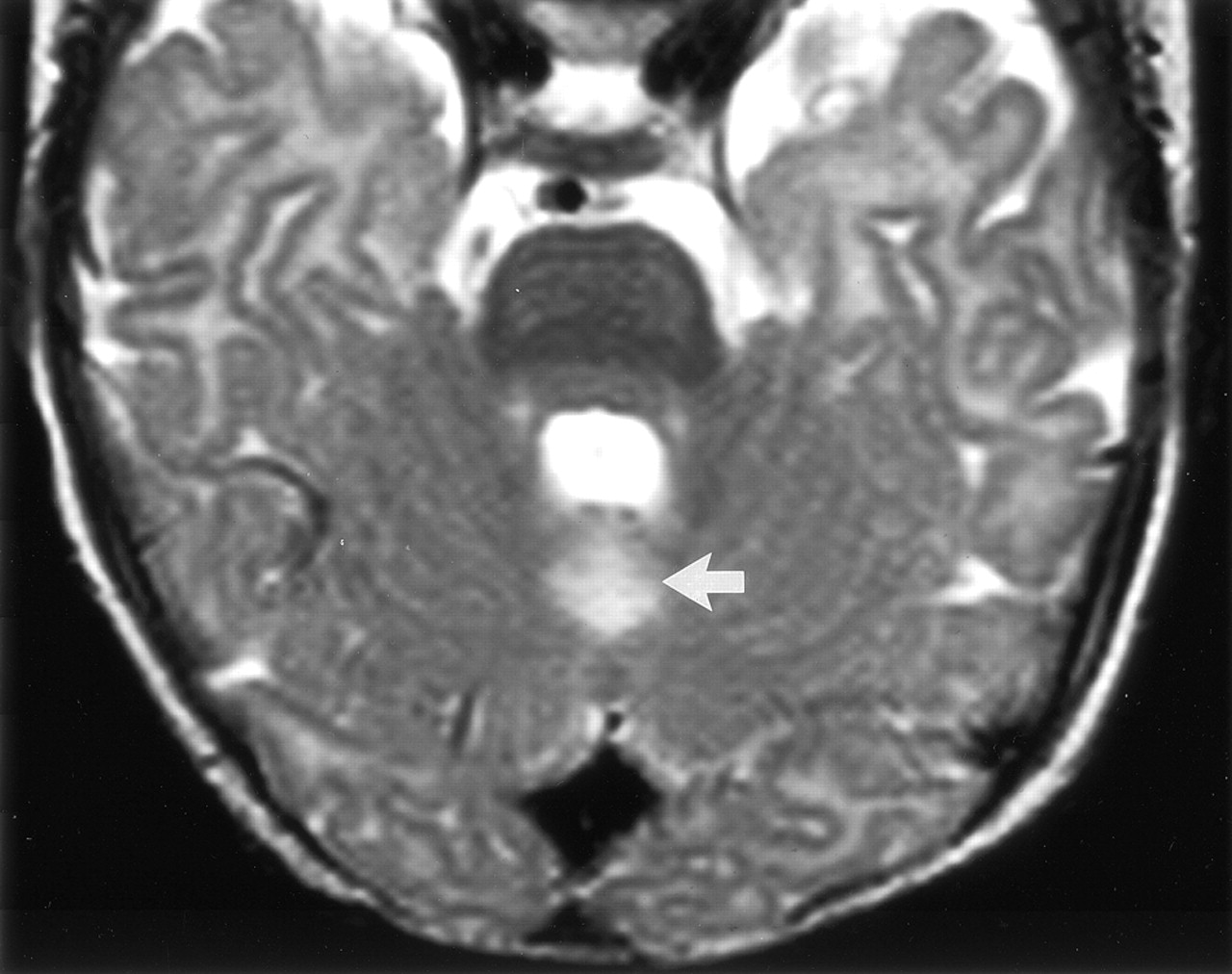
- Fig 3.
Axial view T2-weighted fast spin-echo (3555/112) image obtained through the posterior fossa in a 5-month-old male infant (patient 2), who was born after uterine rupture, shows hyperintensity in the cerebellar vermis immediately behind the fourth ventricle (arrow). Neonatal CT performed on day 3 showed abnormal thalami with normal cortex, whereas MR imaging performed on day 17 showed signal intensity abnormalities in the thalami, hippocampus, and rolandic cortex.
- Fig 4.
Coronal view T2-weighted fast spin-echo (6480/96) MR image of a 20-month-old boy (patient 5) shows hyperintensity with volume loss in the superior cerebellar vermis (arrow). This child had thalamic edema with normal cortex shown by neonatal CT. Other images in this follow-up study showed symmetrical hyperintensity of the ventrolateral thalami, posterior lentiform nuclei, and periventricular white matter extending to the rolandic cortex but with normal cortical gray matter.
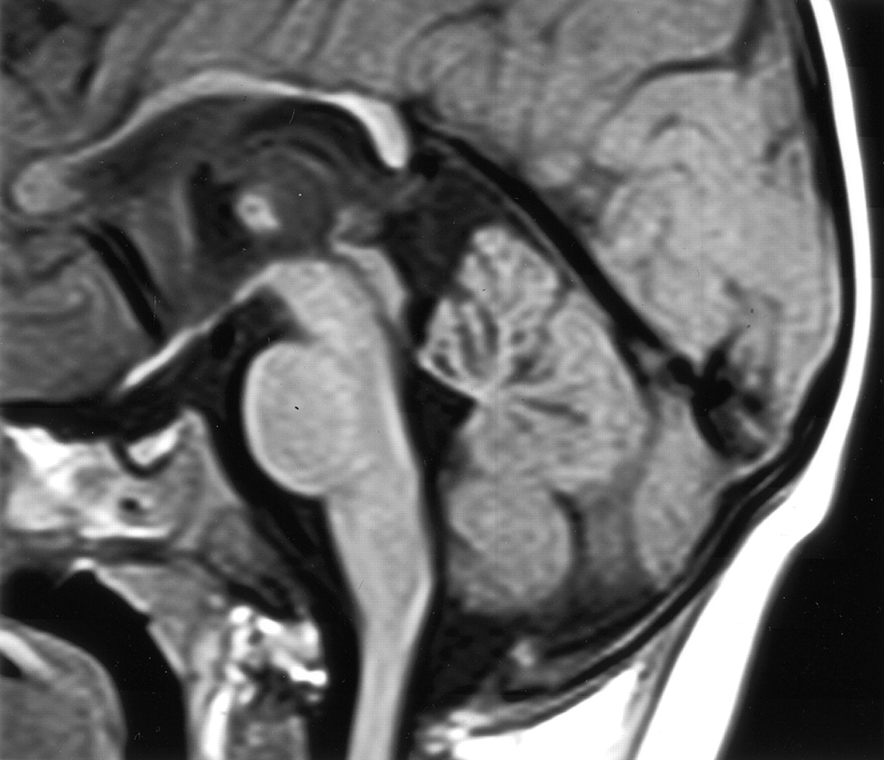
- Fig 5.
Midsagittal view T1-weighted (600/10) MR image of the same patient shown in Figure 2 shows volume loss centrally in the cerebellar vermis. Other findings included abnormal lentiform nuclei, signal intensity changes in the thalami, and white matter with mild atrophy of the rolandic cortex. In the midsagittal view, the corpus callosum appears thin.
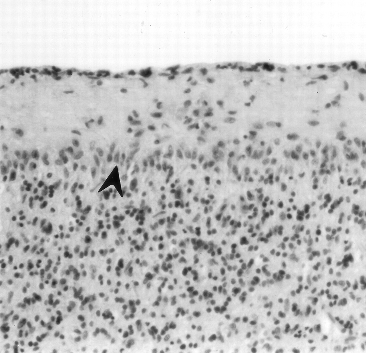
- Fig 6.
Cerebellar vermis of an asphyxiated twin born after cord prolapse. CT performed on day 3 showed thalamic edema without cortical involvement. The neonate died at 16 days old. Photomicrograph of the cerebellum shows loss of Purkinje cells and proliferation of Bergmann glia (arrowhead) (hematoxylin and eosin stain; original magnification, ×100).


Tables
- TABLE 1:
Neonatal and late imaging findings for children who had neonatal thalamic edema with cortical sparing
Patient No. Follow-up Age in Months (CT/MR Imaging) Indications Given for Follow-up Imaging Neonatal CT: Cerebellar Vermis Neonatal CT: Cerebellar Hemispheres Follow-up Imaging: Cerebellar Vermis Follow-up Imaging: Cerebellar Hemispheres Follow-up Basal Ganglia* Follow-up Cerebral Cortex Follow-up Cerebral White Matter 1 NA/19 Perinatal asphyxia Normal ?Edema Superior vermis atrophy Normal Thalami, lentiforms Hippocampi, ?Rolandic Rolandic 2 5/5 Infantile spasms ?Edema Normal Superior vermis atrophy Normal Thalami, lentiforms ?Rolandic Rolandic 3 10/19 Spastic quadriplegia, seizures Normal Normal Superior vermis atrophy Normal Thalami, lentiforms Normal Periventricular 4 NA/24 Neonatal HIE, cerebral palsy Normal Normal Superior vermis atrophy Normal Thalami Normal Normal 5 5/8 Developmental delay, spastic quadriplegia Normal Normal Superior vermis atrophy Normal Thalami, lentiforms Normal Periventricular, Rolandic 6 NA/12 Cerebral palsy ?Edema Normal Normal Normal Thalami, lentiforms Rolandic, ?hippocampi Periventricular, Rolandic 7 NA/15 HIE follow-up Normal Normal Superior vermis atrophy Normal Thalami, lentiforms Rolandic, ?hippocampi Periventricular, Rolandic 8 4/NA Asphyxia follow-up Normal Normal Superior vermis atrophy Normal Thalami Global Global 9 6/NA NA Normal Edema Normal Normal Thalami, lentiforms Normal Periventricular 10 24/NA Developmental delay, cerebral palsy Normal Edema Superior vermis atrophy Normal Thalami Normal Normal 11 21/36 Choreoathetosis Normal Normal Normal Normal Thalami, lentiforms Normal Periventricular 12 NA/27 NA Normal Normal Normal Normal Thalami Rolandic Rolandic Note.—NA indicates not applicable because procedure not performed; ?, equivocal; HIE, hypoxic-ischemic encephalopathy; Global, generalized abnormality. Note that anatomic sites as listed under Follow-up Imaging had one or more of the following: atrophy, abnormal MR imaging signal, or abnormal CT attenuation.
* Caudate nuclei were not separately assessed.
- TABLE 2:
Neonatal and late imaging findings for children who had neonatal thalamic edema with cortical edema
Patient No. Follow-up Age in Months (CT/MR Imaging) Indications Given for Follow-up Imaging Neonatal CT: Cerebellar Vermis Neonatal CT: Cerebellar Hemispheres Follow-up Imaging: Cerebellar Vermis Follow-up Imaging: Cerebellar Hemispheres Follow-up Basal Ganglia* Follow-up Cerebral Cortex Follow-up Cerebral White Matter 13 NA/7 Severe HIE, seizures Normal ?Edema Superior vermis atrophy Normal Thalami, lentiforms Rolandic Global 14 NA/7 Neonatal HIE, infantile spasms Normal ?Edema Normal Normal Lentiforms, ?thalami Global Global 15 13/NA Seizures Normal ?Edema Normal Normal Thalami, lentiforms Global Global 16 13/NA NA Normal Edema Normal Normal Thalami Global asymmetric Global asymmetric 17 5/NA Birth asphyxia Normal Normal Normal Normal Thalami, lentiforms Global Global 18 107/NA Follow-up neonatal IVH and infarction ?Edema Edema Normal Normal Thalami, lentiforms asymmetric Global Global 19 4/NA Follow-up HIE, developmental delay Normal ?Edema Superior vermis atrophy Normal Thalami, ?lentiforms Normal Global 20 13/NA NA Normal Normal Normal Normal Thalami, ?lentiforms Rolandic Global 21 15/NA Seizures, microcephaly, spastic cerebral palsy Normal ?Edema Normal Normal Thalami, lentiforms Global Global 22 80/NA Developmental delay, seizures Normal Normal Superior vermis atrophy ?Atrophy Asymmetric thalami, lentiforms Asymmetric Lt global, Rt Rolandic Asymmetric Lt global 23 5/NA NA Normal ?Edema Normal Normal Asymmetric lentiforms, thalami Asymmetric global Asymmetric global 24 NA/49 Follow-up HIE Normal Normal Normal Normal Normal Posterior Posterior 25 5/NA Follow-up HIE Normal Normal Normal Normal Thalami, lentiforms Asymmetric global Asymmetric global 26 NA/25 Follow-up neonatal HIE seizures Normal Normal Superior vermis atrophy, ?inferior vermis atrophy Normal Thalami, lentiforms Rolandic Global Note.—NA indicates not applicable because procedure not performed; HIE, hypoxic-ischemic encephalopathy; ?, equivocal; IVH, intraventricular hemorrhage; Global, generalized abnormality. Note that anatomic sites as listed under Follow-up Imaging had one or more of the following: atrophy, abnormal MR imaging signal, or abnormal CT attenuation.
* Caudate nuclei were not separately assessed.
In this issue


Jump to section
Related Articles
Cited By...
- Injury to the Cerebellum in Term Asphyxiated Newborns Treated with Hypothermia
- Anatomic Localization of Dyskinesia in Children with "Profound" Perinatal Hypoxic-Ischemic Injury
- Injury to the Developing Cerebellum: Mechanisms and Consequences
- Does perinatal asphyxia impair cognitive function without cerebral palsy?