
Article Figures & Data
Figures
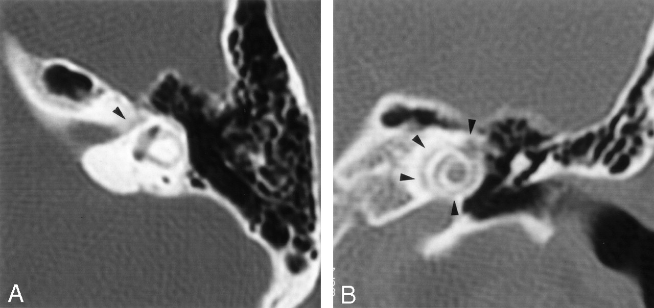
- Fig 1.
Initial axial (A) and direct coronal (B) high-resolution CT scans of the temporal bones through the level of the labyrinthine segment of the facial nerve (axial) and the cochlea (coronal) processed with a bone algorithm (2-mm section thickness; 512 × 512 matrix). The facial nerve canal showed slight irregularities along the labyrinthine segment (arrowhead) but otherwise appeared normal. Bandlike, undermineralized areas around the cochlea can be seen (arrowheads).
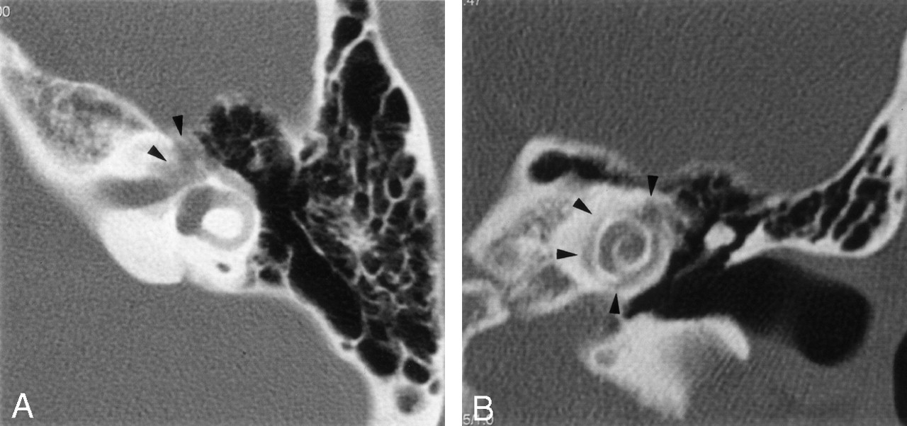
- Fig 2.
Further axial (A) and direct coronal (B) high-resolution CT scans through the level of the labyrinthine segment of the facial nerve (axial) and the cochlea (coronal) obtained 2 years after the initial scans (1-mm section thickness; 512 × 512 matrix). The labyrinthine segment, the geniculate ganglion (arrowheads), and the proximal tympanic segment of the facial nerve canal are severely involved and have indistinct, irregular margins. Progression of demineralization is also demonstrated in pericochlear areas (arrowheads).
- Fig 3.
Axial (A) and coronal (B) T1-weighted gadolinium-enhanced images (TR/TE/NEX, 500/20/3; section thickness, 3 mm) through the level of the distal internal auditory canal (axial) on the left, and the cochlea (coronal) on the right side. Enhancement is depicted in the fundus of the internal auditory canal, along the labyrinthine and proximal tympanic segments of the facial nerve, and in the geniculate ganglion (arrows). Further bandlike, pericochlear contrast enhancement is demonstrated, corresponding to the areas of CT hypoattenuation (arrowheads).


