
Article Figures & Data
Figures
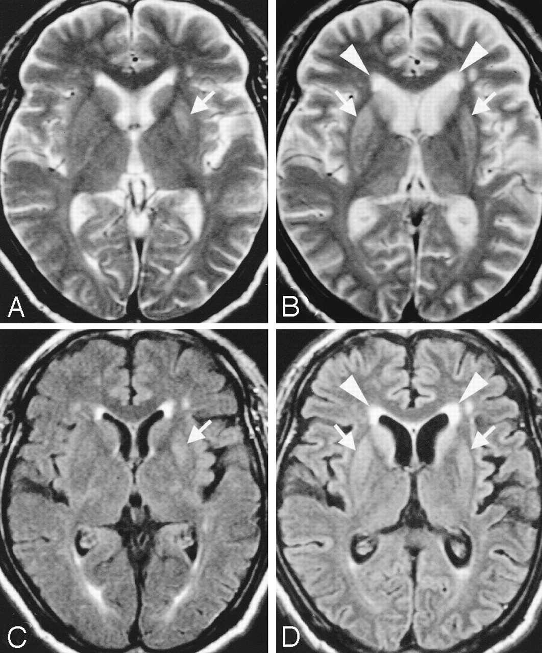
- Fig 1.
Serial axial T2-weighted (A and B) and FLAIR (C and D) images in case 1 obtained 2 (A and C) and 4 (B and D) months after onset of symptoms. FLAIR and T2-weighted images show faint hyperintensity in the left putamen. The finding is more conspicuous on the FLAIR image (C, arrow) than on the T2- weighted image (A, arrow). FLAIR image shows hyperintensities not only in the bilateral striatum (D, arrows) and cerebral cortices, but also in the periventricular white matter (D, arrowheads) with mild cerebral atrophy 4 months after onset. T2-weighted images also show hyperintensities in both the striatum (B, arrows) and periventricular white matter (B, arrowheads), although not as conspicuously as on FLAIR images 4 months after onset. Signal intensity in the cerebral cortex is susceptible to the partial volume artifact with the surrounding CSF on T2-weighted images (A and B).
- Fig 2.
Serial axial T2-weighted MR images from case 4 obtained (A) 3 months, (B) 5 months, (C) 7 months, (D) 9 months, (E) 12 months, and (F) 18 months after onset of symptoms. Slight hyperintensities are shown around the lateral ventricles (B, arrows) 5 months after the onset. The periventricular hyperintensities are gradually extending to the deep and subcortical white matter in parallel with cortical atrophy (C–F).
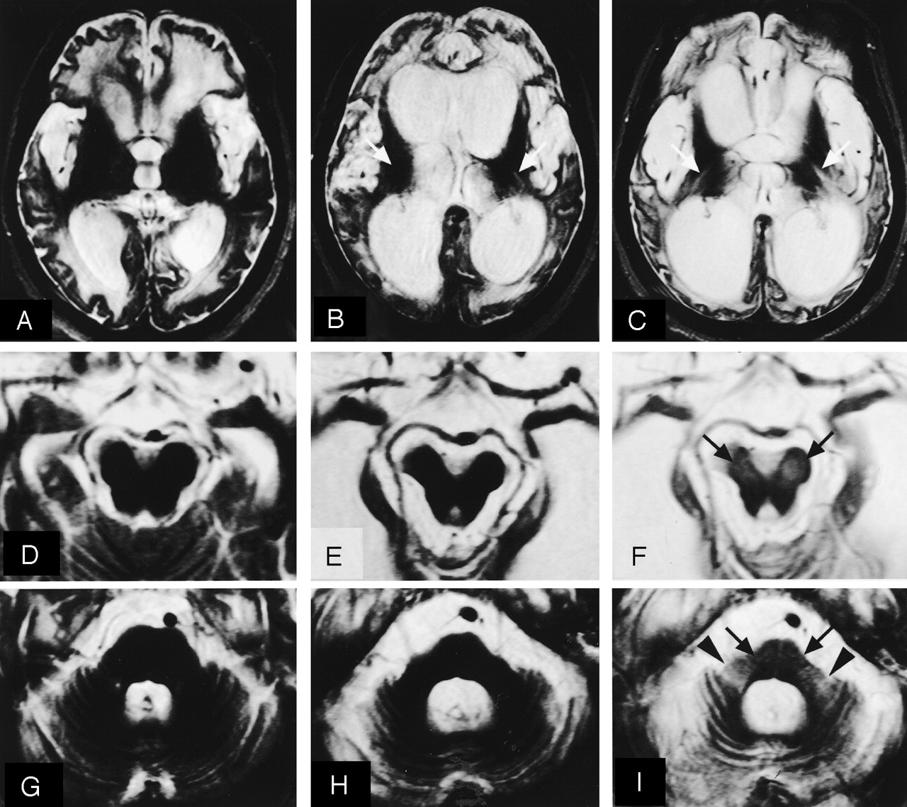
- Fig 3.
Serial axial T2-weighted MR images in case 6 obtained at 11 months (A, D, and G), 18 months (B, E, and H), and 28 months (C, F, and I) after the onset of symptoms. The degree of brain atrophy gradually becomes apparent. Only in this case, T2 hyperintensities are never seen in the basal ganglia at any stage. At 11 months after onset, diffuse T2 hyperintensities in the cerebral white matter are evident, but hyperintensities are not visible in the internal capsules (A). At 18 months, T2 hyperintensities are observed in both internal capsules (B, arrows). At 28 months, high signal intensity of the bilateral internal capsules is apparent (C, arrows). T2 hyperintensities are also visible in both cerebral peduncles (F, arrows), pons (I, arrows), and middle cerebellar peduncles (I, arrowheads).
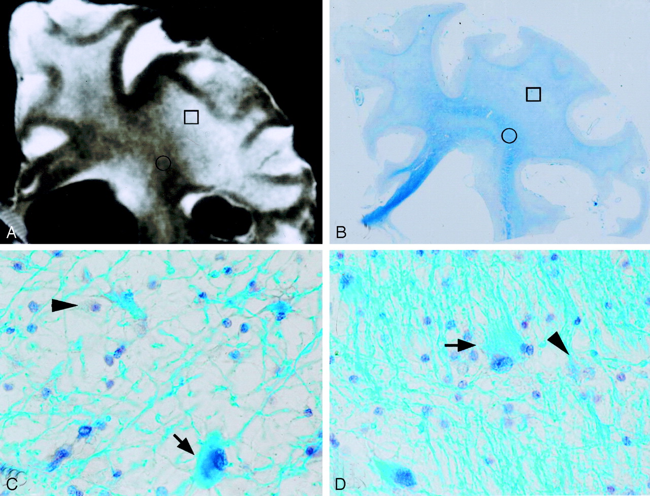
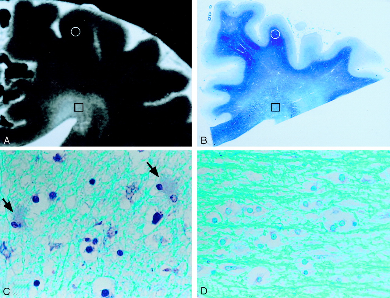
- Fig 4.
A, Postmortem coronal T2-weighted image of the right frontal lobe 22 months after the onset in case 3, showing diffuse hyperintensities in the cerebral white matter. B, Myelin-stained section corresponding to panel A, demonstrating diffuse pallor of the cerebral white matter. C, Histologic specimen of the area of highly increased signal intensity (A and B, squares), revealing a severe loss of myelin and axons, tissue rarefaction, foamy macrophages (arrowhead), and proliferation of gemistocytic astrocytes (arrow). D, Area of moderately increased signal intensity (A and B, circles), showing mild tissue rarefaction. There are also foamy macrophages (arrowhead) and gemistocytic astrocytes (arrow). Because tissue rarefaction and gemistocytic astrocytosis are exceptional features in secondary degeneration, these histologic features indicate primary involvement of the white matter.
- Fig 5.
A, Postmortem coronal T2-weighted image of the right frontal lobe 8 months after the onset of symptoms in case 1, showing hyperintensities around the anterior horn of the lateral ventricle and adjacent deep cerebral white matter. B, Myelin-stained section corresponding to panel A, showing pallor of the cerebral white matter, which is more widespread than the extent of hyperintensities on the MR image. C, Histologic specimen of the area of moderately increased signal intensity (A and B, squares), revealing mild loss of myelin and axons with a few hypertrophic astrocytes (arrows). D, Area of normal signal intensity (A and B, circles), showing no apparent degenerative changes.
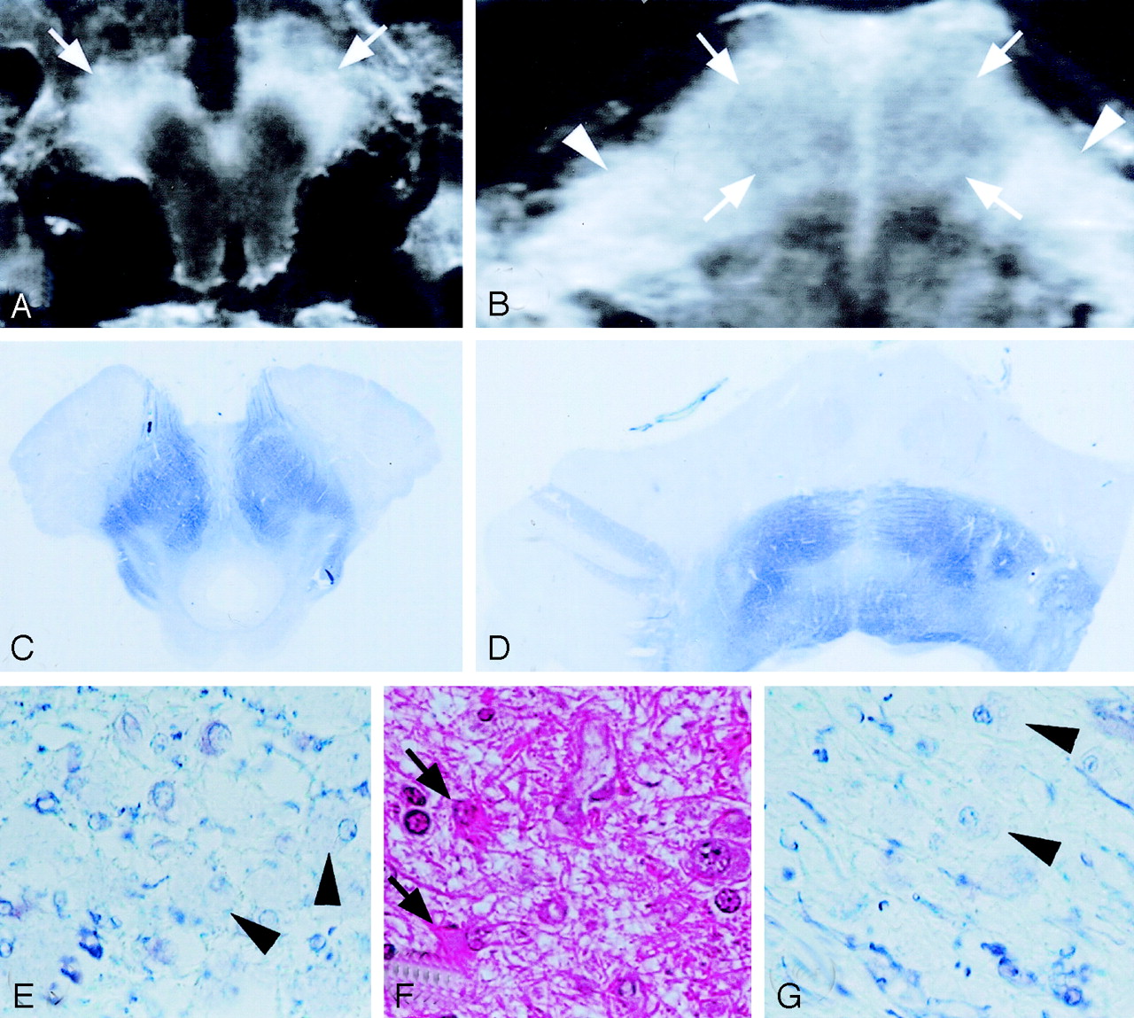
- Fig 6.
A and B, Postmortem axial T2-weighted images of the midbrain (A) and pons (B) 28 months after the onset of symptoms in case 5, showing hyperintensities in the cerebral peduncles (A, arrows), pons (B, arrows), and middle cerebellar peduncles (B, arrowheads) involving the longitudinal fasciculi, pontine nuclei, and pontine transverse fibers. C and D, The myelin-stained sections corresponding to panels A and B demonstrate severe pallor of the cerebral peduncles (C), pons, and middle cerebellar peduncles (D). E, Histologically, the cerebral peduncle shows severe loss of myelin and axons with moderate macrophage infiltration (arrowheads) and fibrillary gliosis, characteristics compatible with secondary degeneration. F, Pontine nuclei reveal neuronal loss and proliferation of hypertrophic astrocytes (arrows). G, Middle cerebellar peduncle shows severe loss of myelin and axons with moderate macrophage infiltration (arrowheads) but no gemistocytic astrocytes, a characteristic compatible with secondary degeneration.


Tables
Summary of clinical and pathologic findings in six cases of pCJD
Case No. Age (y)/Sex Duration of Illness (mo) Clinical Findings (mo after onset) Brain Weight (gs) Histologic Findings (gray matter: spongiform change, neuronal loss and gliosis; white matter: loss of myelin and axons) Cerebral Cortex Cerebral White Matter Internal Capsule Basal Ganglia Thalamus Brain Stem Middle Cerebellar Peduncle 1 66/M 8 Dementia (1); change of character (0); myoclonus (3); ataxia (2); akinetic mutism (4); dysarthria (2); PSD (4) 1210 ++ +–++ − ST: ++ ++ CP: − − GP: ++ PLF: − PN: − PTF: − 2 64 /F 15 Dementia (1); ataxia (0); myoclonus (1); tremor (1); akinetic mutism (2); dystonia (1); PSD 900 +++ +++ − ST: +++ ++ CP: − − GP: ++ PLF: + PN: − PTF: + 3 78 /F 22 Dementia (0); tremor (1); myoclonus (2); diplopia (1); akinetic mutism (3); PSD (2) 740 +++ +++ − ST: +++ ++–+++ CP: − ++ GP: +++ PLF: − PN: + PTF: ++ 4 76 /F 25 Dementia (2); tremor (0); myoclonus (2); hemiplegia (1); akinetic mutism (3); PSD 720 +++ +++ − ST: +++ ++–+++ CP: + + GP: ++ PLF: + PN: + PTF: ++ 5 68 /F 28 Dementia (1); ataxia (0); myoclonus (2); chorea (1); akinetic mutism (2); PSD 650 +++ +++ ++ ST: +++ ++–+++ CP: +++ +++ GP: ++ PLF: +++ PN: +++ PTF: +++ 6 82 /F 30 Dementia (0); gait disturbance (5); myoclonus (5); akinetic mutism (6); PSD 740 +++ +++ ++ ST: +++ ++–+++ CP: +++ +++ GP: ++ PLF: +++ PN: +++ PTF: +++ Note.—PSD, periodic synchronous discharge on electroencephalogram; ST, striatum; GP, globus pallidus; CP, cerebral peduncle; PN, pontine nucleus; PTF, pontine transverse fibers; PLF, pontine longitudinal fibers; +, mild; ++, moderate; +++, marked.


