
Article Figures & Data
Figures
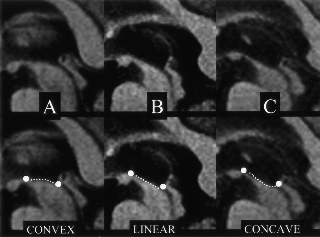
- Fig 1.
Top row, Midsagittal T1-weighted spin-echo sections in PD (A) and PSP (B and C) show the midbrain region. Bottom row, Same images with outlined profiles of the upper midbrain, which appears convex in A, linear (flat) in B, and concave in C.
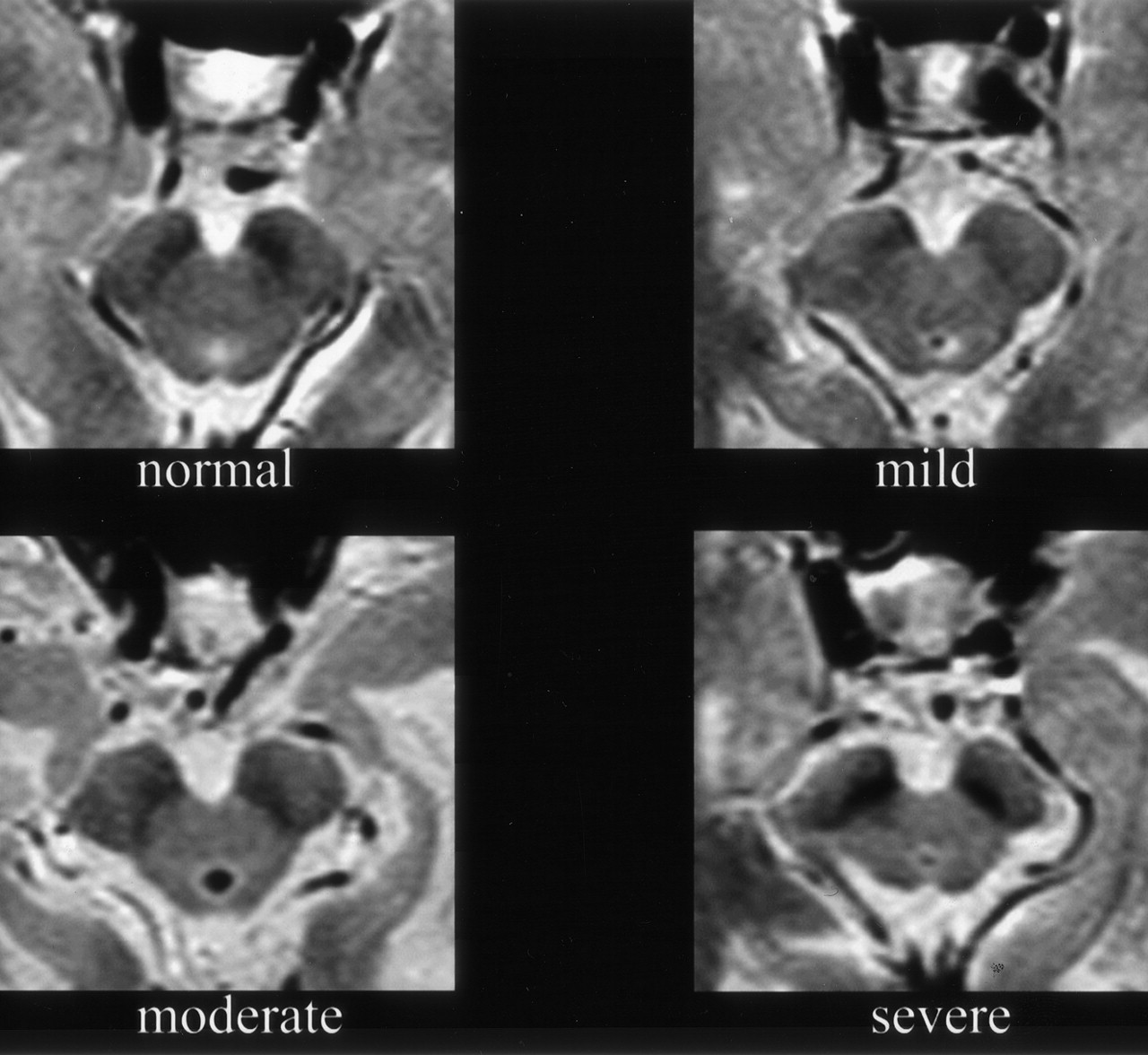
- Fig 2.
Axial, 3-mm-thick, T2-weighted spin-echo sections show different degrees of midbrain atrophy.
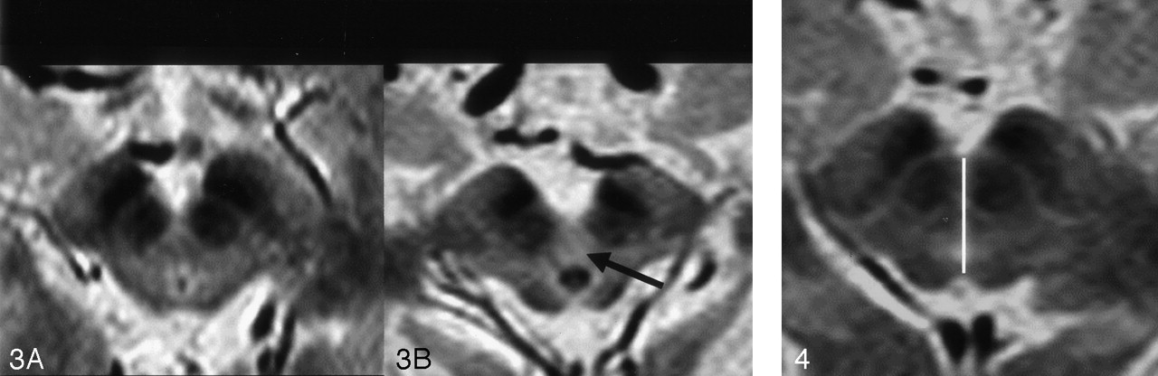
- Fig 3.
Axial, 3-mm-thick, T2-weighted spin-echo sections show no abnormal tegmental hyperintensity (A) and abnormal tegmental hyperintensity (B, arrow).
- Fig 4.
Axial, 3-mm-thick, T2-weighted spin-echo section depicts the method used to measure the AP diameter of the midbrain at the level of the superior colliculus.
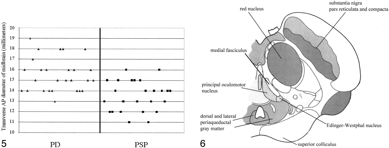
- Fig 5.
Graph illustrates the distribution of midbrain transverse AP diameters for the PSP and PD populations. A threshold value of 12 mm included only seven of 25 patients with PSP.
- Fig 6.
Diagrammatic representation of an axial section of the midbrain at the level of superior colliculus (modified from figure 54 of Duvernoy HM, The Human Brain Stem and Cerebellum, 1995, with permission of Springer-Verlag, Wien). Some of the structures affected in PSP have been reported. Other structures of the dorsal and cranial part of the midbrain affected in PSP (ie, nucleus interstitialis of Cajal and pretectal area) are located on the contiguous cranial section.


Tables
Parameter PD (n = 27) PSP (n = 25) 1) Upper midbrain profile sign Convex 24 8 Linear 3 11 Concave 0 6 2) Global midbrain atrophy Normal 21 8 Mild 6 8 Moderate 0 6 Severe 0 3 3) Tegmental T2 hyperintensity Yes 0 7 No 27 18 4) Putaminal T2 hypointensity Yes 4 6 No 23 19 5) Putaminal proton-density hyperintensity Yes 0 0 No 27 25
In this issue


Jump to section
Related Articles
Cited By...
- Atypical Parkinsonian Syndromes: Structural, Functional, and Molecular Imaging Features
- Neuroimaging of Rapidly Progressive Dementias, Part 1: Neurodegenerative Etiologies
- Differentiation between idiopathic and atypical parkinsonian syndromes using three-dimensional magnetic resonance spectroscopic imaging
- The midbrain to pons ratio: A simple and specific MRI sign of progressive supranuclear palsy