
Article Figures & Data
Figures
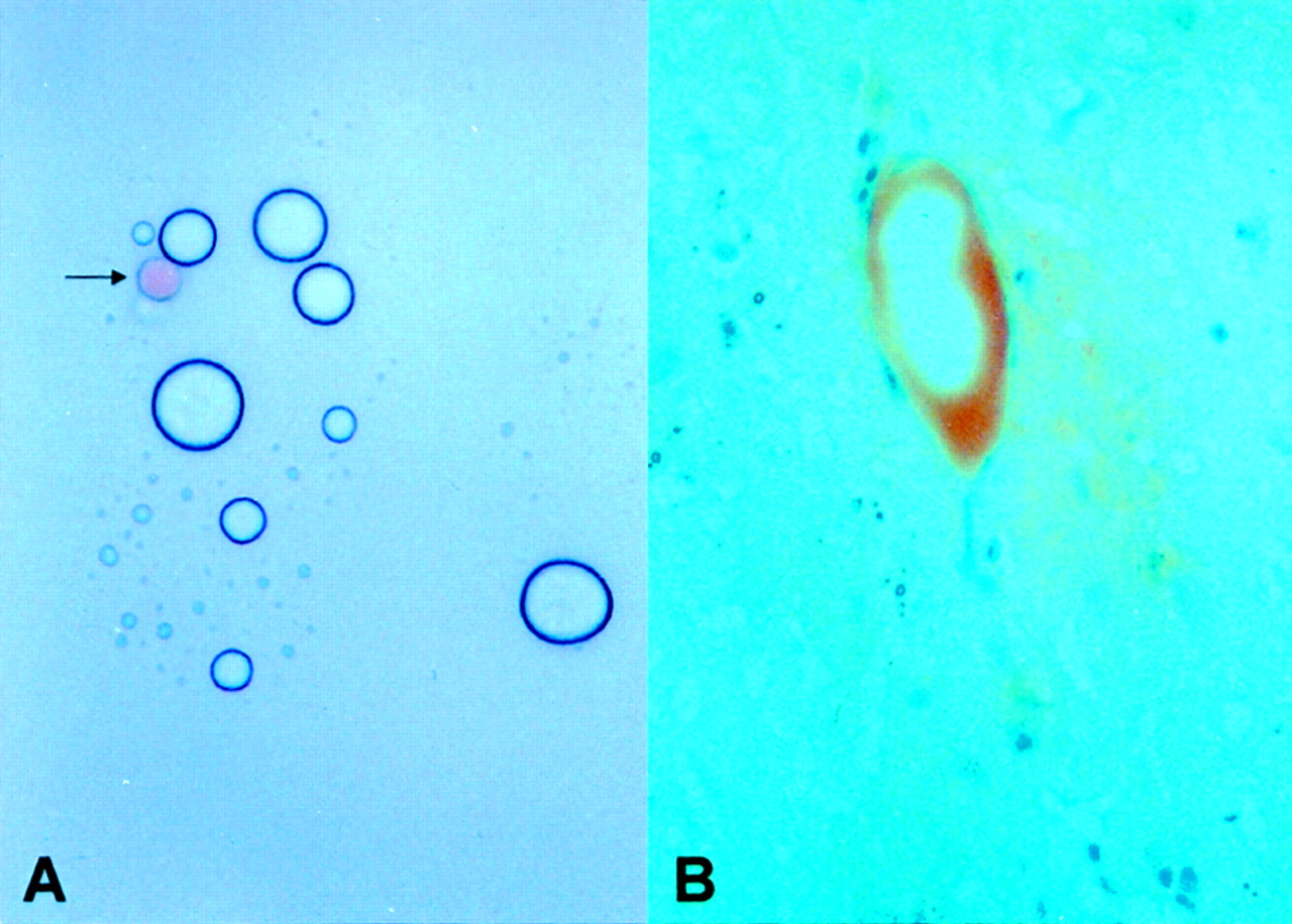
- Fig 1.
Fat globules.
A, Photomicrograph of the fat emulsion. The fat globules are of variable size, mostly less than two times the size of red blood cells (arrow, original magnification ×40).
B, An intravascular fat globule is pinkish-red on oil red O staining (original magnification ×200).
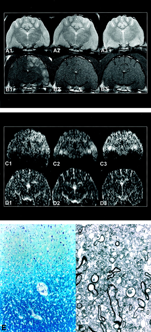
- Fig 2.
Images obtained in a cat in group 4: A indicates T2WIs; B, Gd-enhanced T1WIs; C, DWIs; D, ADC maps; E, light photomicrograph; F, electron photomicrograph. 1 indicates 1 hour after embolization; 2, 1 day; and 3, 7 days. At 1 hour, the embolized lesion in the left hemisphere appears hyperintense in A1, enhanced in B1, mildly hyperintense in C1, and isointense in D1. At 1 day, T2WI hyperintensity (A2) and contrast enhancement (B2) are substantially decreased and not evident at day 7 (A3, B3). After day 1, DWIs (C2, C3) and ADC maps (D2, D3) reveal isointensity of the lesion. In E, Light microscopy of the gray matter (top) and white matter (bottom) shows no evidence of demyelination (Luxol fast blue stain, original magnification ×100). In F, Electron microscopy of the gray matter shows no evidence of neuropil or interstitial swelling (original magnification ×3000).


Tables
Imaging* Hyperintensity Isointensity Hypointensity 1 h (n = 50) T1WI 0 39 11 T2WI 27 23 0 DWI 3 12 0 ADC 0 15 0 1 d (n = 43) T1WI 0 33 10 T2WI 12 31 0 DWI 0 15 0 ADC 1 14 0 4 d (n = 36) T1WI 0 34 2 T2WI 1 35 0 DWI 0 15 0 ADC 0 15 0 1 wk, (n = 29) T1WI 0 29 0 T2WI 0 29 0 DWI 0 15 0 ADC 0 15 0 2 wk (n = 14) T1WI 0 14 0 T2WI 0 15 0 3 wk (n = 7) T1WI 0 7 0 T2WI 0 7 0 Note.—Enhancement on Gd-enhanced T1WIs was as follows: at 1 hour, n = 44; at 1 day, n = 12; at 4 days, n = 1; and at 1, 2, and 3 weeks, n = 0.
* DWI and ADC in group 4 cats (n = 15).
Group Intravascular Fat Globule Defect in Blood-Brain Barrier Perivascular Interstitial Swelling Neuronal Swelling 1 (n = 7) 7 6 4 3 2 (n = 7) 7 5 5 7 3 (n = 7) 7 3 2 1 4 (n = 15) 12 9 2 5 5 (n = 7) 6 2 2 2 6 (n = 7) 6 1 0 0 Total (n = 50) 45 26 15 18
In this issue


Jump to section
Related Articles
Cited By...
- Dynamic MR Imaging Patterns of Cerebral Fat Embolism: A Systematic Review with Illustrative Cases
- Histopathological and biochemical changes following fat embolism with administration of corn oil micelles: A NEW ANIMAL MODEL FOR FAT EMBOLISM SYNDROME
- Clinical and pathological features of fat embolism with acute respiratory distress syndrome