Advertisement

Main menu

  • Home
  • Content
    • Current Issue
    • Accepted Manuscripts
    • Article Preview
    • Past Issue Archive
    • AJNR Case Collection
    • Case of the Week Archive
    • Classic Case Archive
    • Case of the Month Archive
  • Special Collections
    • Spinal CSF Leak Articles (Jan 2020-June 2024)
    • 2024 AJNR Journal Awards
    • Most Impactful AJNR Articles
  • Multimedia
    • AJNR Podcast
    • AJNR Scantastics
    • Video Articles
  • For Authors
    • Submit a Manuscript
    • Author Policies
    • Fast publishing of Accepted Manuscripts
    • Graphical Abstract Preparation
    • Manuscript Submission Guidelines
    • Imaging Protocol Submission
    • Submit a Case for the Case Collection
  • About Us
    • About AJNR
    • Editorial Board
  • More
    • Become a Reviewer/Academy of Reviewers
    • Subscribers
    • Permissions
    • Alerts
    • Feedback
    • Advertisers
    • ASNR Home
  • Other Publications
    • ajnr

User menu

  • Alerts
  • Log in

Search

  • Advanced search
American Journal of Neuroradiology

American Journal of Neuroradiology

ASHNR American Society of Functional Neuroradiology ASHNR American Society of Pediatric Neuroradiology ASSR
  • Alerts
  • Log in

Advanced Search

  • Home
  • Content
    • Current Issue
    • Accepted Manuscripts
    • Article Preview
    • Past Issue Archive
    • AJNR Case Collection
    • Case of the Week Archive
    • Classic Case Archive
    • Case of the Month Archive
  • Special Collections
    • Spinal CSF Leak Articles (Jan 2020-June 2024)
    • 2024 AJNR Journal Awards
    • Most Impactful AJNR Articles
  • Multimedia
    • AJNR Podcast
    • AJNR Scantastics
    • Video Articles
  • For Authors
    • Submit a Manuscript
    • Author Policies
    • Fast publishing of Accepted Manuscripts
    • Graphical Abstract Preparation
    • Manuscript Submission Guidelines
    • Imaging Protocol Submission
    • Submit a Case for the Case Collection
  • About Us
    • About AJNR
    • Editorial Board
  • More
    • Become a Reviewer/Academy of Reviewers
    • Subscribers
    • Permissions
    • Alerts
    • Feedback
    • Advertisers
    • ASNR Home
  • Follow AJNR on Twitter
  • Visit AJNR on Facebook
  • Follow AJNR on Instagram
  • Join AJNR on LinkedIn
  • RSS Feeds

Welcome to the new AJNR, Updated Hall of Fame, and more. Read the full announcements.


AJNR is seeking candidates for the position of Associate Section Editor, AJNR Case Collection. Read the full announcement.


Research ArticleBRAIN

Dynamic Susceptibility Contrast-Enhanced Perfusion and Conventional MR Imaging Findings for Adult Patients with Cerebral Primitive Neuroectodermal Tumors

Meng Law, Khuram Kazmi, Stephan Wetzel, Edwin Wang, Codrin Iacob, David Zagzag, John G. Golfinos and Glyn Johnson
American Journal of Neuroradiology June 2004, 25 (6) 997-1005;
Meng Law
  • Find this author on Google Scholar
  • Find this author on PubMed
  • Search for this author on this site
Khuram Kazmi
  • Find this author on Google Scholar
  • Find this author on PubMed
  • Search for this author on this site
Stephan Wetzel
  • Find this author on Google Scholar
  • Find this author on PubMed
  • Search for this author on this site
Edwin Wang
  • Find this author on Google Scholar
  • Find this author on PubMed
  • Search for this author on this site
Codrin Iacob
  • Find this author on Google Scholar
  • Find this author on PubMed
  • Search for this author on this site
David Zagzag
  • Find this author on Google Scholar
  • Find this author on PubMed
  • Search for this author on this site
John G. Golfinos
  • Find this author on Google Scholar
  • Find this author on PubMed
  • Search for this author on this site
Glyn Johnson
  • Find this author on Google Scholar
  • Find this author on PubMed
  • Search for this author on this site
  • Article
  • Figures & Data
  • Info & Metrics
  • Responses
  • References
  • PDF
Loading

Article Figures & Data

Figures

  • Tables
  • Fig 1.
    • Download figure
    • Open in new tab
    • Download powerpoint
    Fig 1.

    Images of a 58-year-old man (patient 5) with resection-proved PNET.

    A, Contrast-enhanced axial view T1-weighted image (600/14/1) shows a fairly well-defined enhancing mass in the right frontal region. Approximately 70% of the lesion enhanced. Cystic/necrotic components can be seen in the lesion.

    B, Axial view T2-weighted image (3400/119/1) shows some areas of hypointensity within the mass, with a small amount of peritumoral edema and mass effect.

    C, Gradient-echo axial view perfusion MR image (1000/54) and rCBV color overlay map show increased perfusion with a high rCBV of 6.98 and vascular permeability (Ktrans) of 0.00017 s−1.

  • Fig 2.
    • Download figure
    • Open in new tab
    • Download powerpoint
    Fig 2.

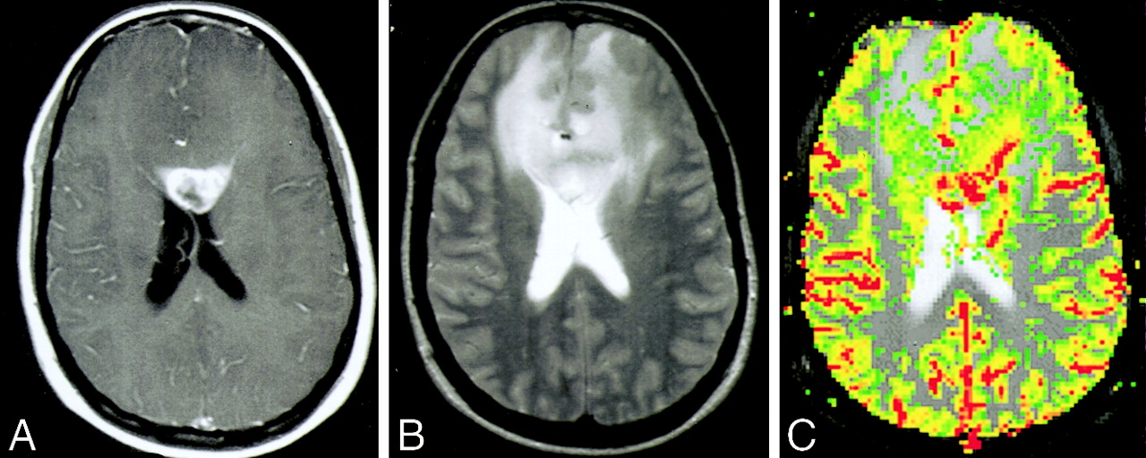
    Images of a 34-year-old woman (patient 11) with biopsy-proved PNET.

    A, Contrast-enhanced axial view T1-weighted image (600/14/1) shows a peripherally enhancing mass in the genu of the corpus callosum. Approximately 50% of the lesion enhanced. Cystic/necrotic components can be seen in the lesion.

    B, Axial view T2-weighted image (3400/119/1) shows some areas of hypointensity within the mass, indicating the increased cellularity of these tumors, with a significant amount of peritumoral edema and mass effect. The signal intensity characteristics and involvement of the corpus callosum would raise the possibility of lymphoma as a differential diagnosis.

    C, Gradient-echo axial view perfusion MR image (1000/54) and rCBV color overlay map show increased perfusion with a high rCBV of 6.82 and vascular permeability (Ktrans) of 0.00034 s−1. Lymphoma would appear as reduced perfusion.

  • Fig 3.
    • Download figure
    • Open in new tab
    • Download powerpoint
    Fig 3.

    Images of a 52-year-old woman with a histologically confirmed grade II/IV glioma (A−C) and a 70-year-old woman with a histologically confirmed grade III/IV glioma (D−F).

    A, Contrast-enhanced axial view T1-weighted image shows a lesion in the right temporoparietal region with low signal intensity and minimal enhancement.

    B, Axial view T2-weighted image shows increase in T2 signal intensity within the lesion, with minimal edema.

    C, Gradient-echo axial view perfusion MR image and rCBV color overlay map show a low rCBV of 1.70, in keeping with a low grade glioma. A thin rim of minimally increased perfusion can be seen at the margin of the lesion. This is compared with the increased rCBV of PNETs shown in Figures 1C and 2C.

    D, Contrast-enhanced axial view T1-weighted image shows a lesion in the right thalamic region with heterogeneous peripheral contrast enhancement and a central cystic/necrotic region.

    E, Axial view T2-weighted image shows increase in T2 signal intensity within the lesion with moderate surrounding edema. The patient also has hydrocephalus and transependymal edema around the ventricles.

    F, Gradient-echo axial view perfusion MR image and rCBV color overlay map show a high rCBV of 3.70, in keeping with a high grade glioma. A thick rind of marked increased perfusion cannot be readily differentiated from the increased rCBV of PNETs shown in Figures 1C and 2C.

  • Fig 4.
    • Download figure
    • Open in new tab
    • Download powerpoint
    Fig 4.

    Photomicrographs of PNETs.

    A, Hematoxylin and eosin stain shows a hypercellular neoplasm. Poorly differentiated, primitive small cells growing in compact sheets, forming a characteristic Homer-Wright rosette (R), can be seen. Tumor cells have round to oval hyperchromatic nuclei with scant cytoplasm. Mitotic figures are also identified (arrow). The increased cellularity explains iso- or hypointensity of many of the lesions on T2-weighted images (original magnification, ×200).

    B, Hematoxylin and eosin stain shows increased vascular channels (arrows) in PNET (original magnification, ×50).

    C, Hematoxylin and eosin stain at higher power shows endothelial hyperplasia (arrows) (original magnification, ×400).

    D, Azocarmine stain highlights the vascular channels and shows proliferation and thickening of the vascular endothelium within the PNET (original magnification, ×100).

Tables

  • Figures
    • View popup
    TABLE 1:

    Conventional MR imaging findings in primitive neuroectodermal tumors

    Patient No.Volume (cm3)T1 SignalT2 SignalEnhancement* (%)EdemaMass EffectNecrotic/CysticLesion DelineationHeterogeneityCalcification
    15 × 3 × 2.5IsoIso50++YesWellYesYes
    22.5 × 2 × 2IsoHypo90−+YesWellYesNo
    36 × 6 × 6HypoIso30+++++YesWellYesNo
    46 × 6 × 6HypoIso80+++YesWellYesNo
    53.5 × 2.5 × 1.5IsoHyper70+++YesWellYesNo
    63.5 × 4 × 3HypoHyper50+++YesIllYesYes
    75 × 3 × 2IsoIso50++YesIllYesNo
    82.5 × 1.5 × 2IsoHyper80−−NoWellNoNo
    93 × 3 × 3IsoHyper90++NoWellYesNo
    107 × 6 × 5IsoIso30+++++YesWellYesYes
    111.5 × 2 × 2IsoHypo50++++YesWellYesNo
    121 × 1 × 1IsoHypo50−−NoWellNoNo
    • Note.—Iso indicates isointense to gray matter; Hypo, hypointense; Hyper, hyperintense; +, mild; ++, moderate; +++, severe.

    • * Enhancement is shown as the percentage of the lesion volume that enhanced.

    • View popup
    TABLE 2:

    Relative cerebral blood volume measurements and vascular permeability measurements in patients with primitive neuroectodermal tumors

    Patient No.Age (y)/SexLocationrCBVSDKtransSD
    128/MTemp/brain stem/cerebellum8.903.00 × 10−04
    237/FThalamus/brain stem3.335.70 × 10−03
    342/MRight frontal4.289.20 × 10−03
    468/MBifrontal3.971.20 × 10−03
    558/MRight frontal6.981.70 × 10−04
    652/MRight frontal4.344.80 × 10−05
    746/MRight temporal5.438.40 × 10−03
    846/FPineal5.115.20 × 10−03
    983/FLeft parietal2.725.90 × 10−04
    1042/MTemporoparietal2.283.35 × 10−05
    1134/FBifrontal6.823.40 × 10−04
    1222/MSuprasellar2.912.00 × 10−03
    Mean46.504.761.990.00330.0035
    • Note.—M indicates male; F, female; Temp, temporal; rCBV, relative cerebral blood volume; Ktrans, vascular permeability (s−1).

    • View popup
    TABLE 3:

    Comparison of relative cerebral blood volume and vascular permeability measurements in patients with primitive neuroectodermal tumors with low grade gliomas (grade II/IV) and high grade gliomas (grade III/IV and IV/V)

    Tumor TyperCBVSDKtransSD
    PNET (n = 12)4.761.990.00330.0035
    Low grade glioma (n = 30)2.141.670.00050.001
    P value<.0005<.05
    High grade glioma (n = 55)5.823.570.00160.003
    P value0.530.19
    • Note.—rCBV indicates relative cerebral blood volume; Ktrans, vascular permeability (s−1); PNET, primitive neuroectodermal tumor.

PreviousNext
Back to top

In this issue

American Journal of Neuroradiology: 25 (6)
American Journal of Neuroradiology
Vol. 25, Issue 6
1 Jun 2004
  • Table of Contents
  • Index by author
Advertisement
Print
Download PDF
Email Article

Thank you for your interest in spreading the word on American Journal of Neuroradiology.

NOTE: We only request your email address so that the person you are recommending the page to knows that you wanted them to see it, and that it is not junk mail. We do not capture any email address.

Enter multiple addresses on separate lines or separate them with commas.
Dynamic Susceptibility Contrast-Enhanced Perfusion and Conventional MR Imaging Findings for Adult Patients with Cerebral Primitive Neuroectodermal Tumors
(Your Name) has sent you a message from American Journal of Neuroradiology
(Your Name) thought you would like to see the American Journal of Neuroradiology web site.
CAPTCHA
This question is for testing whether or not you are a human visitor and to prevent automated spam submissions.
Cite this article
Meng Law, Khuram Kazmi, Stephan Wetzel, Edwin Wang, Codrin Iacob, David Zagzag, John G. Golfinos, Glyn Johnson
Dynamic Susceptibility Contrast-Enhanced Perfusion and Conventional MR Imaging Findings for Adult Patients with Cerebral Primitive Neuroectodermal Tumors
American Journal of Neuroradiology Jun 2004, 25 (6) 997-1005;

Citation Manager Formats

  • BibTeX
  • Bookends
  • EasyBib
  • EndNote (tagged)
  • EndNote 8 (xml)
  • Medlars
  • Mendeley
  • Papers
  • RefWorks Tagged
  • Ref Manager
  • RIS
  • Zotero
0 Responses
Respond to this article
Share
Bookmark this article
Dynamic Susceptibility Contrast-Enhanced Perfusion and Conventional MR Imaging Findings for Adult Patients with Cerebral Primitive Neuroectodermal Tumors
Meng Law, Khuram Kazmi, Stephan Wetzel, Edwin Wang, Codrin Iacob, David Zagzag, John G. Golfinos, Glyn Johnson
American Journal of Neuroradiology Jun 2004, 25 (6) 997-1005;
del.icio.us logo Twitter logo Facebook logo Mendeley logo
  • Tweet Widget
  • Facebook Like
  • Google Plus One

Jump to section

  • Article
    • Abstract
    • Methods
    • Results
    • Discussion
    • Conclusion
    • Footnotes
    • References
  • Figures & Data
  • Info & Metrics
  • Responses
  • References
  • PDF

Related Articles

  • No related articles found.
  • PubMed
  • Google Scholar

Cited By...

  • MRI Characteristics of Ependymoblastoma: Results from 22 Centrally Reviewed Cases
  • Effects of Microvascular Permeability Changes on Contrast-Enhanced T1 and Pharmacokinetic MR Imagings After Ischemia
  • Dynamic contrast-enhanced imaging techniques: CT and MRI
  • Crossref
  • Google Scholar

This article has not yet been cited by articles in journals that are participating in Crossref Cited-by Linking.

More in this TOC Section

  • Fast Contrast-Enhanced 4D MRA and 4D Flow MRI Using Constrained Reconstruction (HYPRFlow): Potential Applications for Brain Arteriovenous Malformations
  • Quiet PROPELLER MRI Techniques Match the Quality of Conventional PROPELLER Brain Imaging Techniques
  • Predictors of Reperfusion in Patients with Acute Ischemic Stroke
Show more Brain

Similar Articles

Advertisement

Indexed Content

  • Current Issue
  • Accepted Manuscripts
  • Article Preview
  • Past Issues
  • Editorials
  • Editors Choice
  • Fellow Journal Club
  • Letters to the Editor

Cases

  • Case Collection
  • Archive - Case of the Week
  • Archive - Case of the Month
  • Archive - Classic Case

Special Collections

  • Special Collections

Resources

  • News and Updates
  • Turn around Times
  • Submit a Manuscript
  • Author Policies
  • Manuscript Submission Guidelines
  • Evidence-Based Medicine Level Guide
  • Publishing Checklists
  • Graphical Abstract Preparation
  • Imaging Protocol Submission
  • Submit a Case
  • Become a Reviewer/Academy of Reviewers
  • Get Peer Review Credit from Publons

Multimedia

  • AJNR Podcast
  • AJNR SCANtastic
  • Video Articles

About Us

  • About AJNR
  • Editorial Board
  • Not an AJNR Subscriber? Join Now
  • Alerts
  • Feedback
  • Advertise with us
  • Librarian Resources
  • Permissions
  • Terms and Conditions

American Society of Neuroradiology

  • Not an ASNR Member? Join Now

© 2025 by the American Society of Neuroradiology All rights, including for text and data mining, AI training, and similar technologies, are reserved.
Print ISSN: 0195-6108 Online ISSN: 1936-959X

Powered by HighWire