
Article Figures & Data
Figures
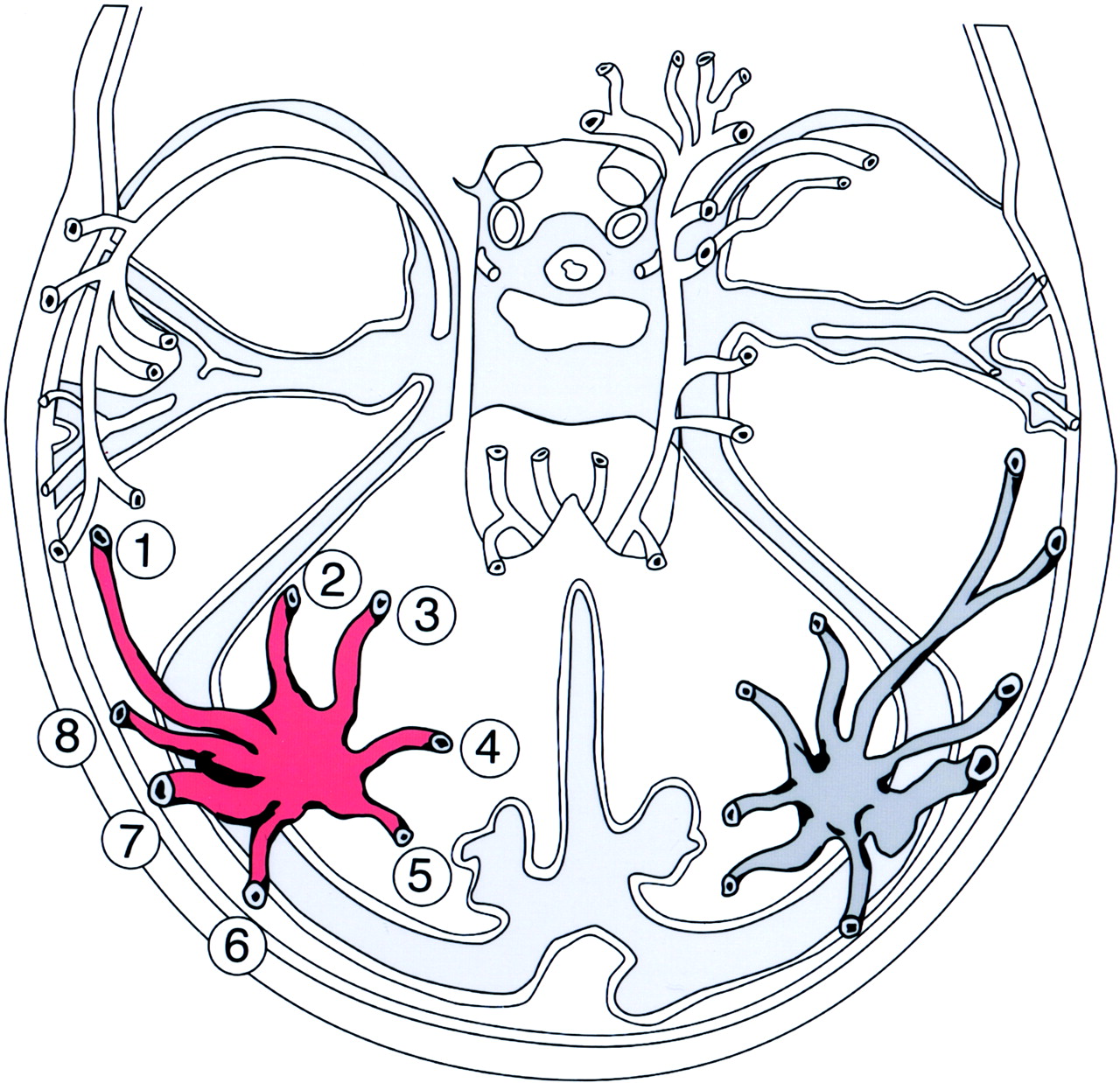
- Fig 1.
Direct superior of the LTS. 1, anterior temporal vein; 2, anterior temporobasal vein; 3, middle temporobasal vein; 4, posterior temporobasal vein; 5, occipitobasal vein; 6, posterior temporal vein; 7, vein of Labbé; 8, middle temporal vein.
- Fig 2.
LTS venous configuration.
A, Type I, venous candelabra.
B, Type II, multiple independent veins.
C, Type III, venous lakes within tentorium (14).
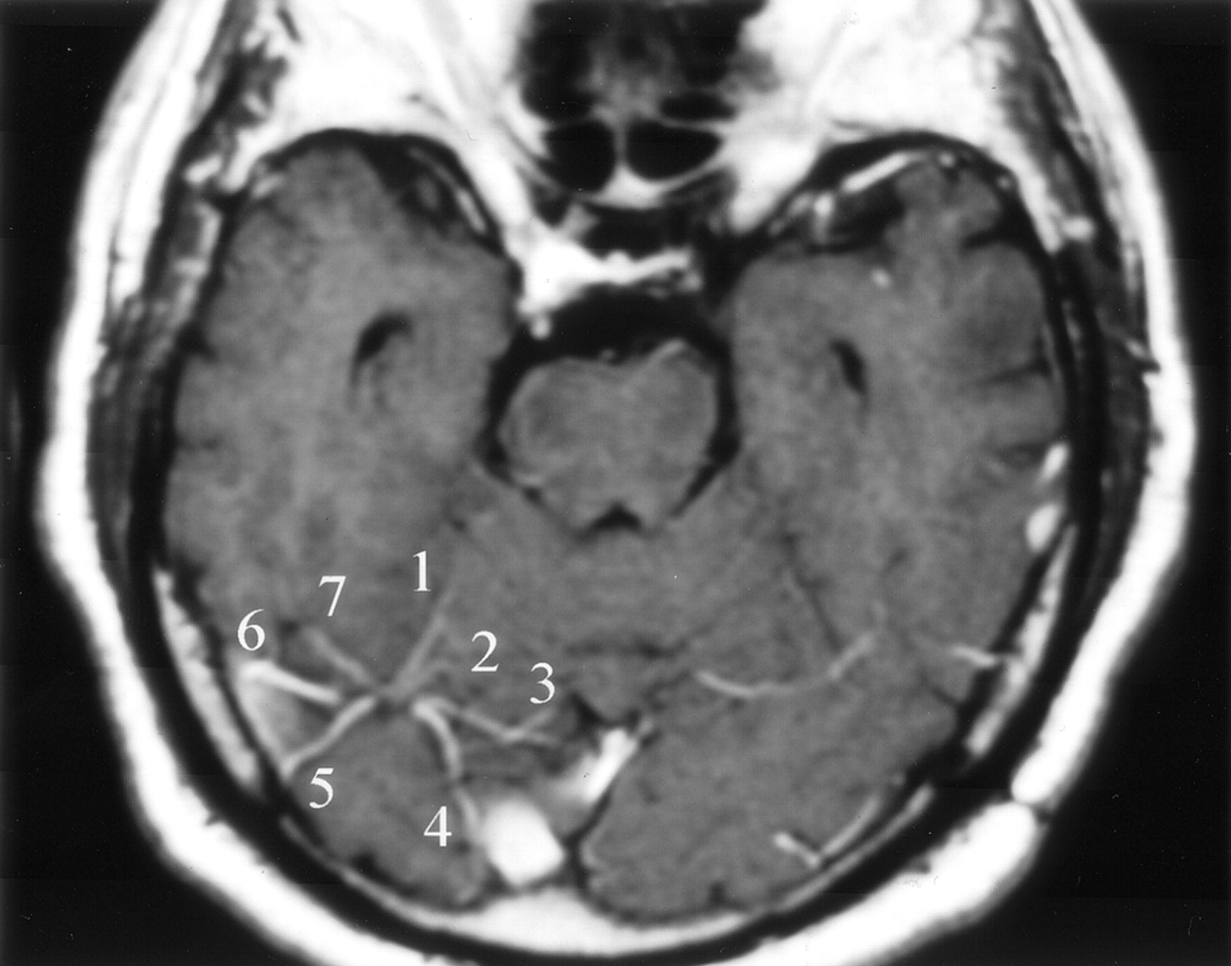
- Fig 3.
Type I LTS. Axial contrast-enhanced T1-weighted MR image obtained in a 37-year-old man shows several branches of the right LTS. 1, anterior temporobasal vein; 2, middle temporobasal vein; 3, posterior tomporobasal vein; 4, occiptiobasal vein; 5, posterior temporal vein; 6, vein of Labbé; 7, middle temporal vein. Left LTS is not well depicted on this image.
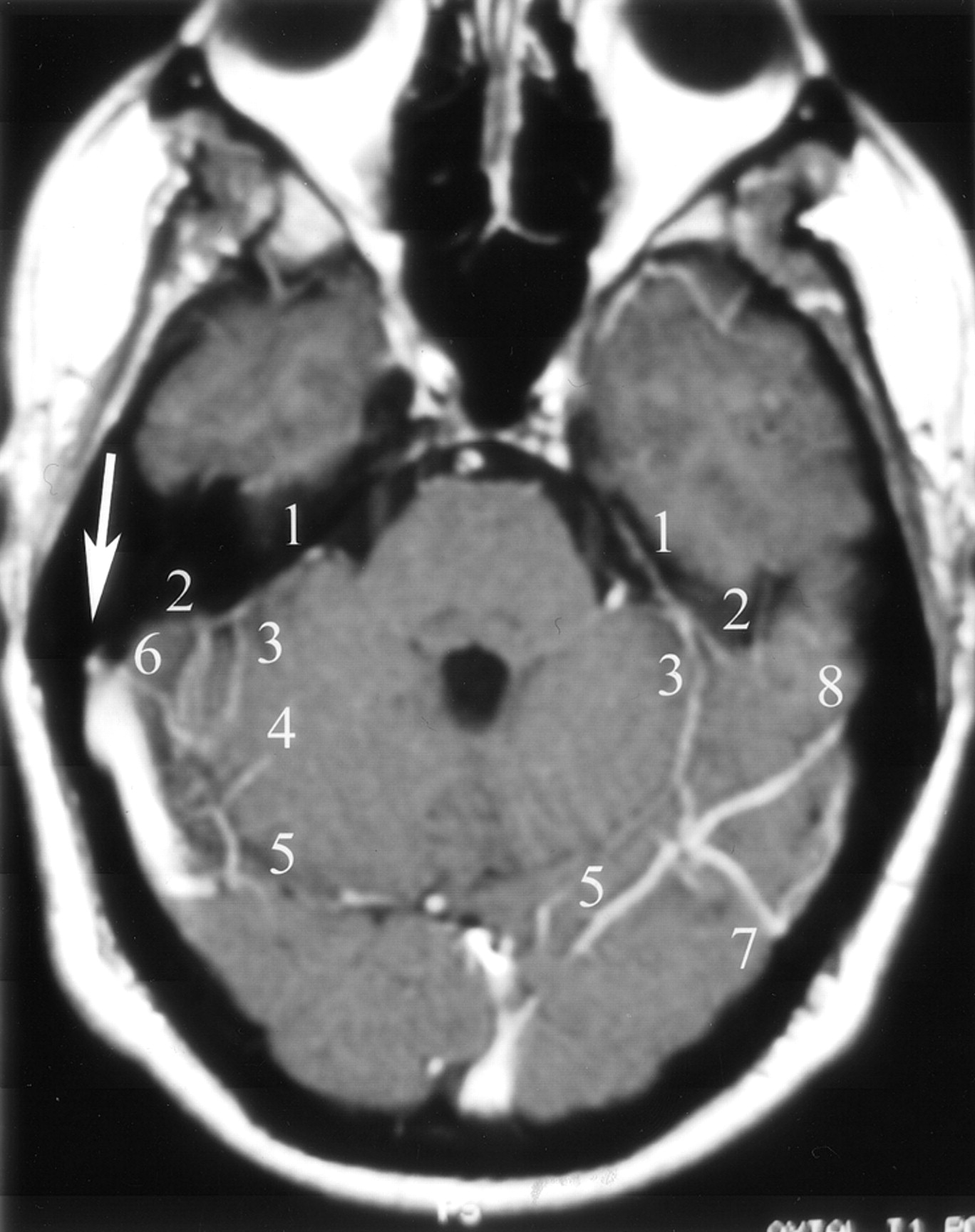
- Fig 4.
Types I and II LTS. Axial contrast-enhanced T1-weighted MR image obtained in a 55-year-old man with a type I LTS on the left and a type II LTS on the right. Right vein of Labbé terminates into the transverse sinus (arrow). 1, superior petrosal sinus; 2, anterior temporobasal vein; 3, middle temporobasal vein; 4, posterior temporobasal vein; 5, occipitobasal vein; 6, middle temporal vein; 7, posterior temporal vein; 8, vein of Labbé.
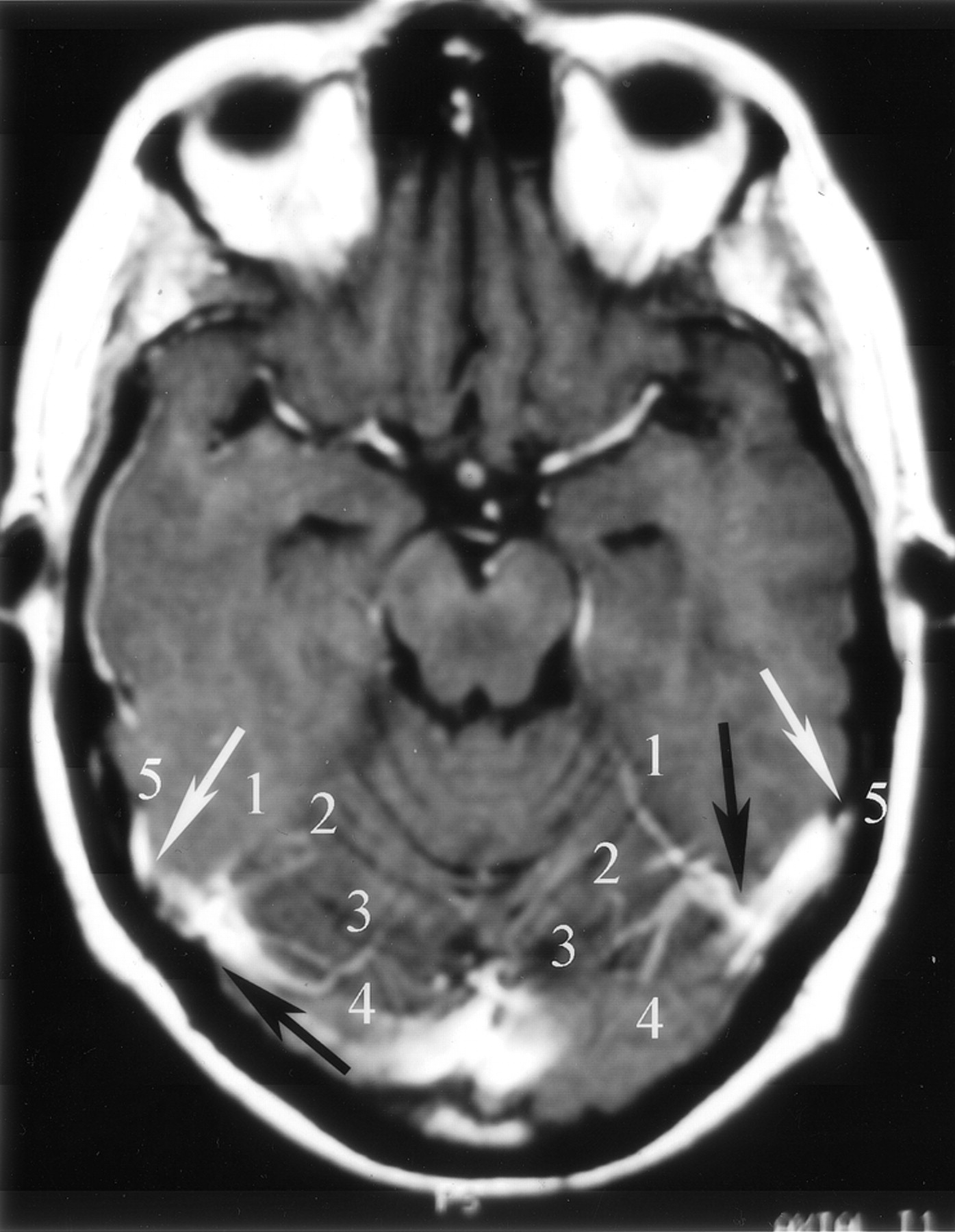
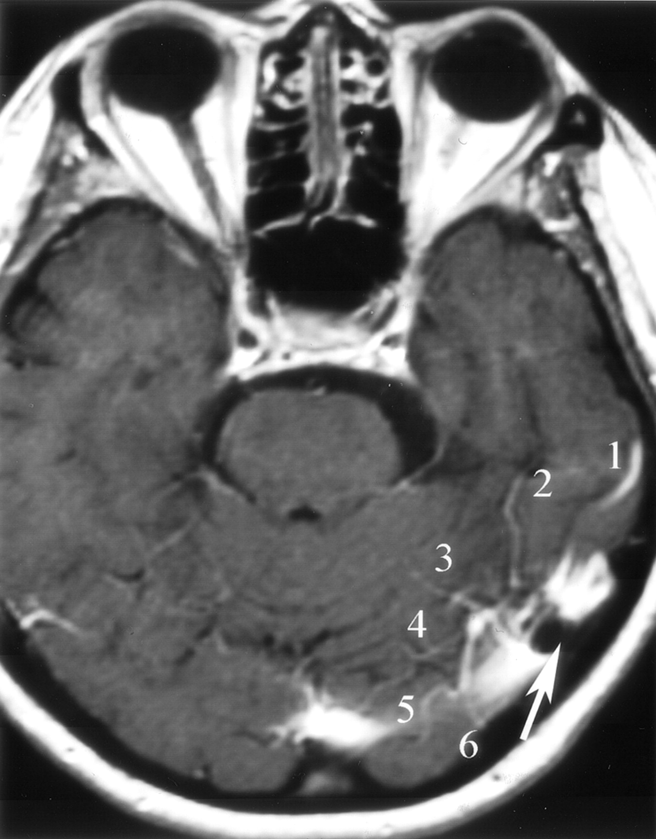
- Fig 5.
Types II and III LTS. Axial contrast-enhanced T1-weighted MR image obtained in a 34-year-old woman shows a type II LTS on the left and a type III LTS on the right side (black arrows). The veins of Labbé are draining separately into the transverse-sigmoid sinus junction (white arrows). 1, superior petrosal sinus; 2, middle temporobasal vein; 3, posterior temporobasal vein; 4, occipitobasal vein; 5, vein of Labbé.
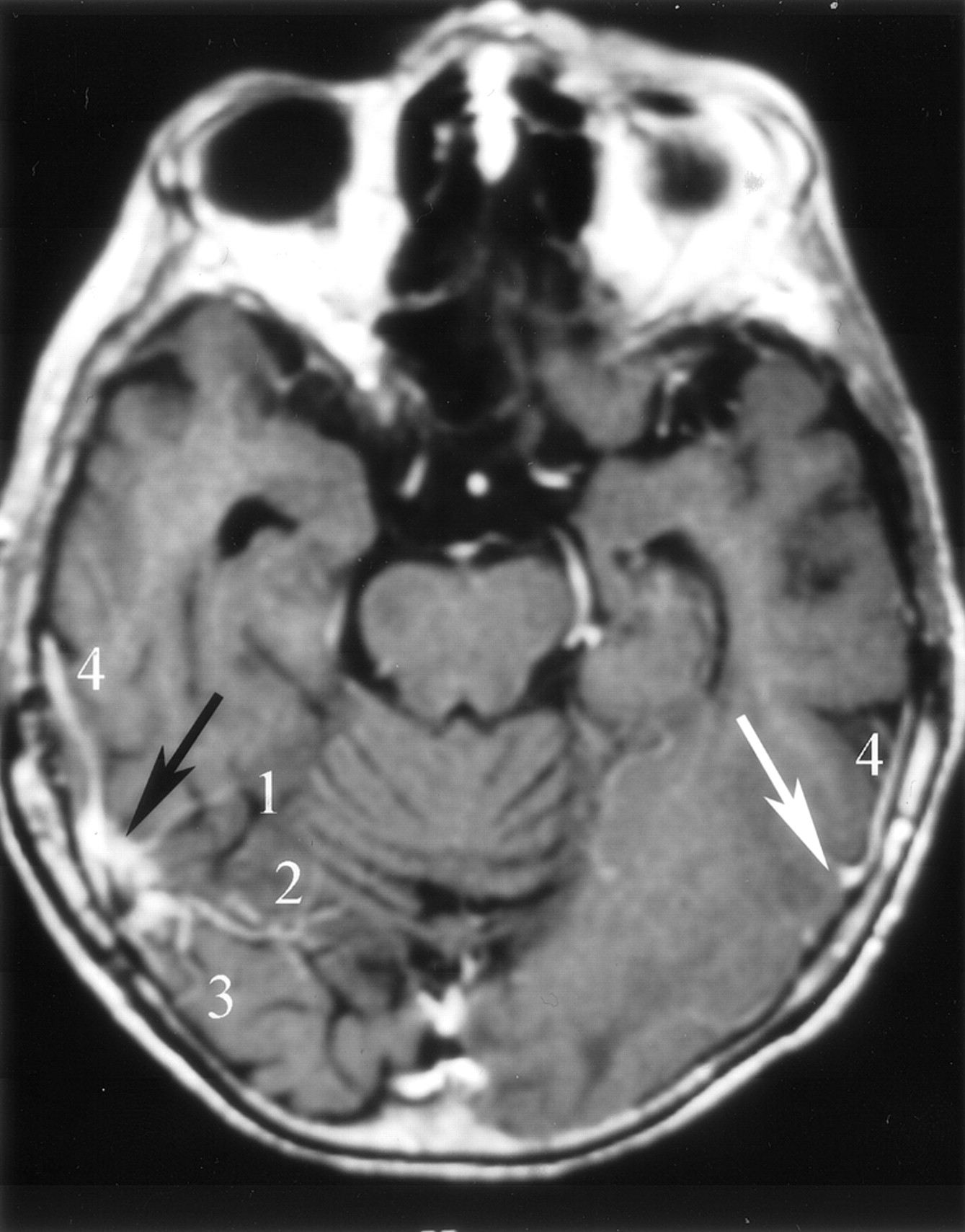
- Fig 6.
Type III LTS. Axial contrast-enhanced T1-weigthed MR image obtained in a 57-year-old man depicts “venous lakes” of a type III LTS (black arrow) and the left vein of Labbé just before it drains into the transverse sinus (white arrow). 1, middle temporobasal vein; 2, posterior temporobasal vein; 3, posterior temporal vein; 4, vein of Labbé.
- Fig 7.
Anterior temporal vein and anterior temporobasal vein common trunk. Axial contrast-enhanced T1-weighted MR image obtained in a 45-year-old woman with a type I LTS on the right side. Anterior temporal and anterior temporobasal branches form a trunk (arrow) before draining into the LTS. 1, anterior temporobasal vein; 2, posterior temporal vein; 3, vein of Labbé; 4, anterior temporal vein.
- Fig 8.
Vein of Labbé termination into the LTS. Axial contrast-enhanced T1-weighted MR image obtained in a 39-year-old man shows the VL draining into a type I LTS (arrow). 1, anterior temporobasal vein; 2, middle temporobasal vein; 3, posterior temporobasal vein; 4, occipitobasal vein; 5, posterior temporal vein; 6, vein of Labbé.
- Fig 9.
Termination of the vein of Labbé into the transverse sinus. Axial contrast-enhanced T1-weighted MR image obtained in a 42-year-old woman shows the right vein of Labbé (arrow) as it drains into the transverse-sigmoid sinus junction. 1, vein of Labbé; 2, middle temporobasal vein.
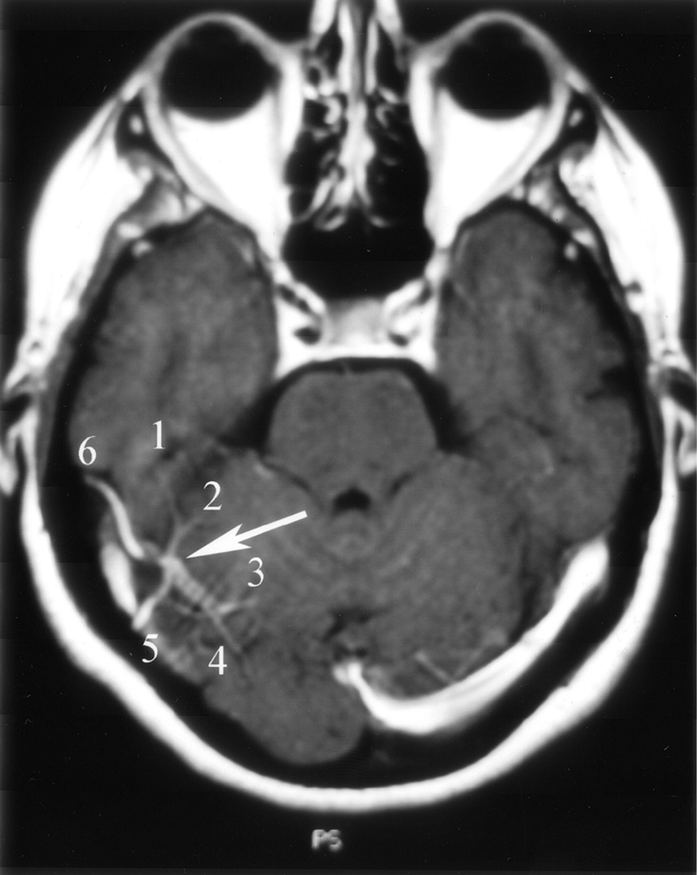
- Fig 10.
Arachnoid granulation in type III LTS. Axial contrast-enhanced T1-weighted MR image obtained in a 61-year-old woman shows multiple venous tributaries of a type III LTS. Note the close relationship between the vein of Labbé and the arachnoid granulation (arrow). 1, vein of Labbé; 2, anterior temporobasal vein; 3, middle temporoabasal vein; 4, posterior temporobasal vein; 5, occipitobasal vein; 6, posterior temporal vein.
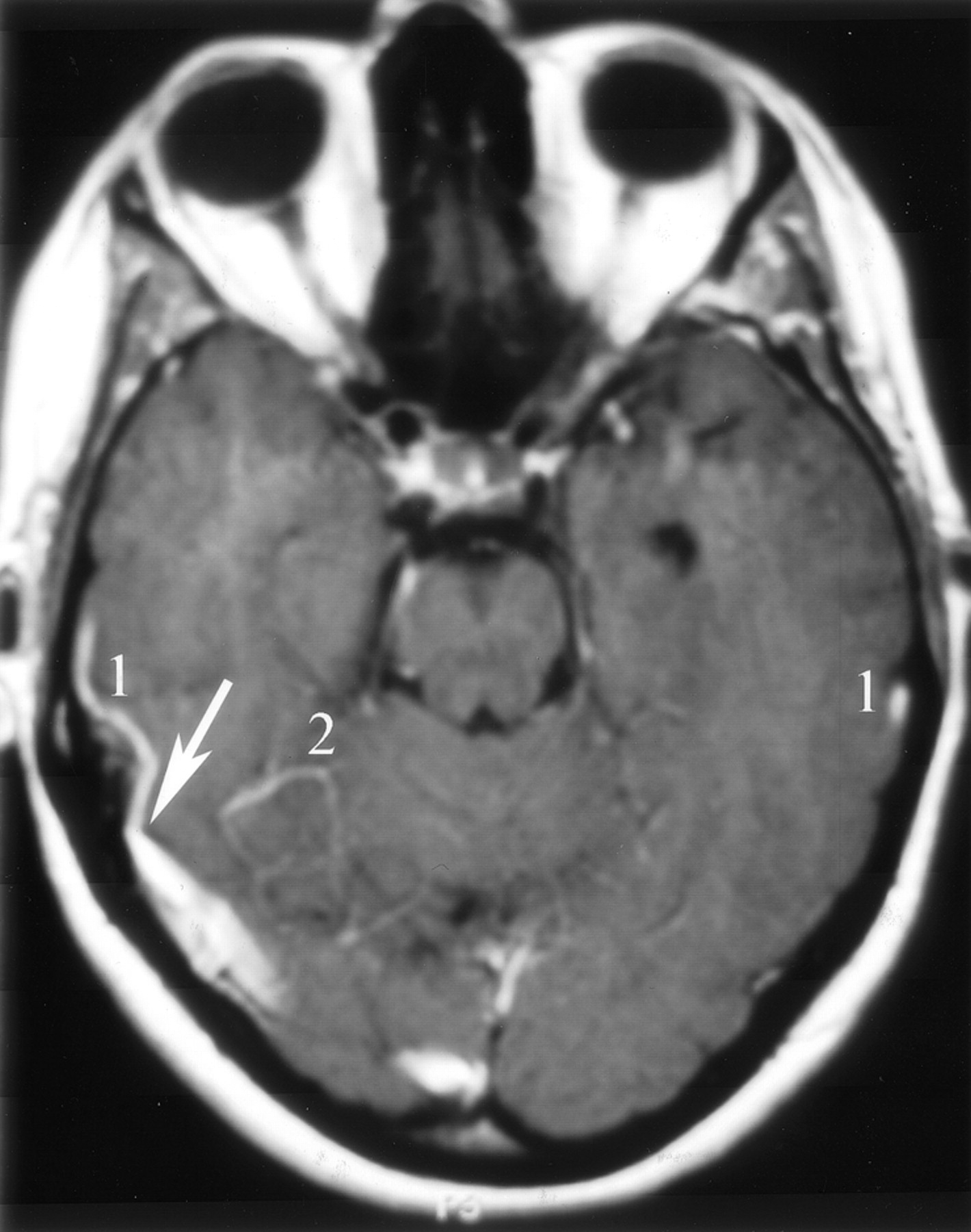
- Fig 11.
Tentorial meningioma displacing a type III LTS. Axial T1-weighted contrast-enhanced MR image obtained in a 64-year-old man shows a posterior fossa meningioma (asterisk) compressing the transverse sinus and distorting the nearby LTS (arrow). Note that the transverse sinuses are codominant and that the confluence of sinuses is widely patent. 1, superior petrosal sinus; 2, anterior temporobasal vein; 3, vein of Labbé; 4, occipitobasal vein.
- Fig 12.
Sphenopetrosal sinus in a patient with a petroclival meningioma.
A, Left common carotid artery injection, venous phase image obtained in a 61-year-old woman who presented with persistent headache reveals sphenotemporal sinus (arrowhead) draining into the transverse-sigmoid sinus junction (arrow).
B−D, Axial contrast-enhanced T1-weighted MR images again show a left petroclival meningioma (asterisks) and sphenopetrosal sinuses (arrows) emptying into transverse-sigmoid sinus junction, which is better seen on the right side (arrowhead). 1, superior petrosal sinus; 2, vein of Labbé; 3, occipitobasal vein; 4, posterior temporal vein; 5, middle temporal vein.


Tables
- TABLE 1:
Methodology for identifying the lateral tentorial sinus by type and configuration according to Guppy et al (14) (Fig 2)
LTS Type Configuration I Branches converge to form a single trunk II Multiple independent draining veins III Draining veins form venous lakes in the tentorium - TABLE 2:
Occurrence by type of lateral tentorial sinuses on contrast-enhanced T1-weighted MR images of 55 patients
LTS Type Side Right Left n (%) n (%) I 16 29 14 25.5 II 12 22 10 18 III 17 31 20 36.5 Indeterminate 5 9 10 18 Not visualized 5 9 1 2 Total 55 100 55 100 - TABLE 3:
Visualization of the lateral tentorial sinus branches on contrast-enhanced T1-weighted MR images of 55 patients (excluding posterior temporobasal and occipitobasal veins)
Lateral Tentorial Sinus Branch* Right Lobe Left Lobe ATBV and ATV trunk 30 29 MTBV 25 26 PTV 27 27 MTV 28 26 VL termination 47 47 LTS 23 30 Transverse sinus 12 10 Indeterminate 12 7 Not visualized 8 8 Note.—ATBV indicates anterior temporobasal vein; ATV, anterior temporal vein; MTBV, middle temporobasal vein; PTV, posterior temporal vein; MTV, middle temporal vein; VL, vein of Labbé; LTS, lateral tentorial sinus.
* Posterior temporobasal vein and occipitobasal vein not included in assessment because of poor visibility.


