Advertisement

Main menu

  • Home
  • Content
    • Current Issue
    • Accepted Manuscripts
    • Article Preview
    • Past Issue Archive
    • AJNR Case Collection
    • Case of the Week Archive
    • Classic Case Archive
    • Case of the Month Archive
  • Special Collections
    • Spinal CSF Leak Articles (Jan 2020-June 2024)
    • 2024 AJNR Journal Awards
    • Most Impactful AJNR Articles
  • Multimedia
    • AJNR Podcast
    • AJNR Scantastics
    • Video Articles
  • For Authors
    • Submit a Manuscript
    • Author Policies
    • Fast publishing of Accepted Manuscripts
    • Graphical Abstract Preparation
    • Manuscript Submission Guidelines
    • Imaging Protocol Submission
    • Submit a Case for the Case Collection
  • About Us
    • About AJNR
    • Editorial Board
  • More
    • Become a Reviewer/Academy of Reviewers
    • Subscribers
    • Permissions
    • Alerts
    • Feedback
    • Advertisers
    • ASNR Home
  • Other Publications
    • ajnr

User menu

  • Alerts
  • Log in

Search

  • Advanced search
American Journal of Neuroradiology

American Journal of Neuroradiology

ASHNR American Society of Functional Neuroradiology ASHNR American Society of Pediatric Neuroradiology ASSR
  • Alerts
  • Log in

Advanced Search

  • Home
  • Content
    • Current Issue
    • Accepted Manuscripts
    • Article Preview
    • Past Issue Archive
    • AJNR Case Collection
    • Case of the Week Archive
    • Classic Case Archive
    • Case of the Month Archive
  • Special Collections
    • Spinal CSF Leak Articles (Jan 2020-June 2024)
    • 2024 AJNR Journal Awards
    • Most Impactful AJNR Articles
  • Multimedia
    • AJNR Podcast
    • AJNR Scantastics
    • Video Articles
  • For Authors
    • Submit a Manuscript
    • Author Policies
    • Fast publishing of Accepted Manuscripts
    • Graphical Abstract Preparation
    • Manuscript Submission Guidelines
    • Imaging Protocol Submission
    • Submit a Case for the Case Collection
  • About Us
    • About AJNR
    • Editorial Board
  • More
    • Become a Reviewer/Academy of Reviewers
    • Subscribers
    • Permissions
    • Alerts
    • Feedback
    • Advertisers
    • ASNR Home
  • Follow AJNR on Twitter
  • Visit AJNR on Facebook
  • Follow AJNR on Instagram
  • Join AJNR on LinkedIn
  • RSS Feeds

Welcome to the new AJNR, Updated Hall of Fame, and more. Read the full announcements.


AJNR is seeking candidates for the position of Associate Section Editor, AJNR Case Collection. Read the full announcement.


OtherBRAIN

Pericallosal Artery Ectasia with Associated Stenosis

Will Beringer and Joseph Alenghat
American Journal of Neuroradiology August 2004, 25 (7) 1197-1198;
Will Beringer
  • Find this author on Google Scholar
  • Find this author on PubMed
  • Search for this author on this site
Joseph Alenghat
  • Find this author on Google Scholar
  • Find this author on PubMed
  • Search for this author on this site
  • Article
  • Figures & Data
  • Info & Metrics
  • Responses
  • References
  • PDF
Loading

Abstract

Summary: We report a rare case of bilateral pericallosal artery ectasia with stenosis in a 49-year-old man complaining of frontal headache. CT showed a serpiginous calcified interhemispheric fissure lesion with contrast enhancement. Imaging of this lesion was pursued with MR and MR angiography; however, conventional cerebral angiography was needed to make a definitive diagnosis.

Dilatation, elongation, and tortuosity of the vertebrobasilar and distal internal carotid arteries are frequently encountered. Ectasia in branches of the anterior cerebral arteries without lipoma, however, is rarely seen (1–3). We report a case of ectasia of the pericallosal arteries and discuss its characteristic CT, MR imaging, and cerebral angiographic findings.

Case Report

A normotensive 49-year-old man with no medical or surgical history and negative findings on neurologic and funduscopic examinations underwent CT for intermittent frontal headache that was unresponsive to 1 week of conservative treatment. CT revealed a hyperattenuated, interhemispheric-fissure lesion that enhanced after intravenous contrast agent administration. This lesion was suspicious for a calcified arteriovenous malformation (AVM) or dural fistula. To better characterize this lesion, the patient underwent MR imaging and MR angiography (MRA) studies. MR images showed calcifications and abnormal flow voids in the interhemispheric fissure closely following the corpus callosum without causing edema or mass effect. Upon contrast agent administration, these flow voids enhanced. MRA did not adequately demonstrate the distal anterior cerebral artery branches, although no other vascular lesions were seen.

To better characterize the nature of this vascular lesion, complete cerebral angiography was performed. It revealed bilateral, torturous pericallosal arteries without early venous drainage. The left pericallosal artery was noticeably stenotic immediately proximal to the region of ectasia. The carotid bifurcation and internal and external carotid and vertebrobasilar arteries were completely normal. No evidence of atherosclerotic disease, fibromuscular dysplasia, or vasculitis was seen. An MR imaging study of the cervical spine and CSF analysis were performed to search for a cause of the persistent frontal headaches. Serum analysis and echocardiography were performed to rule out collagen vascular diseases. No abnormality was found. The next day, the patient reported notable improvement in his frontal headaches despite no change in treatment. He was discharged and has not had recurrence of his headaches after several months of neurologic follow-up (Figs 1–4).

Fig 1.
  • Download figure
  • Open in new tab
  • Download powerpoint
Fig 1.

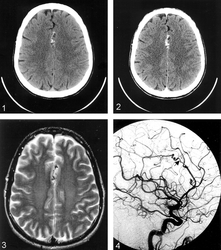
CT scan without contrast enhancement shows calcified interhemispheric lesion.

Fig 2.
  • Download figure
  • Open in new tab
  • Download powerpoint
Fig 2.

Postcontrast CT scan shows enhancing interhemispheric lesion.

Fig 3.
  • Download figure
  • Open in new tab
  • Download powerpoint
Fig 3.

Axial T2-weighted MR image shows interhemispheric flow voids.

Fig 4.
  • Download figure
  • Open in new tab
  • Download powerpoint
Fig 4.

Left lateral carotid angiogram. Arrow indicates region of stenosis and pericallosal ectasia.

Discussion

The torturous and elongated nature of this vascular anomaly fit the description of previously described cases of pericallosal arterial ectasia and dolichoectasia of the anterior cerebral atrteries (1–3). Dolicoectasia is more frequently encountered in the vertebrobasilar system, but it has also been described to exist in the middle and anterior cerebral arteries (4–6). Vertebrobasilar dolichoectasia can present with seizure, headache hydrocephalus, ataxia, dementia, stroke, and several cranial nerve deficits (7). These findings have been attributed to direct compression of intracranial structures and infarcts (7, 8). Anterior cerebral artery dolichoectasia has been associated with headaches, seizure, visual field defects, and mania (1–3, 5). There has been one report of subarachnoid hemorrhage caused by rupture of an A1-segment dolichoectatic vessel (6).

Initially, this case was suspicious for dural fistula or AVM owing to findings of calcification at CT and flow voids at MR imaging. Although vertebrobasilar artery dolichoectasia can be visualized using MR imaging and MRA, this technique cannot be relied upon for imaging distal anterior circulation lesions (4).

It was only by cerebral angiography that we were able to determine that the interhemispheric vascular anomaly seen using CT and MR imaging was indeed ectatic pericallosal arteries rather than an AVM or dural fistula. With angiography, the anatomy and flow dynamics of the entire intracranial and extracranial carotid and vertebrobasilar arteries could be studied. Although fibromuscular dysplasia has been reported in the A1 segment of the anterior cerebral artery, no findings suggestive of fibromuscular dysplasia were seen (9). No atherosclerotic plaques were noted in any arteries of the head or neck.

Acquired dolichoectatic intracranial arteries have been attributed to arteriosclerosis, hypertension, septic emboli, lipomas, and trauma (10). Congenital dolicoectasia is thought to be caused by defects in the arterial wall or migrational abnormalities (2). The histologic charateristics of these vessels frequently show hyaline degeneration of muscular layers and abrupt gaps in the internal elastic membrane (5, 10, 11). Accordingly, patients with polycystic kidney disease and Marfan syndrome have an increased incidence of dolichoectasia (12). Cases with concurrent medial frontal lobe thickening have been reported as well, which suggests a neuronal migration abnormality along the anterior cerebral artery (2).

Dilated and torturous intracranial arteries can be calcified and harbor atherosclerotic plaques. These areas of focal atherosclerosis and calcification can be caused by turbulent forces against the endothelium. In histologic studies, dolichoectatic arteries have been shown to have multiple, abrupt gaps of the internal elastic membrane where tissue of the media bulges through forming plaquelike excrecences into the arterial lumen (5). Because atherosclerotic stenosis and vessel-wall defects appear similar angiographically, it is unknown whether our patient’s stenotic left pericallosal artery is caused by a focal defect in the arterial wall or formation of an atherosclerotic plaque.

The cause of bilateral pericallosal artery ectasia with unilateral focal stenosis in this patient is not determined. It is possible this lesion is congenital, because the patient has no history of intracranial infection, trauma, or atherosclerosis, and no lipoma was visualized at MR imaging. Because the headaches resolved with no real intervention and have not recurred after several months of follow-up, the patient’s headaches may have been an incidental finding.

We hope this case reminds readers that this condition may mimic the CT and MR imaging findings of the AVM. Cerebral angiography may be needed for definitive diagnosis until enough experience is accumulated on the CT and MR imaging appearance of this lesion.

References

  1. ↵
    Tsukamoto Y, Nakata H, Soejima T, et al. Bilateral pericallosal artery ectasia. Neuroradiology 1985;27:271–274
    PubMed
  2. ↵
    Doran S, Deveikis J, Chandler W. Dolichoectasia of the anterior cerebral arteries in an adolescent. AJNR Am J Neuroradiol 1995;16:1548–1550
    Abstract
  3. ↵
    Thompson J, Weinstein P, Simmons C. Cerebral artery dolichoectasia with seizure. J Neurosurg44:509–512
  4. ↵
    Levine R, Turaski P, Grist T. Basilar artery dolichoectasia: review of the literature and six patients studied with magnetic resonance angiography. J Neuroimaging 1995;5:164–170
    PubMed
  5. ↵
    Sacks J, Lindenburg R. Dolicho-ectatic intracranial arteries: symptomatology and pathogenesis of arterial elongation and distention. Johns Hopkins Med J 1969;125:95–106
    PubMed
  6. ↵
    Goldstein S, Tibbs P. Recurrent subarachnoid hemorrhage complicating cerebral arterial ectasia. J Neurosurg 1981;55:139–142
    PubMed
  7. ↵
    Smoker W, Corbett J, Gentry L, et al. High-resolution computed tomography of the basilar artery: 2 vertebrobasilar dolichoectasia: clinical-pathologic correlation and review. AJNR Am J Neuroradiol 1986;7:61–72
    Abstract/FREE Full Text
  8. ↵
    Kryst-Widzgowska T, Kozlowski P, Binkiewicz M, et al. The angiographic and scintigraphic picture of dolichoectasia of the anterior cerebral artery. Eur J Nucl Med 1980;5:387–389
    PubMed
  9. ↵
    Nomura S, Yamashita K, Kato S, et al. Childhood subarachnoid hemorrhage associated with fibromuscular dysplasia. Childs Nerv Syst 2001;7:419–422
  10. ↵
    Wallace S, Jaffe M. Cerebral arterial ectasia with saccular aneurysms. Radiology 1967;88:90–93
    CrossRefPubMed
  11. ↵
    Hulten-Gyllensten I-L, Lofstedt S, von Reis G. Observations on generalized arteriectasis. Acta Med Scand 1959;163:125–130
    PubMed
  12. ↵
    Schievink W, Torres V, Wiebers D, Houston J. Intracranial arterial dolichoectasia in autosomal dominant polycystic kidney disease. J Am Soc Nephrol 1997;8:1298–1303
    Abstract
  • Received June 15, 2003.
  • Accepted after revision November 18, 2003.
  • Copyright © American Society of Neuroradiology
View Abstract
PreviousNext
Back to top

In this issue

American Journal of Neuroradiology: 25 (7)
American Journal of Neuroradiology
Vol. 25, Issue 7
1 Aug 2004
  • Table of Contents
  • Index by author
Advertisement
Print
Download PDF
Email Article

Thank you for your interest in spreading the word on American Journal of Neuroradiology.

NOTE: We only request your email address so that the person you are recommending the page to knows that you wanted them to see it, and that it is not junk mail. We do not capture any email address.

Enter multiple addresses on separate lines or separate them with commas.
Pericallosal Artery Ectasia with Associated Stenosis
(Your Name) has sent you a message from American Journal of Neuroradiology
(Your Name) thought you would like to see the American Journal of Neuroradiology web site.
CAPTCHA
This question is for testing whether or not you are a human visitor and to prevent automated spam submissions.
Cite this article
Will Beringer, Joseph Alenghat
Pericallosal Artery Ectasia with Associated Stenosis
American Journal of Neuroradiology Aug 2004, 25 (7) 1197-1198;

Citation Manager Formats

  • BibTeX
  • Bookends
  • EasyBib
  • EndNote (tagged)
  • EndNote 8 (xml)
  • Medlars
  • Mendeley
  • Papers
  • RefWorks Tagged
  • Ref Manager
  • RIS
  • Zotero
0 Responses
Respond to this article
Share
Bookmark this article
Pericallosal Artery Ectasia with Associated Stenosis
Will Beringer, Joseph Alenghat
American Journal of Neuroradiology Aug 2004, 25 (7) 1197-1198;
del.icio.us logo Twitter logo Facebook logo Mendeley logo
  • Tweet Widget
  • Facebook Like
  • Google Plus One

Jump to section

  • Article
    • Abstract
    • Case Report
    • Discussion
    • References
  • Figures & Data
  • Info & Metrics
  • Responses
  • References
  • PDF

Related Articles

  • No related articles found.
  • PubMed
  • Google Scholar

Cited By...

  • No citing articles found.
  • Crossref
  • Google Scholar

This article has not yet been cited by articles in journals that are participating in Crossref Cited-by Linking.

More in this TOC Section

  • Progression of Microstructural Damage in Spinocerebellar Ataxia Type 2: A Longitudinal DTI Study
  • Usefulness of Quantitative Susceptibility Mapping for the Diagnosis of Parkinson Disease
  • Evaluating the Effects of White Matter Multiple Sclerosis Lesions on the Volume Estimation of 6 Brain Tissue Segmentation Methods
Show more Brain

Similar Articles

Advertisement

Indexed Content

  • Current Issue
  • Accepted Manuscripts
  • Article Preview
  • Past Issues
  • Editorials
  • Editors Choice
  • Fellow Journal Club
  • Letters to the Editor

Cases

  • Case Collection
  • Archive - Case of the Week
  • Archive - Case of the Month
  • Archive - Classic Case

Special Collections

  • Special Collections

Resources

  • News and Updates
  • Turn around Times
  • Submit a Manuscript
  • Author Policies
  • Manuscript Submission Guidelines
  • Evidence-Based Medicine Level Guide
  • Publishing Checklists
  • Graphical Abstract Preparation
  • Imaging Protocol Submission
  • Submit a Case
  • Become a Reviewer/Academy of Reviewers
  • Get Peer Review Credit from Publons

Multimedia

  • AJNR Podcast
  • AJNR SCANtastic
  • Video Articles

About Us

  • About AJNR
  • Editorial Board
  • Not an AJNR Subscriber? Join Now
  • Alerts
  • Feedback
  • Advertise with us
  • Librarian Resources
  • Permissions
  • Terms and Conditions

American Society of Neuroradiology

  • Not an ASNR Member? Join Now

© 2025 by the American Society of Neuroradiology All rights, including for text and data mining, AI training, and similar technologies, are reserved.
Print ISSN: 0195-6108 Online ISSN: 1936-959X

Powered by HighWire