
Article Figures & Data
Figures
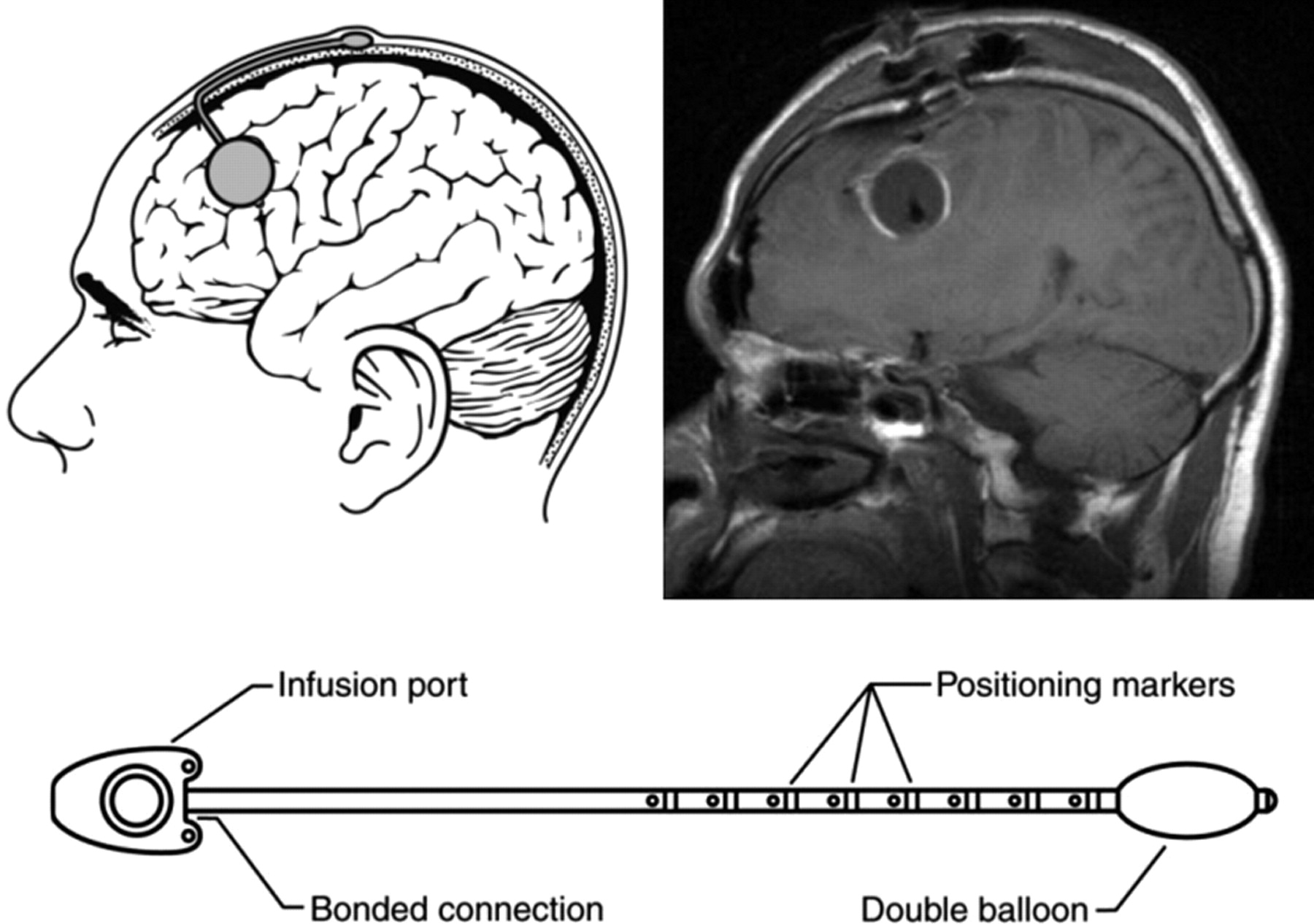
- Fig 1.
GSRTS device. Upper left, Diagram shows the balloon, subcutaneous tube, and port. Upper right, Corresponding sagittal nonenhanced T1-weighted MR image (TR/TE/NEX=500/12/1) obtained 24 hours after tumor resection and implantation shows the balloon surrounded by a thin, hyperintense peripheral zone corresponding to a thin layer of blood. Central hypointense area corresponds to the tube. Visualization of neighboring tissues is good. Bottom, Diagram shows components of the device.
- Fig 2.
CT scan obtained <48 hours after device implantation shows the attenuating balloon filled with iodinated contrast material, with artifacts obscuring tissues around it.
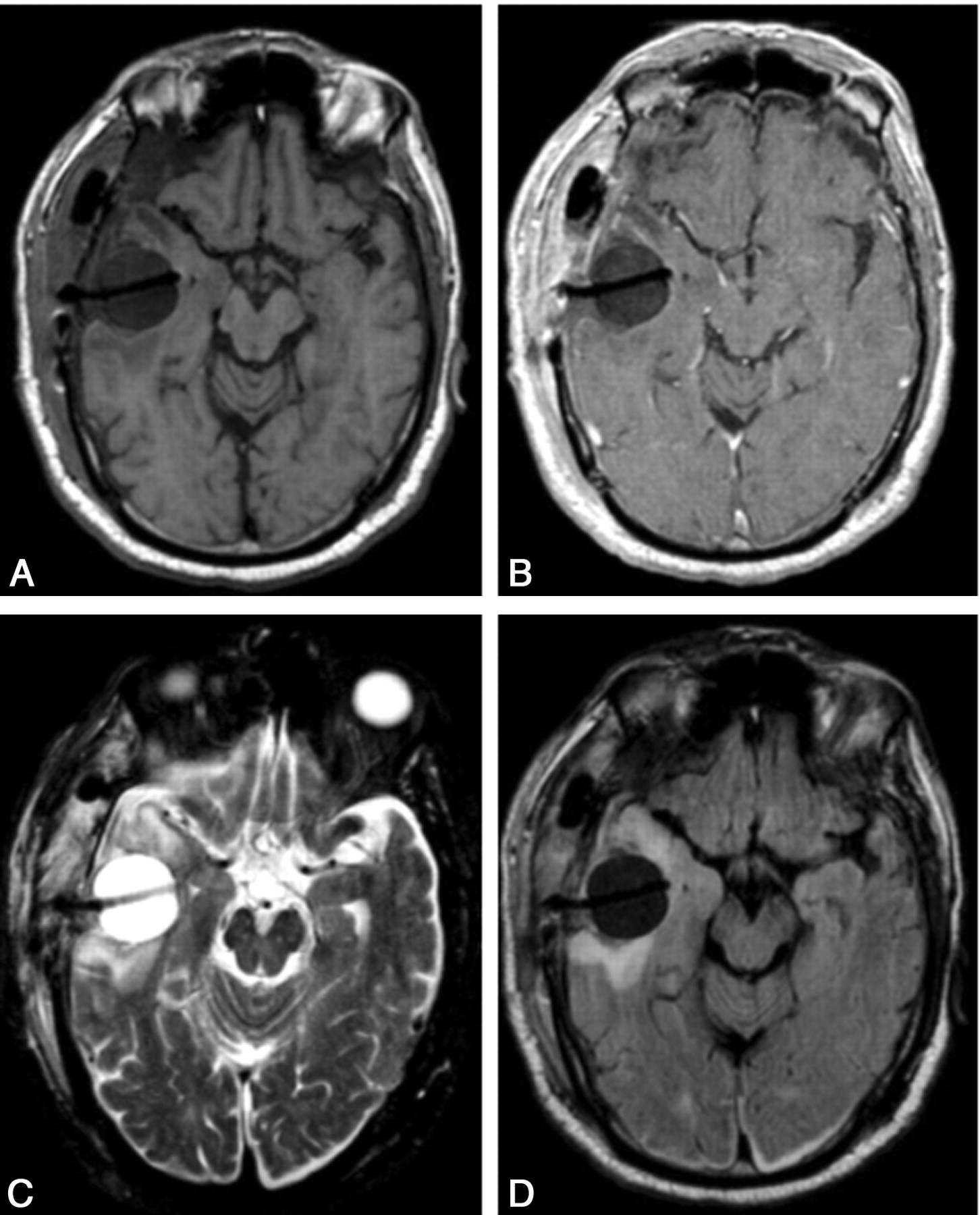
- Fig 3.
MR images obtained <48 hours after surgery.
A, Axial nonenhanced T1-weighted image (500/12/1) shows the hypointense balloon and central shaft (tube), with good visualization of surrounding tissues.
B, Axial contrast-enhanced T1-weighted image (500/12/1) shows the well-defined borders of the device and surroundingtissue, associated with thin enhancement of the surgical bed.
C, Corresponding axial T2-weighted image (5050/99/1) shows the hyperintense balloon. There is a small amount of ill-defined hyperintensity in adjacent tissues.
D, Corresponding axial FLAIR image (8000/110/1) shows the hyperintensity in the tissues around the balloon better. These areas are most likely residual vasogenic tumor edema and postsurgical changes.
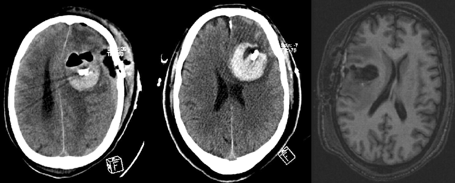
- Fig 4.
Images from the three patients in whom the devices were removed before radioactive unloading. Left, CT scan shows mass effect and bleeding around the device after acute hyponatremia. Center, CT scan shows substantial hemorrhage in the surgical cavity in this patient with coagulopathy. Right, 3D gradient-echo T1-weighted image (3.5/7/1) shows blood products in the surgical cavity and an increased distance between the edges of the balloon and surrounding tissues; this was considered a lack of conformance.
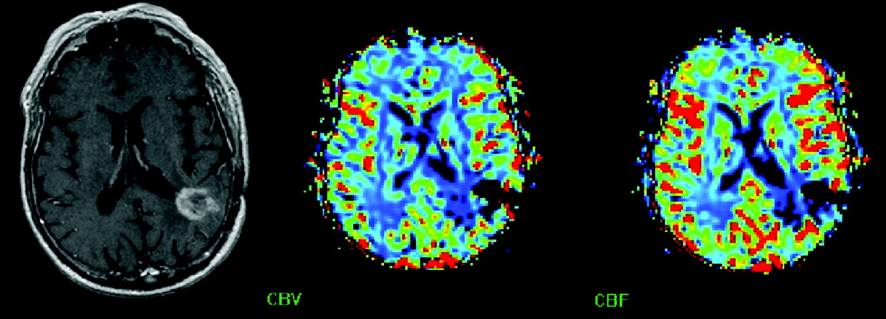
- Fig 5.
Perfusion maps after device removal in a patient treated for a brain metastasis. Left, Axial contrast-enhanced T1-weighted image (500/12/1) shows an irregular ringlike area of enhancement at the resection site. Center and right, Color perfusion maps show low rCBV and rCBF in the site of the lesion. At biopsy, no tumor was found.
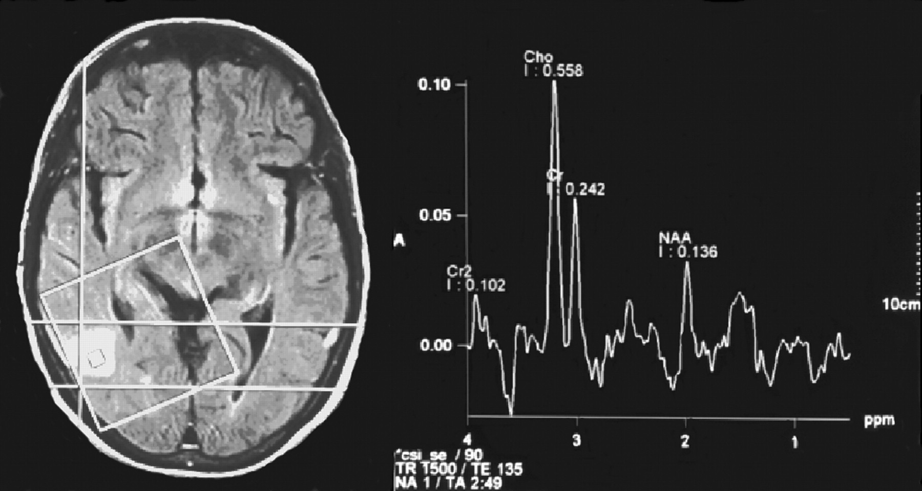
- Fig 6.
Results of MR spectroscopy after the device was removed in the treatment of anaplastic astrocytoma. Left, Axial contrast-enhanced T1-weighted localizer image shows the voxel. Right, Long-TE (135-ms) spectrum from the voxel shows an elevated Cho peak and a low NAA level.
- Fig 7.
Progressive signal-intensity abnormality after device removal. Left, Axial T2-weighted image (5050/99/1) at 1 month after removal shows minimal, somewhat ill-defined, hyperintensity in the right temporal lobe. Right, Corresponding axial T2-weighted image (5050/99/1) 5 months later shows progressive hyperintensity and mass effect; however, only radionecrosis was found at pathologic analysis.


