Advertisement

Main menu

  • Home
  • Content
    • Current Issue
    • Accepted Manuscripts
    • Article Preview
    • Past Issue Archive
    • AJNR Case Collection
    • Case of the Week Archive
    • Classic Case Archive
    • Case of the Month Archive
  • Special Collections
    • Spinal CSF Leak Articles (Jan 2020-June 2024)
    • 2024 AJNR Journal Awards
    • Most Impactful AJNR Articles
  • Multimedia
    • AJNR Podcast
    • AJNR Scantastics
    • Video Articles
  • For Authors
    • Submit a Manuscript
    • Author Policies
    • Fast publishing of Accepted Manuscripts
    • Graphical Abstract Preparation
    • Manuscript Submission Guidelines
    • Imaging Protocol Submission
    • Submit a Case for the Case Collection
  • About Us
    • About AJNR
    • Editorial Board
  • More
    • Become a Reviewer/Academy of Reviewers
    • Subscribers
    • Permissions
    • Alerts
    • Feedback
    • Advertisers
    • ASNR Home
  • Other Publications
    • ajnr

User menu

  • Alerts
  • Log in

Search

  • Advanced search
American Journal of Neuroradiology

American Journal of Neuroradiology

ASHNR American Society of Functional Neuroradiology ASHNR American Society of Pediatric Neuroradiology ASSR
  • Alerts
  • Log in

Advanced Search

  • Home
  • Content
    • Current Issue
    • Accepted Manuscripts
    • Article Preview
    • Past Issue Archive
    • AJNR Case Collection
    • Case of the Week Archive
    • Classic Case Archive
    • Case of the Month Archive
  • Special Collections
    • Spinal CSF Leak Articles (Jan 2020-June 2024)
    • 2024 AJNR Journal Awards
    • Most Impactful AJNR Articles
  • Multimedia
    • AJNR Podcast
    • AJNR Scantastics
    • Video Articles
  • For Authors
    • Submit a Manuscript
    • Author Policies
    • Fast publishing of Accepted Manuscripts
    • Graphical Abstract Preparation
    • Manuscript Submission Guidelines
    • Imaging Protocol Submission
    • Submit a Case for the Case Collection
  • About Us
    • About AJNR
    • Editorial Board
  • More
    • Become a Reviewer/Academy of Reviewers
    • Subscribers
    • Permissions
    • Alerts
    • Feedback
    • Advertisers
    • ASNR Home
  • Follow AJNR on Twitter
  • Visit AJNR on Facebook
  • Follow AJNR on Instagram
  • Join AJNR on LinkedIn
  • RSS Feeds

Welcome to the new AJNR, Updated Hall of Fame, and more. Read the full announcements.


AJNR is seeking candidates for the position of Associate Section Editor, AJNR Case Collection. Read the full announcement.


Research ArticleHEAD AND NECK

Warthin Tumor of the Parotid Gland: Diagnostic Value of MR Imaging with Histopathologic Correlation

Mitsuaki Ikeda, Ken Motoori, Toyoyuki Hanazawa, Yuichiro Nagai, Seiji Yamamoto, Takuya Ueda, Hiroyuki Funatsu and Hisao Ito
American Journal of Neuroradiology August 2004, 25 (7) 1256-1262;
Mitsuaki Ikeda
  • Find this author on Google Scholar
  • Find this author on PubMed
  • Search for this author on this site
Ken Motoori
  • Find this author on Google Scholar
  • Find this author on PubMed
  • Search for this author on this site
Toyoyuki Hanazawa
  • Find this author on Google Scholar
  • Find this author on PubMed
  • Search for this author on this site
Yuichiro Nagai
  • Find this author on Google Scholar
  • Find this author on PubMed
  • Search for this author on this site
Seiji Yamamoto
  • Find this author on Google Scholar
  • Find this author on PubMed
  • Search for this author on this site
Takuya Ueda
  • Find this author on Google Scholar
  • Find this author on PubMed
  • Search for this author on this site
Hiroyuki Funatsu
  • Find this author on Google Scholar
  • Find this author on PubMed
  • Search for this author on this site
Hisao Ito
  • Find this author on Google Scholar
  • Find this author on PubMed
  • Search for this author on this site
  • Article
  • Figures & Data
  • Info & Metrics
  • Responses
  • References
  • PDF
Loading

Article Figures & Data

Figures

  • Tables
  • Fig 1.
    • Download figure
    • Open in new tab
    • Download powerpoint
    Fig 1.

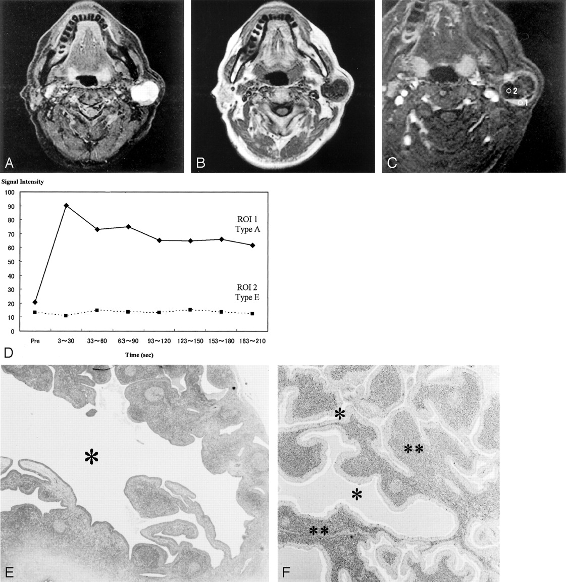
    Images of a Warthin tumor in the left parotid gland of a 70-year-old man.

    A, STIR image (4000/30), obtained in the axial plane, shows the tumor with moderate-to-high signal intensity. The high-signal-intensity area is a cystic lesion (*); the area showed no enhancement on contrast-enhanced images (see panel C, region of interest 2).

    B, T1-weighted image (400/9), obtained in the axial plane, shows a hypointense tumor.

    C, Fat suppression contrast-enhanced T1-weighted image (300/20), obtained in the coronal plane, shows solid (region of interest 1) and cystic (region of interest 2) tumor in the inferior pole of the parotid gland.

    D, Signal intensity graph shows that the washout ratio of the solid component was 41%. The cystic region shows no enhancement (type E). The ADC values of the solid and cystic components were 0.96 × 10⁻³ mm2/s and 2.74 × 10⁻³ mm2/s, respectively. The ADC value of the spinal cord was 1.02 × 10⁻³ mm2/s.

    E, Axial section of the specimen shows solid and large cystic components (*). The large cyst lost its contents.

    F, Solid component has slitlike or dendriform spaces (*) lined with papillary proliferation of bilayered oncocytic epithelia, with supporting stroma composed largely of lymphoid tissue (**). The small slitlike cysts are filled with proteinous secretion.

  • Fig 2.
    • Download figure
    • Open in new tab
    • Download powerpoint
    Fig 2.

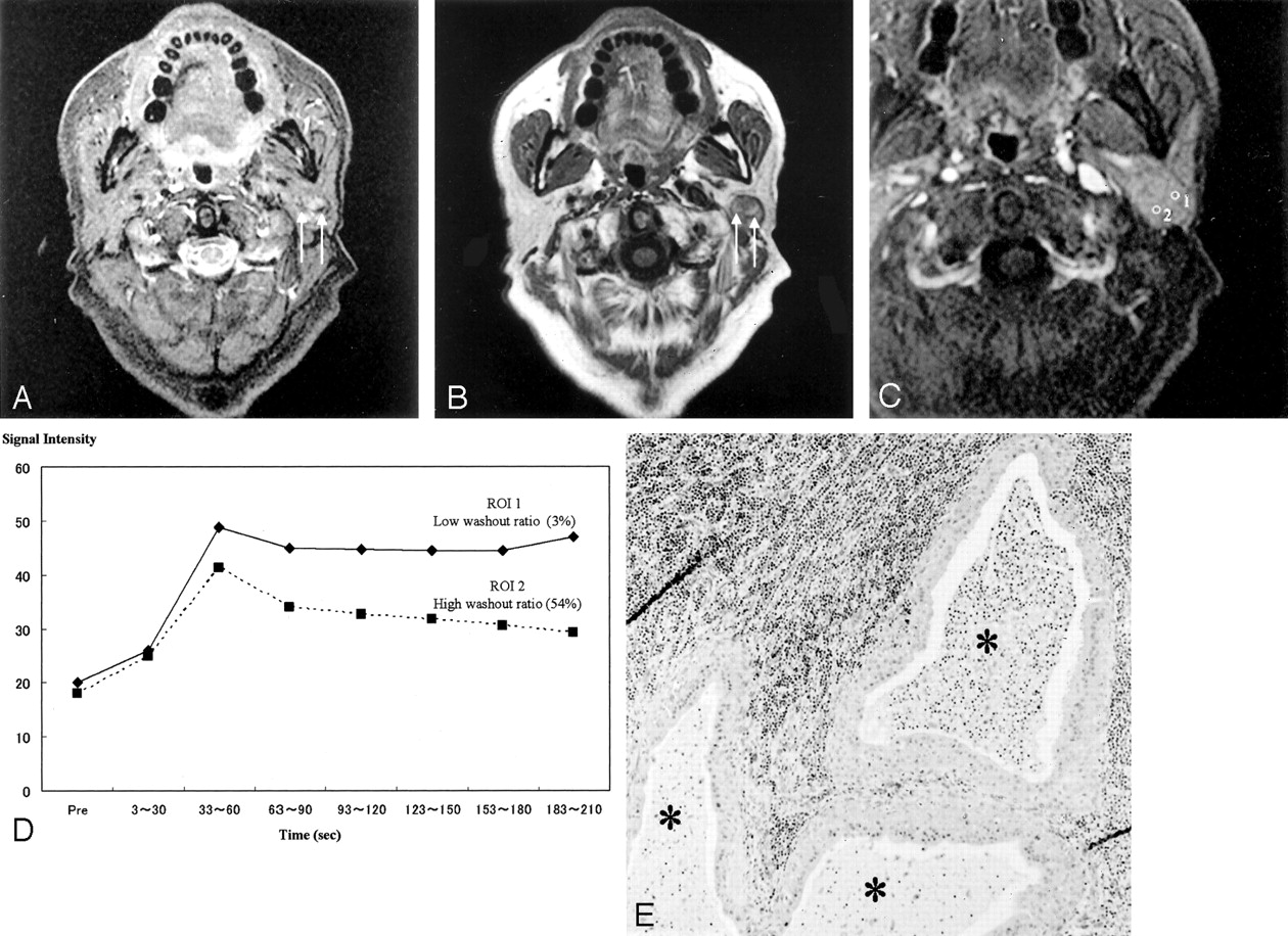
    Images of a Warthin tumor in the left parotid gland of a 61-year-old man.

    A, STIR image (4000/30), obtained in the axial plane, shows the tumor to be iso- to hypointense to the parotid gland. Characteristically small low-signal-intensity foci (arrows) can be detected. The margin of the tumor also has hypointense foci.

    B, T1-weighted image (400/9), obtained in the axial plane, shows the tumor to have the isointensity of muscle and relatively high-signal-intensity areas, whereas the STIR image shows low-signal-intensity areas (arrows). The margin of the tumor also is hypointense on T1-weighted images.

    C, Axial dynamic contrast-enhanced image shows all areas of this tumor to have type B perfusion curves.

    D, Signal intensity graph shows that foci that showed hypointensity on the STIR image and relatively high signal intensity on the T1-weighted image (region of interest 1) had a low washout ratio (3%) and the other region (region of interest 2) had a high washout ratio (54%).

    E, Axial section of the specimen shows cysts containing proteinous fluid with inflammatory cells (*).

Tables

  • Figures
    • View popup
    TABLE 1:

    Histopathology diagnoses of salivary gland tumors

    DiagnosisNo. of Lesions
    Benign (n = 19)
     Warthin tumor19
    Malignant (n = 17)
     Mucoepidermoid carcinoma1
     Acinic cell adenocarcinoma4
     Adenoid cystic carcinoma2
     Salivary duct carcinoma5
     Squamous cell carcinoma1
     Basal cell adenocarcinoma1
     Carcinoma ex pleomorphic adenoma3
    • View popup
    TABLE 2:

    Time intensity curves of tumors as shown by dynamic MR imaging

    Case No.Curve Types
    ABC1C2C3C4DE
    WA 1+ (41–50)+
    WA 2+ (39–50)+
    WA 3+ (22–38)
    WA 4+ (50–58)+
    WA 5+ (3–54)
    WA 6+ (45)
    WA 7+ (39)
    WA 8+ (40–52)
    WA 9+ (97)
    WA10+ (45)
    WA11+ (52)
    WA12+ (22–32)
    WA13+ (50)+ (29)
    WA14+ (22–47)+
    WA15+ (18)+
    WA16+ (73–102)
    WA17+ (45)+ (0)+
    WA18+ (46–61)
    WA19+ (11–55)
    AC 1+ (22)
    AC 2+ (25)
    AC 3+ (20)+
    AC 4+ (18–24)+ (4.4)
    ME 1+ (38)+ (11)
    ACC1+ (15)+ (0)
    ACC2+ (17)+ (0)+
    SD 1+ (11)+ (6)+
    SD 2+ (0)
    SD 3+ (12)+
    SD 4+ (0)
    SD 5+ (16–27)+ (0)
    SQ 1+ (18–35)
    BC 1+ (16)+ (5)+ (0)
    CEP1+ (0)
    CEP2+ (6)+ (0)+
    CEP3+ (25)+ (4)+
    • Note.—WA indicates Warthin tumor; AC, acinic cell adenocarcinoma; ME, mucoepidermoid carcinoma; ACC, adenoid cystic carcinoma; SD, salivary duct carcinoma; SQ, squamous cell carcinoma; BC, basal cell adenocarcinoma; CEP, carcinoma ex pleomorphic adenoma. Washout ratio, shown in parentheses, is expressed as a percentage.

PreviousNext
Back to top

In this issue

American Journal of Neuroradiology: 25 (7)
American Journal of Neuroradiology
Vol. 25, Issue 7
1 Aug 2004
  • Table of Contents
  • Index by author
Advertisement
Print
Download PDF
Email Article

Thank you for your interest in spreading the word on American Journal of Neuroradiology.

NOTE: We only request your email address so that the person you are recommending the page to knows that you wanted them to see it, and that it is not junk mail. We do not capture any email address.

Enter multiple addresses on separate lines or separate them with commas.
Warthin Tumor of the Parotid Gland: Diagnostic Value of MR Imaging with Histopathologic Correlation
(Your Name) has sent you a message from American Journal of Neuroradiology
(Your Name) thought you would like to see the American Journal of Neuroradiology web site.
CAPTCHA
This question is for testing whether or not you are a human visitor and to prevent automated spam submissions.
Cite this article
Mitsuaki Ikeda, Ken Motoori, Toyoyuki Hanazawa, Yuichiro Nagai, Seiji Yamamoto, Takuya Ueda, Hiroyuki Funatsu, Hisao Ito
Warthin Tumor of the Parotid Gland: Diagnostic Value of MR Imaging with Histopathologic Correlation
American Journal of Neuroradiology Aug 2004, 25 (7) 1256-1262;

Citation Manager Formats

  • BibTeX
  • Bookends
  • EasyBib
  • EndNote (tagged)
  • EndNote 8 (xml)
  • Medlars
  • Mendeley
  • Papers
  • RefWorks Tagged
  • Ref Manager
  • RIS
  • Zotero
0 Responses
Respond to this article
Share
Bookmark this article
Warthin Tumor of the Parotid Gland: Diagnostic Value of MR Imaging with Histopathologic Correlation
Mitsuaki Ikeda, Ken Motoori, Toyoyuki Hanazawa, Yuichiro Nagai, Seiji Yamamoto, Takuya Ueda, Hiroyuki Funatsu, Hisao Ito
American Journal of Neuroradiology Aug 2004, 25 (7) 1256-1262;
del.icio.us logo Twitter logo Facebook logo Mendeley logo
  • Tweet Widget
  • Facebook Like
  • Google Plus One

Jump to section

  • Article
    • Abstract
    • Methods
    • Results
    • Discussion
    • Conclusion
    • Acknowledgments
    • References
  • Figures & Data
  • Info & Metrics
  • Responses
  • References
  • PDF

Related Articles

  • No related articles found.
  • PubMed
  • Google Scholar

Cited By...

  • Diffusion weighted magnetic resonance imaging in the diagnosis of parotid masses. Preliminary results
  • Evaluating Instantaneous Perfusion Responses of Parotid Glands to Gustatory Stimulation Using High-Temporal-Resolution Echo-Planar Diffusion-Weighted Imaging
  • CT and Ultrasound Features of Basal Cell Adenoma of the Parotid Gland: A Report of 22 Cases with Pathologic Correlation
  • Diagnosing common parotid tumours with magnetic resonance imaging including diffusion-weighted imaging vs fine-needle aspiration cytology: a comparative study
  • Diffusion-Weighted Echo-Planar MR Imaging of Primary Parotid Gland Tumors: Is a Prediction of Different Histologic Subtypes Possible?
  • Current controversies in the management of Warthin tumour
  • Crossref
  • Google Scholar

This article has not yet been cited by articles in journals that are participating in Crossref Cited-by Linking.

More in this TOC Section

  • Correlation of Apparent Diffusion Coefficient at 3T with Prognostic Parameters of Retinoblastoma
  • Parathyroid Lesions: Characterization with Dual-Phase Arterial and Venous Enhanced CT of the Neck
  • Efficacy of Diffusion-Weighted Imaging for the Differentiation between Lymphomas and Carcinomas of the Nasopharynx and Oropharynx: Correlations of Apparent Diffusion Coefficients and Histologic Features
Show more Head and Neck

Similar Articles

Advertisement

Indexed Content

  • Current Issue
  • Accepted Manuscripts
  • Article Preview
  • Past Issues
  • Editorials
  • Editors Choice
  • Fellow Journal Club
  • Letters to the Editor

Cases

  • Case Collection
  • Archive - Case of the Week
  • Archive - Case of the Month
  • Archive - Classic Case

Special Collections

  • Special Collections

Resources

  • News and Updates
  • Turn around Times
  • Submit a Manuscript
  • Author Policies
  • Manuscript Submission Guidelines
  • Evidence-Based Medicine Level Guide
  • Publishing Checklists
  • Graphical Abstract Preparation
  • Imaging Protocol Submission
  • Submit a Case
  • Become a Reviewer/Academy of Reviewers
  • Get Peer Review Credit from Publons

Multimedia

  • AJNR Podcast
  • AJNR SCANtastic
  • Video Articles

About Us

  • About AJNR
  • Editorial Board
  • Not an AJNR Subscriber? Join Now
  • Alerts
  • Feedback
  • Advertise with us
  • Librarian Resources
  • Permissions
  • Terms and Conditions

American Society of Neuroradiology

  • Not an ASNR Member? Join Now

© 2025 by the American Society of Neuroradiology All rights, including for text and data mining, AI training, and similar technologies, are reserved.
Print ISSN: 0195-6108 Online ISSN: 1936-959X

Powered by HighWire