Advertisement

Main menu

  • Home
  • Content
    • Current Issue
    • Accepted Manuscripts
    • Article Preview
    • Past Issue Archive
    • AJNR Case Collection
    • Case of the Week Archive
    • Classic Case Archive
    • Case of the Month Archive
  • Special Collections
    • Spinal CSF Leak Articles (Jan 2020-June 2024)
    • 2024 AJNR Journal Awards
    • Most Impactful AJNR Articles
  • Multimedia
    • AJNR Podcast
    • AJNR Scantastics
    • Video Articles
  • For Authors
    • Submit a Manuscript
    • Author Policies
    • Fast publishing of Accepted Manuscripts
    • Graphical Abstract Preparation
    • Manuscript Submission Guidelines
    • Imaging Protocol Submission
    • Submit a Case for the Case Collection
  • About Us
    • About AJNR
    • Editorial Board
  • More
    • Become a Reviewer/Academy of Reviewers
    • Subscribers
    • Permissions
    • Alerts
    • Feedback
    • Advertisers
    • ASNR Home
  • Other Publications
    • ajnr

User menu

  • Alerts
  • Log in

Search

  • Advanced search
American Journal of Neuroradiology

American Journal of Neuroradiology

ASHNR American Society of Functional Neuroradiology ASHNR American Society of Pediatric Neuroradiology ASSR
  • Alerts
  • Log in

Advanced Search

  • Home
  • Content
    • Current Issue
    • Accepted Manuscripts
    • Article Preview
    • Past Issue Archive
    • AJNR Case Collection
    • Case of the Week Archive
    • Classic Case Archive
    • Case of the Month Archive
  • Special Collections
    • Spinal CSF Leak Articles (Jan 2020-June 2024)
    • 2024 AJNR Journal Awards
    • Most Impactful AJNR Articles
  • Multimedia
    • AJNR Podcast
    • AJNR Scantastics
    • Video Articles
  • For Authors
    • Submit a Manuscript
    • Author Policies
    • Fast publishing of Accepted Manuscripts
    • Graphical Abstract Preparation
    • Manuscript Submission Guidelines
    • Imaging Protocol Submission
    • Submit a Case for the Case Collection
  • About Us
    • About AJNR
    • Editorial Board
  • More
    • Become a Reviewer/Academy of Reviewers
    • Subscribers
    • Permissions
    • Alerts
    • Feedback
    • Advertisers
    • ASNR Home
  • Follow AJNR on Twitter
  • Visit AJNR on Facebook
  • Follow AJNR on Instagram
  • Join AJNR on LinkedIn
  • RSS Feeds

Welcome to the new AJNR, Updated Hall of Fame, and more. Read the full announcements.


AJNR is seeking candidates for the position of Associate Section Editor, AJNR Case Collection. Read the full announcement.


OtherPEDIATRICS

Pre- and Postnatal MR Imaging of Hypothalamic Hamartomas Associated with Arachnoid Cysts

Timothy N. Booth, Charles Timmons, Kenneth Shapiro and Nancy K. Rollins
American Journal of Neuroradiology August 2004, 25 (7) 1283-1285;
Timothy N. Booth
  • Find this author on Google Scholar
  • Find this author on PubMed
  • Search for this author on this site
Charles Timmons
  • Find this author on Google Scholar
  • Find this author on PubMed
  • Search for this author on this site
Kenneth Shapiro
  • Find this author on Google Scholar
  • Find this author on PubMed
  • Search for this author on this site
Nancy K. Rollins
  • Find this author on Google Scholar
  • Find this author on PubMed
  • Search for this author on this site
  • Article
  • Figures & Data
  • Info & Metrics
  • Responses
  • References
  • PDF
Loading

Abstract

Summary: We describe two cases of hypothalamic hamartoma associated with arachnoid cysts. One case was initially documented on prenatal MR images. Because of the rarity of the association and resultant distortion in regional anatomy, the solid component of the mass may be overlooked. This would certainly be true when using lower-resolution diagnostic studies such as fetal MR imaging. The lesion could also be confused with a cystic tumor such as pilocytic astrocytoma. Thorough evaluation is required in patients with precocious puberty, gelastic seizures, and the presence of a suprasellar arachnoid cyst.

Hypothalamic hamartomas are benign tumors consisting of ectopic neural tissue presumably congenital in origin. A hypothalamic hamartoma is typically a solid, nonenhancing mass that has similar MR signal intensity characteristics to those of gray matter (1–3). Associated cystic components have been described but are rare (4–7). We report two patients with hypothalamic hamartomas associated with arachnoid cysts, including one with prenatal MR imaging. This confirms that these lesions form in utero. To the best of our knowledge, prenatal depiction by MR imaging has not been reported.

Case Reports

Case 1

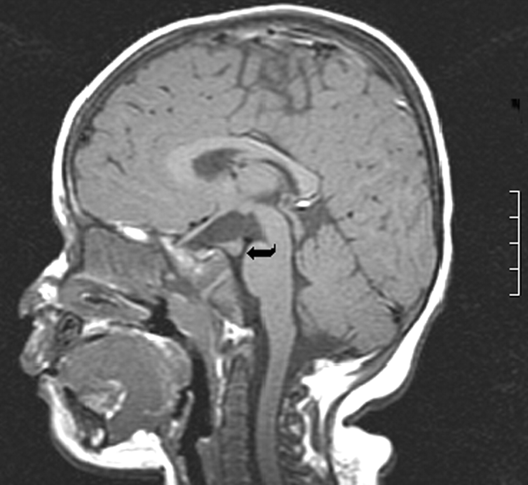
A 16-month-old female patient presented with vaginal bleeding. Physical examination revealed accelerated sexual maturation (Tanner stage II) with breast development and pubic hair. MR imaging done at 1.5 T by using standard T1-, T2-, and gadolinium-enhanced T1-weighted sequences showed a solid lesion that was nearly isointense relative to cortex on all images and did not enhance. The nodule projected into the prepontine cistern. There was an associated cyst, which appeared to be arising from the floor of the third ventricle (Fig 1). No additional intracranial abnormalities were present. Depo-lupron was administered to halt further development of secondary sexual characteristics. The nodule was felt to be remote from the floor of the third ventricle, so surgical resection was attempted. Pathologic analysis revealed scattered neurons and normal glial cells in the solid portion of the tumor consistent with a hypothalamic hamartoma. Histologic evaluation of the cyst wall showed glial and meningeal components.

Fig 1.
  • Download figure
  • Open in new tab
  • Download powerpoint
Fig 1.

Case 1. Nonenhanced sagittal T1-weighted image (460/11/2 [TR/TE/NEX]). A solid-appearing nodule projects into the prepontine cistern on the right (curved arrow). The nodule is isointense relative to cortex. No enhancement is present after contrast medium administration (not shown).

Case 2

A 20-year-old gravida 1, para 1 female patient was referred for fetal MR imaging after sonography showed an enlarged head circumference, ventriculomegaly, and a midline defect. Fetal MR imaging with heavily T2-weighted single-shot spin-echo (SE) sequences revealed an anterior suprasellar soft tissue lesion with cysts in the suprasellar and prepontine cisterns as well as extension into both middle cranial fossae (Fig 2A and B). The patient was delivered at term by cesarean section and was neurologically intact. Head circumference was enlarged at 38.5 cm (95th percentile). Postnatal MR imaging was performed at 1 month by using T1-weighted SE, T2-weighted fast SE, gadolinium-enhanced T1-weighted SE, and diffusion-weighted images. Extension of extra-axial fluid collections of high signal intensity, which were presumably arachnoid cysts, extended into both middle cranial fossa and elevated the floor of the third ventricle. The third and lateral ventricles were enlarged. The solid portion of the mass was suprasellar, isointense relative to cortex, and demonstrated no enhancement after gadolinium administration (Fig 2C). No additional intracranial malformations were visible, and the corpus callosum and pituitary gland appeared normal. Surgery was performed to decompress the cystic portion of the mass while biopsy of the solid component was carried out. Pathologic analysis revealed scattered neurons and normal glial cells within the solid portion of the mass. No cellular atypia was noted, and the MIB-1 index was low. The cyst wall consisted of meninges on one side and glial cells on the other.

Fig 2.
  • Download figure
  • Open in new tab
  • Download powerpoint
Fig 2.

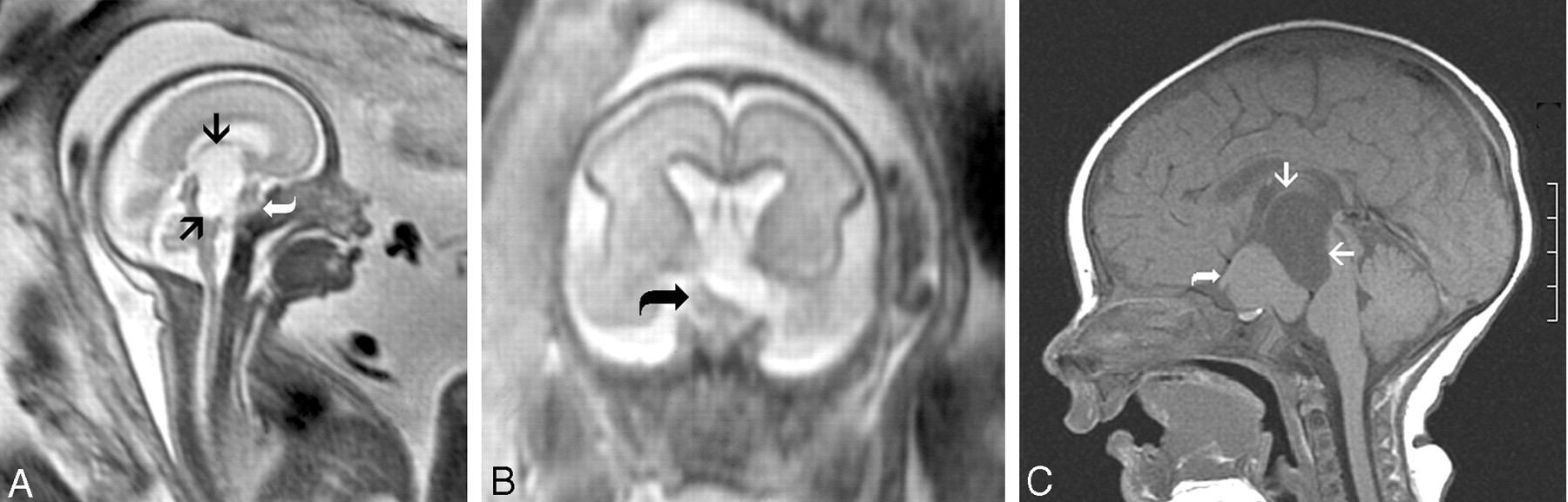
Case 2. Pre- (A and B) and postnatal (C) MR images.

A and B, Sagittal and coronal single-shot rapid acquisition SE images (∞/60–80). A mass isointense relative to cortex is present within the suprasellar cistern (curved arrow). High-signal-intensity fluid-filled cysts project superiorly and posteriorly (arrows). There is elevation of the floor of the third ventricle and compression of the midbrain and pons.

C, Nonenhanced sagittal T1-weighted image (450/11/2). The nodule is isointense relative to cortex and lies in the suprasellar cistern (curved arrow). Associated fluid-filled cysts are of high signal intensity (arrows). No enhancement is present after contrast medium administration (not shown).

Discussion

A hypothalamic hamartoma arises from the region of the tuber cinereum and is commonly associated with isosexual precocious puberty. Efferent pathways that allow modulation of gonadotropins and contain releasing hormones and secretory granules are present within the floor of the third ventricle (8). Precocious puberty may either result from a physical pertubation of inhibitory pathways by the hamartoma or, more likely, are a direct neurosecretory process of the hamartoma itself (9). Although MR imaging findings are usually normal in patients with isosexual precocious puberty, a hypothalamic hamartoma is the most common identifiable abnormality. Differential diagnosis includes hypothalamic and chiasmatic glioma, germ cell tumor, arachnoid cyst in the absence of a hamartoma, and isolated hydrocephalus (10, 11). Only one of our patients presented with central isosexual precocious puberty.

Seizure activity, most commonly described as gelastic, is the other common clinical presentation in children with a hypothalamic hamartoma. Theories for the pathogenesis of seizures are either connections with the limbic system and subsequent propagation of the seizure activity or possibly the presence of associated malformations (12). Neither of our patients had seizures. In the literature, one patient with a hamartoma and arachnoid cyst did present with seizures of temporal lobe origin (6).

This report describes the first case of a hypothalamic hamartoma seen by use of fetal MR imaging. This directly confirms that hamartomas form in utero before 26 weeks’ gestation. Because of the known association with other congenital anomalies of the central nervous system, hypothalamic hamartoma is thought to form as early as 4 weeks and may in fact be part of a developmental anomaly spectrum (12, 13). Two theories have been proposed to explain the coexistence of an arachnoid cyst and a hypothalamic hamartoma. The formation of the subarachnoid space begins at 14 weeks with the intracellular space expanding as the meninx privativa degenerates. The space is first seen ventral to the mesoencephalon, which would include the suprasellar cistern (14). The presence of the hamartoma may disrupt the formation of the subarachnoid space leading to the formation of an arachnoid cyst. Alternatively, arachnoid or meningeal tissue could be within the hamartoma, resulting in the formation of an associated arachnoid cyst (4, 6).

MR imaging has been proved to be the optimal imaging study for evaluating the presence of hypothalamic hamartomas. The lesions are isointense on T1-weighted images and iso- to more commonly slightly hyperintense on T2-weighted images and do not enhance (1–3). The presence of increased T2 signal intensity and progressive growth may suggest malignant transformation or, more likely, a different tumor. Hypothalamic hamartomas are usually small and may be sessile or pedunculated. Pedunculated and smaller lesions tend to present with precocious puberty, whereas sessile and larger lesions are more commonly associated with seizures (15). MR spectroscopy has shown increased concentration of myo-inositol and relatively normal n-acetylaspartate and choline levels (16).

Atypical features of hypothalamic hamartomas have been reported in the literature and include giant hamartomas, lipomatous content, cystic necrosis, and prominent T2 prolongation (17–20). An arachnoid cyst is rarely reported in association with a hypothalamic hamartoma with, to the best of our knowledge, only four previous cases in the literature (4–7). The development of a hypothalamic hamartoma has been closely associated with duplication of the pituitary gland. The presence of a hamartoma has been attributed to abnormal splitting of the rostral notochord, which explains the association with pituitary duplication and the duplicated floor of the third ventricle and aqueduct. Early insults may lead to abnormal development of the facial structures (13). Normal pituitary glands and facial development were present in both our patients. Additional associated congenital abnormalities of the olfactory tracts, corpus callosum, cerebral cortex, and posterior fossa have been described (14, 21, 22).

The presence of prominent cysts associated with a hamartoma can lead to significant alteration in regional anatomy and incorrect preoperative diagnosis, especially in the absence of a typical clinical presentation. The presumptive diagnosis of hamartoma associated with arachnoid cysts should be made when MR imaging features of the solid component are typical for a hypothalamic hamartoma. These features should be sought in any patient presenting with precocious puberty or seizures and the presence of a suprasellar arachnoid cyst. Even in the absence of a typical clinical presentation, as was the case in one of our patients, an arachnoid cyst may be associated with a hamartoma. Arachnoid cysts and their association with precocious puberty are well documented in the literature. We suggest careful evaluation of every suprasellar arachnoid cyst for the presence of associated an associated hamartoma, particularly in prenatal MR imaging.

Footnotes

  • Presented at the 41st Annual Meeting of the American Society of Neuroradiology, April 28–May 2, 2003; Washington, D.C.

References

  1. ↵
    Hahn FJ, Leibrock LG, Huseman CA, Makos MM. The MR appearance of hypothalamic hamartoma. Neuroradiology 1988;30:65–68
    CrossRefPubMed
  2. Boyko OB, Curnes JT, Oakes WJ, Burger PC. Hamartomas of the tuber cinereum: CT, MR, and pathologic findings. AJNR Am J Neuroradiol 1991;12:309–314
    Abstract/FREE Full Text
  3. ↵
    Burton EM, Ball WS, Crone K, Dolan LM. Hamartoma of the tuber cinereum: a comparison of MR and CT findings in four cases. AJNR Am J Neuroradiol 1989;10:497–501
    Abstract/FREE Full Text
  4. ↵
    Nishio S, Morioka T, Hamada Y, et al. Hypothalamic hamartoma associated with an arachnoid cyst. J Clin Neurosci 2001;8:46–48
    CrossRefPubMed
  5. Isaki T, Nakatani S, Yoshimine T, et al. Asymptomatic hypothalamic hamartoma associated with an arachnoid cyst [in Japanese]. Neurol Med Chir (Tokyo) 1996;36:725–728
    CrossRefPubMed
  6. ↵
    Goda M, Tashima A, Isono M, et al. A case of hypothalamic hamartoma associated with arachnoid cyst. Childs Nerv Syst 1999;15:490–492
    CrossRefPubMed
  7. ↵
    Paillas JE, Roger J, Toga M, et al. Hamartoma of the hypothalmus: clinical, radiological, and histological study [in French]. Résultats de l’exerèse Rev Neurol (Paris) 1969;120:177–194
  8. ↵
    Carpenter MB. The Hypothalmus. Core Text of Neuroanatomy. 4th ed. Baltimore: Wiliams & Wilkins,1991 :297–324
  9. ↵
    Jung H, Ojeda SR. Pathogenesis of precocious puberty in hypothalamic hamartoma. Horm Res 2002;57:31–34
  10. ↵
    Reith KG, Comite F, Dwyer AJ, et al. CT of cerebral abnormalities in precocious puberty. AJR Am J Roentgenol 1987;148:1231–1238
    PubMed
  11. ↵
    Balagura S, Shulman, Sobel EH. Precocious puberty of cerebral origin. Surg Neurol 1979;11:315–326
    PubMed
  12. ↵
    Diebler C, Ponsot G. Hamartomas of the tuber cinereum. Neuroradiology 1983;25:93–101
    CrossRefPubMed
  13. ↵
    Kollias SS, Ball WS, Prenger EC. Review of the embryologic development of the pituitary gland and report of a case of hypothalamic duplication detected by MRI. Neuroradiology 1995;37:3–12
    CrossRefPubMed
  14. ↵
    McLone DG. The subarachnoid space: a review. Childs Brain 1980;6:113–130
    PubMed
  15. ↵
    Arita K, Ikawa F, Kurisu K, et al. The relationship between magnetic resonance imaging findings and clinical manifestations of hypothalamic hamartoma. J Neurosurg 1999;91:212–220
    PubMed
  16. ↵
    Martin DD, Seeger U, Ranke MB, Grodd W. MR imaging and spectroscopy of a tuber cinereum hamartoma in a patient with growth hormone deficiency and hypogonadotropic hypogonadism. AJNR Am J Neuroradiol 2003;24:1177–1180
    Abstract/FREE Full Text
  17. ↵
    Prasad S, Shah J, Patkar D, et al. Giant hypothalamic hamartoma with cystic change: report of two cases and review of the literature. Neuroradiology 2000;42:648–650
    CrossRefPubMed
  18. Tonami H, Higashi K, Okamoto K, et al. Report of changing signal intensity of follow-up MRI in a case of hypothalamic hamartoma. J Comput Assist Tomogr 2001;25:130–132
    PubMed
  19. Sharma MC, Gaikwad S, Mahapatra AK, et al. Hypothalamic hamartoma: report of a case with unusual histologic features. Am J Surg Pathol 1998;22:1538–1541
    PubMed
  20. ↵
    Hubbard AM. MR imaging of large hypothalamic hamartomas in two infants. AJNR Am J Neuroradiol 1989;10:1277
    FREE Full Text
  21. ↵
    Naidich TP, Zimmerman RA. Primary brain tumors in children. Semin Roentgenol 1984;19:100–114
    CrossRefPubMed
  22. ↵
    Tsugu H, Fukushima T, Nagashima T, et al. Hypothalamic hamartoma associated with multiple congenital abnormalities: two patients and a review of reported cases. Pediatr Neurosurg 1998;29:290–296
    CrossRefPubMed
  • Received August 28, 2003.
  • Accepted after revision November 18, 2003.
  • Copyright © American Society of Neuroradiology
View Abstract
PreviousNext
Back to top

In this issue

American Journal of Neuroradiology: 25 (7)
American Journal of Neuroradiology
Vol. 25, Issue 7
1 Aug 2004
  • Table of Contents
  • Index by author
Advertisement
Print
Download PDF
Email Article

Thank you for your interest in spreading the word on American Journal of Neuroradiology.

NOTE: We only request your email address so that the person you are recommending the page to knows that you wanted them to see it, and that it is not junk mail. We do not capture any email address.

Enter multiple addresses on separate lines or separate them with commas.
Pre- and Postnatal MR Imaging of Hypothalamic Hamartomas Associated with Arachnoid Cysts
(Your Name) has sent you a message from American Journal of Neuroradiology
(Your Name) thought you would like to see the American Journal of Neuroradiology web site.
CAPTCHA
This question is for testing whether or not you are a human visitor and to prevent automated spam submissions.
Cite this article
Timothy N. Booth, Charles Timmons, Kenneth Shapiro, Nancy K. Rollins
Pre- and Postnatal MR Imaging of Hypothalamic Hamartomas Associated with Arachnoid Cysts
American Journal of Neuroradiology Aug 2004, 25 (7) 1283-1285;

Citation Manager Formats

  • BibTeX
  • Bookends
  • EasyBib
  • EndNote (tagged)
  • EndNote 8 (xml)
  • Medlars
  • Mendeley
  • Papers
  • RefWorks Tagged
  • Ref Manager
  • RIS
  • Zotero
0 Responses
Respond to this article
Share
Bookmark this article
Pre- and Postnatal MR Imaging of Hypothalamic Hamartomas Associated with Arachnoid Cysts
Timothy N. Booth, Charles Timmons, Kenneth Shapiro, Nancy K. Rollins
American Journal of Neuroradiology Aug 2004, 25 (7) 1283-1285;
del.icio.us logo Twitter logo Facebook logo Mendeley logo
  • Tweet Widget
  • Facebook Like
  • Google Plus One

Jump to section

  • Article
    • Abstract
    • Case Reports
    • Discussion
    • Footnotes
    • References
  • Figures & Data
  • Info & Metrics
  • Responses
  • References
  • PDF

Related Articles

  • No related articles found.
  • PubMed
  • Google Scholar

Cited By...

  • A Case Series of X-Linked Deafness-2 with Sensorineural Hearing Loss, Stapes Fixation, and Perilymphatic Gusher: MR Imaging and Clinical Features of Hypothalamic Malformations
  • Crossref
  • Google Scholar

This article has not yet been cited by articles in journals that are participating in Crossref Cited-by Linking.

More in this TOC Section

  • Comparison of Image Quality and Radiation Dose in Pediatric Temporal Bone CT Using Photon-Counting Detector CT and Energy-Integrating Detector CT
  • SyMRI & MR Fingerprinting in Brainstem Myelination
  • Dandy-Walker Phenotype with Brainstem Involvement
Show more Pediatrics

Similar Articles

Advertisement

Indexed Content

  • Current Issue
  • Accepted Manuscripts
  • Article Preview
  • Past Issues
  • Editorials
  • Editors Choice
  • Fellow Journal Club
  • Letters to the Editor

Cases

  • Case Collection
  • Archive - Case of the Week
  • Archive - Case of the Month
  • Archive - Classic Case

Special Collections

  • Special Collections

Resources

  • News and Updates
  • Turn around Times
  • Submit a Manuscript
  • Author Policies
  • Manuscript Submission Guidelines
  • Evidence-Based Medicine Level Guide
  • Publishing Checklists
  • Graphical Abstract Preparation
  • Imaging Protocol Submission
  • Submit a Case
  • Become a Reviewer/Academy of Reviewers
  • Get Peer Review Credit from Publons

Multimedia

  • AJNR Podcast
  • AJNR SCANtastic
  • Video Articles

About Us

  • About AJNR
  • Editorial Board
  • Not an AJNR Subscriber? Join Now
  • Alerts
  • Feedback
  • Advertise with us
  • Librarian Resources
  • Permissions
  • Terms and Conditions

American Society of Neuroradiology

  • Not an ASNR Member? Join Now

© 2025 by the American Society of Neuroradiology All rights, including for text and data mining, AI training, and similar technologies, are reserved.
Print ISSN: 0195-6108 Online ISSN: 1936-959X

Powered by HighWire