
Article Figures & Data
Figures
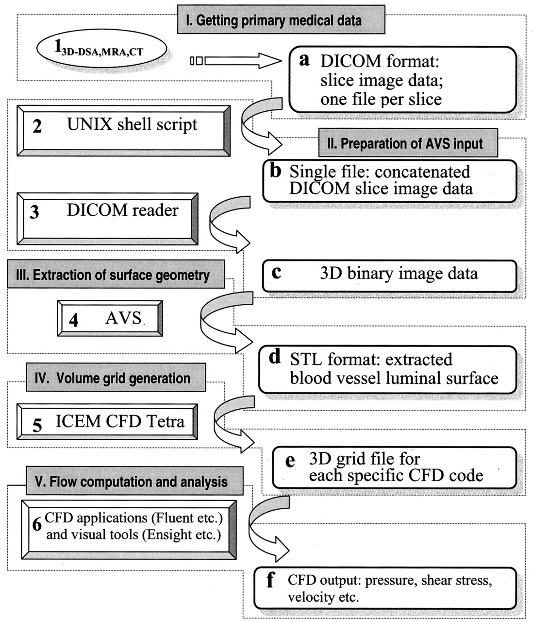
- Fig 1.
Flowchart of patient-specific segmentation, volume grid generation and blood flow analysis shows software tools (1–6) and output formats (a–f). I–V denote major stages of the procedure.
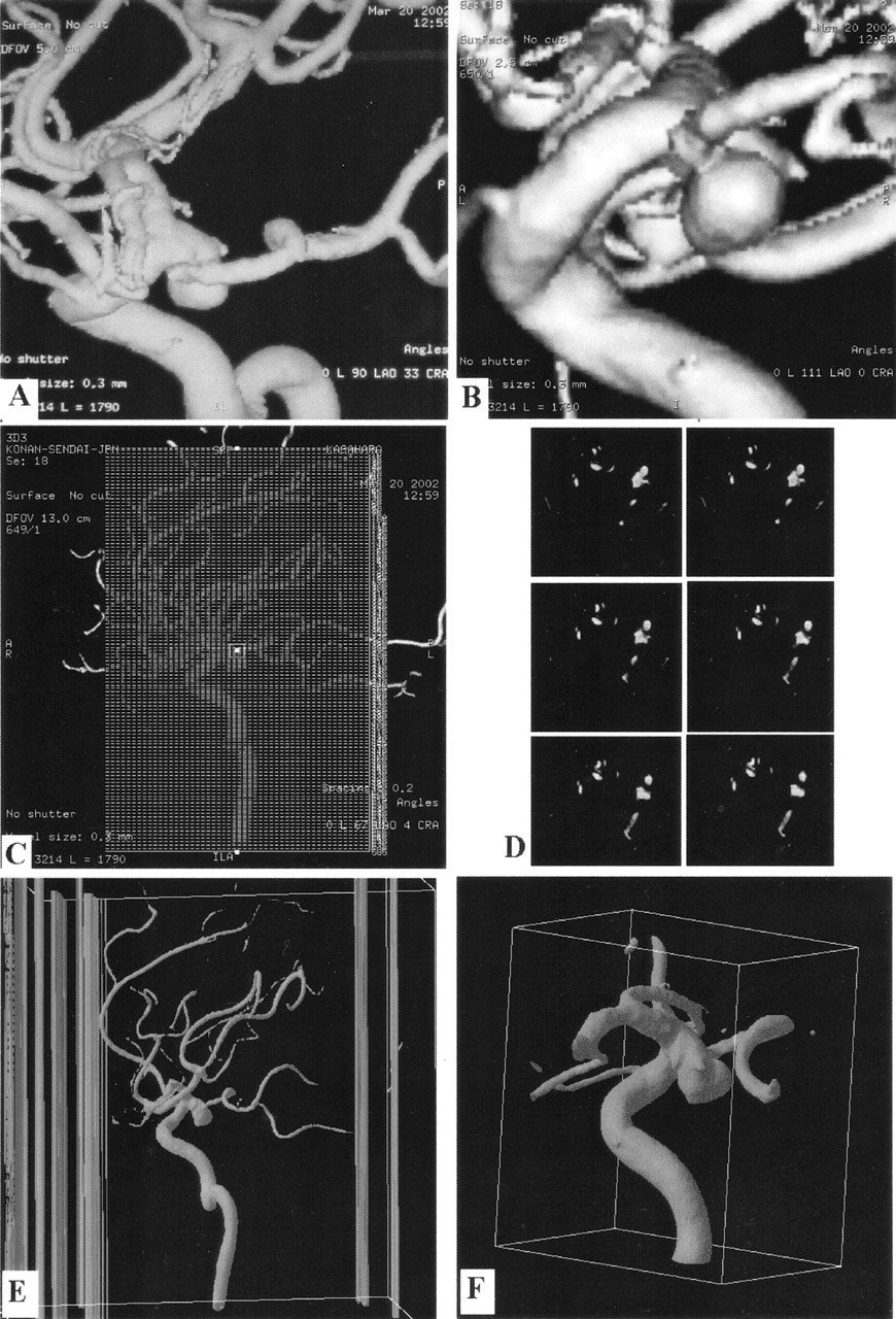
- Fig 2.
Data transformation from an angiographic image into a computational grid model.
A and B, 3D DSA images (different views) of a posterior communicating artery aneurysm.
C, Typical setup for reformatting of the angiographic image into 585 secondary DICOM sections in a 13-cm displayed field of view.
D, Sequential secondary DICOM sections. The respective files are concatenated before processing with DICOM reader (X) MedCon.
E, 3D gray-scale raster image produced by (X) MedCon and imported by AVS/Express. Columns are patient data and image parameters still present in the file.
F, Result of image manipulations with AVS/Express. ROI is cut out and the luminal vascular surface is identified with a gray-scale isosurface value of 1500. This ROI includes the aneurysm and its small branching vessel.
G, AVS/Express STL file containing the surface mesh is imported by using the ICEM CFD Mesh Editor and Tetra grid generator. These tools allow us to further remove unnecessary parts, manually repair and smooth the surface mesh, close inflow and outflow boundaries, and generate the volume grid.
H, Final computational tetrahedral grid model for blood flow simulation with Fluent or another suitable software.
I, Typical instant streamlines colored by velocity (in m/s) show that the entering bloodstream hits the aneurysm wall at the angiographically determined rupture area (arrow).
J, Typical instant wall shear-stress distribution (Pa) shows a local shear-stress maximum where the aneurysm ruptured (arrow).
K, Arterial-phase 2D DSA image shows escape of a linear stream of contrast agent into the subarachnoid space (arrow).
L, Arterial-phase 2D DSA image obtained during endovascular intervention shows escape of the coils through the aneurysm rupture (arrow).
- Fig 3.
Examples of four vessels reconstructed from MRA image and CTA data.
A, MRA data of four vessels with basilar top aneurysm (arrow).
B, Surface grid reconstruction of MRA image data. Note the inferior quality of this grid, as compared with that obtained from 3D DSA and multiple defects in the vessel walls.
C, Surface grid reconstruction (STL file) from CTA of four vessels of a normal circle of Willis. Note the reconstructed skull base requiring manual removal during mesh generation.
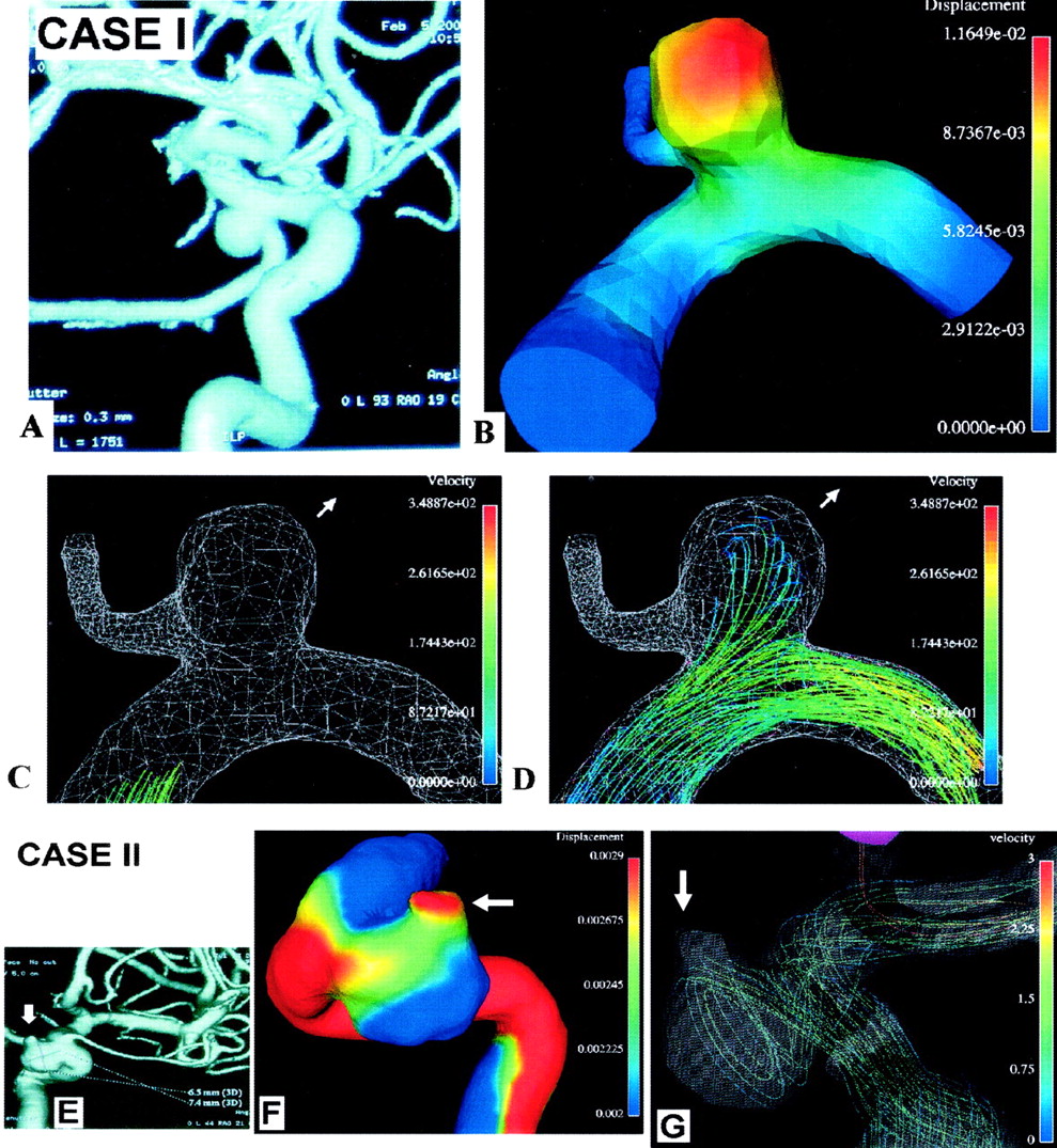
- Fig 4.
Computations for grid models with moving walls. Case I: A, 3D DSA image of a nonruptured internal carotid–posterior communicating aneurysm. B, Displacement (in mm) of the aneurysm wall at the systolic peak. Red is maximal displacement. C and D, Movie frames illustrate deformation of the aneurysm from early systole (C) to midsystolic peak (D). Arrow indicates the direction of movement (and possible future growth). Bloodstream enters the aneurysm, hits its wall, decelerates (note change in color; velocity in cm/s), and exerts pressure resulting in maximal displacement. Case II: E, 3D DSA image of a nonruptured carotid cave aneurysm. F, Displacement of the aneurysm wall at the systolic peak. Red is maximal displacement at the nonruptured aneurysmal bleb (arrow). G, Typical instant streamlines show that the entering bloodstream hits the aneurysm wall at the angiographically determined bleb (arrow), exerting pressure that results in maximal displacement.
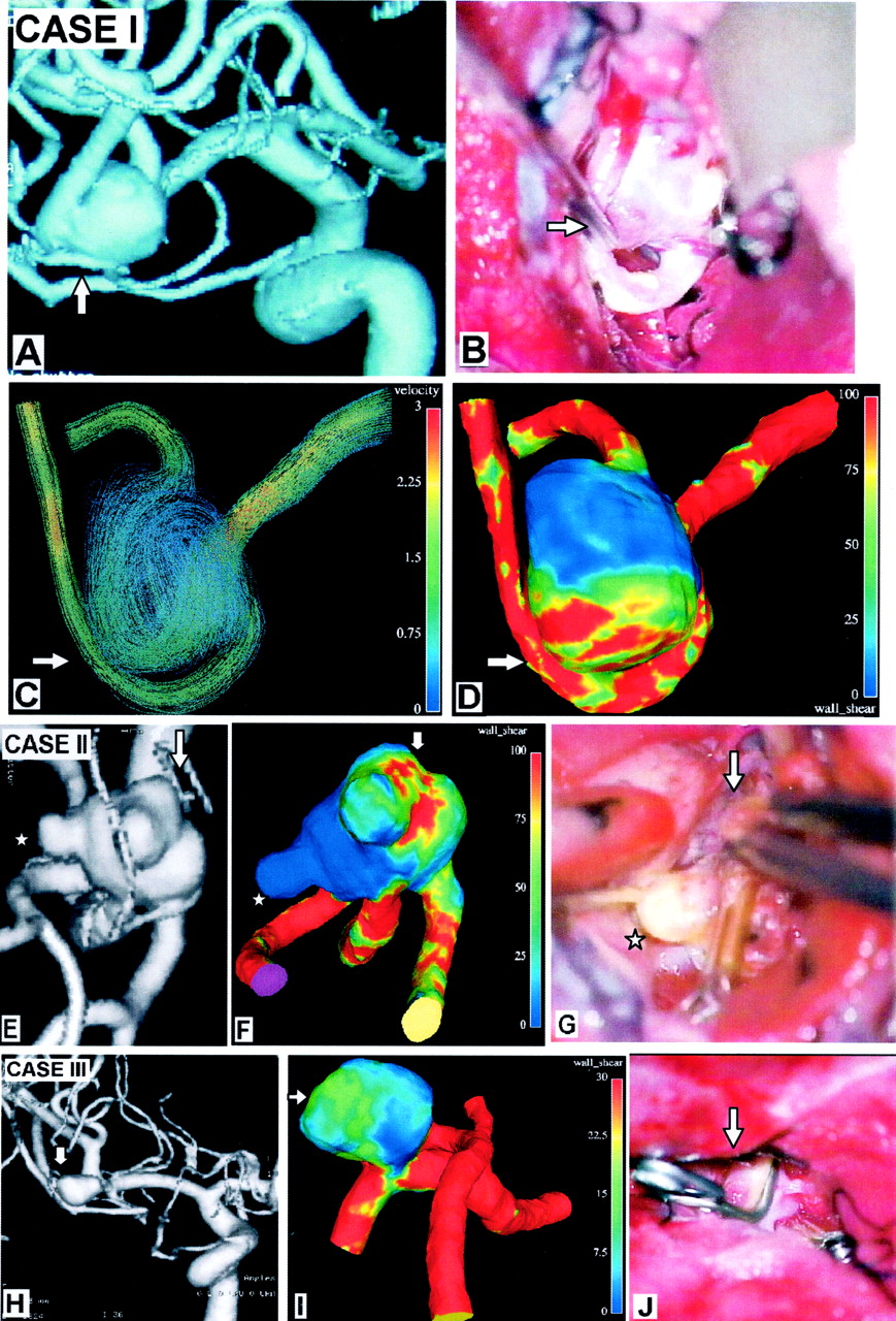
- Fig 5.
Computational results for grid models with rigid walls compared with operative photos and 3D DSA images. Case I: A, 3D DSA shows a ruptured middle cerebral artery (MCA) bifurcation aneurysm. B, Operative photo shows the aneurysm and a curved branch coming from it, the lower trunk of the MCA. Note a surgical instrument pointing to the ruptured zone on the aneurysm wall (arrow) under the genu of the lower trunk of the MCA. C, This zone faces the blood stream entering the aneurysm (arrow, velocity in m/s). D, Wall exposed to high shear stress (arrow, units of Pa). Case II: E, 3D DSA of a ruptured MCA bifurcation aneurysm with multiple blebs. F, Typical instant wall-shear-stress distribution (Pa) shows a local shear-stress maximum where the aneurysm ruptured (arrow in F). Largest bleb (star) in the low-shear-stress area of the aneurysm wall. G, At surgery, this was not ruptured and had an intact wall. Dome blebs in the high shear stress area (arrow) in front of the coming bloodstream were the ruptured ones and were tentatively clipped (arrow). Case III: H, 3D DSA image of a small, ruptured MCA bifurcation aneurysm. I, Typical instant wall-shear-stress distribution shows a local shear-stress maximum where the aneurysm ruptured (arrow). 3D orientations of the aneurysm are similar in I and J, an operative photo showing the aneurysm in the sylvian fissure after proximal clip placement of the MCA and a thin-walled, ruptured area (arrow) where the aneurysm had maximum local shear stress.


