
Abstract
Summary: Two carotid ophthalmic artery aneurysms with incorporation of the artery into the neck were referred for endovascular assessment. Temporary balloon occlusion at the aneurysm neck was performed in an attempt to assess the adequacy of collateral flow to the retina. During inflation, the patients reported visual deterioration that resolved upon deflation, which indicates that collateral blood flow was insufficient. The patients were referred for surgical clipping to ensure preservation of the ophthalmic artery.
Temporary balloon occlusion (TBO) of the internal carotid artery (ICA) is a well-accepted technique to evaluate collateral flow in patients with large or complex ICA aneurysms in whom arterial sacrifice or prolonged temporary occlusion of the ICA might occur during surgical or endovascular therapy (1). Although surgical clipping has been the standard of care for most intracranial aneurysms in the past, endovascular treatment has evolved rapidly and is becoming integrated into the assessment and treatment paradigms for intracranial aneurysms. The decision to clip or coil depends on many factors, including the patient’s vascular anatomy, aneurysm morphology, and collateral circulation (2).
In this report, we describe the use of TBO to occlude temporarily both the ICA and the ophthalmic artery (OpthA) in an attempt to test the adequacy of collateral supply to the retina. Two patients with carotid-ophthalmic aneurysms presented for endovascular assessment. Angiography demonstrated that the origin of the OpthA arose from the base of the aneurysm in both cases. The potential risk of OpthA occlusion from coiling was tested with TBO. The patients reported visual deterioration, which was attributed to retinal ischemia from poor collateral flow. The patients were referred for surgical clipping.
Case Reports
Case 1
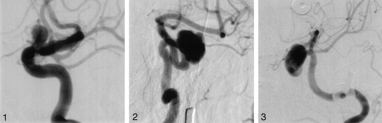
A 56-year-old female patient was referred for endovascular assessment and possible coiling of an asymptomatic carotid-ophthalmic aneurysm. CT angiography (CTA) of the anterior circulation confirmed the presence of an aneurysm, measuring 5.2 mm across the neck and 7 mm in length, with the OpthA arising from the ventral aspect of the neck (Fig 1). Before TBO, the patient’s visual acuity was tested by using a hand-held eye chart at a 14-inch distance. Pre-TBO acuity for both eyes was 20/30. Following routine cervical and cerebral diagnostic angiography, a 6F Envoy guide catheter (Cordis Endovascular Systems, Miami, FL) was placed in the left ICA. The procedure was performed with the patient under conscious sedation by use of intravenous fentanyl and Versed (Roche Pharmaceuticals, Nutley, NJ) and monitored by the attending physician. After heparin anticoagulation and by using biplane roadmap imaging, a 4 × 20 mm Hyperglide balloon was advanced into the intracranial ICA and placed across the neck of the carotid-ophthalmic aneurysm, which included the OpthA origin. The balloon was inflated, and occlusion of the ICA was documented by hand injection. During 5.5 minutes of occlusion, vision in the left eye deteriorated from 20/30 to 20/70, while vision in the right eye remained at 20/30. The balloon was deflated, and visual acuity in the left eye returned to 20/30 over the next 10 minutes. The results of the TBO were regarded as an indication of insufficient collateral flow and increased risk of visual loss if the OpthA were to occlude during coiling. The patient was subsequently referred for surgical clipping.

Case 1. ICA angiogram. Lateral view, clearly showing the OpthA arising from the neck of the aneurysm.
Case 2
A 60-year-old female patient with a history of headaches was found to have a left paraclinoid-cavernous aneurysm during workup for a headache. She was referred for endovascular assessment and possible coiling. CTA along with carotid-cerebral arteriography confirmed the presence of the aneurysm measuring 6 mm across the neck and 13 × 11 mm in greatest coronal dimensions, with the OpthA arising from the superior aspect of the neck (Figs 2 and 3). Pre-TBO visual acuity was measured to be 20/100 in the left eye. After guide catheter placement and heparinization, by using biplane roadmap imaging, a 4 × 20 mm Hyperglide balloon (Micro Therapeutics Inc., Irvine, CA) was advanced into the intracranial ICA and placed across the neck of the carotid-ophthalmic aneurysm, including the aneurysm and the OpthA origin (documented by hand injection). The procedure was performed under conscious sedation by use of intravenous fentanyl and Versed and monitored by the attending physician. During balloon inflation of 2.5 minutes’ duration, visual acuity dropped to 20/400. The balloon was deflated, and vision returned to pretest levels over the course of the next 2 hours. Again, this was considered indicative of inadequate collateral development and a sufficient indication to recommend clipping with preservation of the OpthA over endovascular coiling.

Case 2. ICA angiogram. Magnified anteroposterior view, showing the aneurysm clearly and the OpthA in relation to it.

Case 2. ICA angiogram. Magnified lateral view, showing the aneurysm and the OpthA.
Discussion
In 1911, Matas (3) described temporary arterial occlusion by manual compression of the common carotid artery to determine tolerance for permanent arterial occlusion. Serbinenko introduced the concept of endovascular arterial occlusion by using small endovascular balloons in the early 1970s. His novel method of endovascular arterial occlusion has since been widely adopted and remains the current foundation for endovascular arterial sacrifice and temporary arterial test occlusion (3). Current indications for TBO include conditions wherein permanent ICA occlusion may be used for treatment. These include cases of large complex ICA aneurysms or pseudoaneurysms, cranial and cervical neoplasms with ICA involvement, uncontrollable hemorrhage related to trauma, infection, or neoplasm, and carotid cavernous fistulas that may not be treatable without sacrifice of the parent artery (3).
Although the standard of care for intracranial aneurysms in the past has been conventional neurosurgical clipping (2, 4), advances in endovascular therapy are challenging established assessment and treatment strategies. As the endovascular approach proliferates, appropriate case selection and risk aversion strategies will be important for optimal patient care. Factors that help determine the appropriateness of endovascular therapy include aneurysm neck and dome morphology, aneurysm location, vessel incorporation into the neck of the aneurysm, and medical stability of the patient.
The risk of visual loss with acute occlusion of the OpthA is approximately 10% (5), because a rich network of collateral supply exists between the orbital external carotid branches and OpthA branches (6). Hence, when required for treating regional vascular disease, OpthA sacrifice proximal to the retinal and ciliary artery origins remains a viable option, with 90% of patients expected to remain asymptomatic (7).
There are very few reports of the use of TBO for combined occlusion of the OpthA and the ICA. There is a reported case of isolated monocular blindness occurring during a carotid occlusion test on a patient with an anomalous (cavernous) origin of the OpthA (8). By contrast, there is a report of three cases of symptomatic giant aneurysms of the supraclinoid ICA, manifesting as cranial nerve palsy and impaired visual acuity, wherein combined parent artery and OpthA occlusion was carried out to test collateral flow before treatment by permanent occlusion (7). The occlusion tests were negative, as expected, because of well-developed collaterals, which are the norm. There is also mention in the literature of ocular ischemic syndrome (ipsilateral orbital pain and progressive monocular visual loss) developing during carotid balloon occlusion testing, resulting from total or partial replacement of oxygenated blood within the OpthA due to rapid saline infusion (9).
In both of our cases, the OpthA arose from the base of a carotid-ophthalmic aneurysm with endovascular coiling carrying a high risk of OpthA occlusion. To further stratify the patients’ risk for visual loss, we applied the principles of TBO and interrogated the effectiveness of collateral flow. The literature tells us that 90% of patients will have adequate collaterals and will tolerate occlusion of the origin of the OpthA, with preservation of vision. On the basis of this premise, our hypothesis was that our two patients would have an excellent chance of tolerating permanent occlusion while maintaining vision, thus allowing us to proceed with endovascular treatment of the aneurysms. After pre-TBO visual acuity measurements, a TBO was performed to target the neck of the aneurysm to ensure temporary occlusion of the ophthalmic origin. To our surprise, the patients’ visual acuity ipsilateral to the TBO decreased during inflation and recovered shortly after deflation, which suggests that collateral supply to the retina was insufficient to maintain oxygenation and prevent ischemia. Our interpretation of this finding was that endovascular treatment with unavoidable occlusion of the OpthA would not be tolerated and would lead to visual loss. On the basis of this interpretation, direct visualization and preservation of the OpthA was thought to be the safest approach to preserve visual acuity, so surgical clipping was recommended.
The Snellen chart is the most common tool for the measurement of visual acuity in ophthalmic practice. It does, however, exhibit some design flaws that can lead to a measurement error (test-retest variability) (10). However, a change in visual acuity needed to be assessed with our patients in the supine position immediately before and during occlusion of the OpthA origin. In this clinical setting, the handheld Snellen eye chart was the most practical and accurate test available to us by which this could be accomplished. Existing LogMAR charts are not yet adopted widely in routine clinical practice. Care was taken to address some of the factors that have been shown to reduce reliability of testing with a Snellen chart (11). Testing was carried out in optimal lighting, at an exact measured distance and by the same clinician to minimize test-retest variation and increase reliability. We believe that the change in visual acuity seen in both patients was significant enough to warrant the belief that the collateral flow to the retina was inadequate.
The question of whether it is possible to apply the principles of TBO on the basis of the brain vascular model can also arise when it comes to assessment of ophthalmic vasculature and retinal perfusion. Can the failure of both patients to tolerate occlusion of their OpthA be attributed to chance, or was the duration of ophthalmic occlusion insufficient for assessment of collateral flow? In other words, would tolerance have developed over a longer period of occlusion, because of acute enlargement of collateral vessels? Injection of the external carotid to determine retrograde flow in the ophthalmic could provide further confirmation. In addition, although unlikely, the possibility of transient visual loss from inadvertent thromboembolism to the OpthA cannot be ruled out. The technique applied in these two cases suffers from the fact that a selective angiogram of the ipsilateral ECA was not obtained during test balloon occlusion to document the presence or absence of collaterals. If retrograde filling of the OpthA had been seen, it may have convinced us to maintain the occlusion longer and to test visual acuity over a longer period of time. The use of high-resolution video flourescein angiography with scanning laser ophthalmoscopy for assessment of retinal macro- and microcirculation has been reported in the literature (12). In the future, perhaps this or other techniques used in retinal vascular assessment can aid, along with a subjective test of vision, to permit an accurate interpretation of the results of temporary occlusion.
Conclusion
It is technically feasible to occlude temporarily flow to the OpthA concurrent with TBO of the ICA. The decision to proceed with endovascular treatment of an aneurysm involving the OpthA origin can be influenced by whether the patient is able to tolerate permanent occlusion of the OpthA origin while maintaining vision. We think that further research regarding this topic is both interesting and important for patient care, as more and more patients are undergoing endovascular treatment of intracranial aneurysms.
References
- Received October 7, 2003.
- Accepted after revision January 5, 2004.
- Copyright © American Society of Neuroradiology
In this issue


Jump to section
Related Articles
Cited By...
- Endovascular treatment of unruptured ophthalmic artery aneurysms: clinical usefulness of the balloon occlusion test in predicting vision outcomes after coil embolization
- Endovascular Treatment of Ophthalmic Artery Aneurysms: Assessing Balloon Test Occlusion and Preservation of Vision in Coil Embolization