Advertisement

Main menu

  • Home
  • Content
    • Current Issue
    • Accepted Manuscripts
    • Article Preview
    • Past Issue Archive
    • AJNR Case Collection
    • Case of the Week Archive
    • Classic Case Archive
    • Case of the Month Archive
  • Special Collections
    • Spinal CSF Leak Articles (Jan 2020-June 2024)
    • 2024 AJNR Journal Awards
    • Most Impactful AJNR Articles
  • Multimedia
    • AJNR Podcast
    • AJNR Scantastics
    • Video Articles
  • For Authors
    • Submit a Manuscript
    • Author Policies
    • Fast publishing of Accepted Manuscripts
    • Graphical Abstract Preparation
    • Manuscript Submission Guidelines
    • Imaging Protocol Submission
    • Submit a Case for the Case Collection
  • About Us
    • About AJNR
    • Editorial Board
  • More
    • Become a Reviewer/Academy of Reviewers
    • Subscribers
    • Permissions
    • Alerts
    • Feedback
    • Advertisers
    • ASNR Home
  • Other Publications
    • ajnr

User menu

  • Alerts
  • Log in

Search

  • Advanced search
American Journal of Neuroradiology

American Journal of Neuroradiology

ASHNR American Society of Functional Neuroradiology ASHNR American Society of Pediatric Neuroradiology ASSR
  • Alerts
  • Log in

Advanced Search

  • Home
  • Content
    • Current Issue
    • Accepted Manuscripts
    • Article Preview
    • Past Issue Archive
    • AJNR Case Collection
    • Case of the Week Archive
    • Classic Case Archive
    • Case of the Month Archive
  • Special Collections
    • Spinal CSF Leak Articles (Jan 2020-June 2024)
    • 2024 AJNR Journal Awards
    • Most Impactful AJNR Articles
  • Multimedia
    • AJNR Podcast
    • AJNR Scantastics
    • Video Articles
  • For Authors
    • Submit a Manuscript
    • Author Policies
    • Fast publishing of Accepted Manuscripts
    • Graphical Abstract Preparation
    • Manuscript Submission Guidelines
    • Imaging Protocol Submission
    • Submit a Case for the Case Collection
  • About Us
    • About AJNR
    • Editorial Board
  • More
    • Become a Reviewer/Academy of Reviewers
    • Subscribers
    • Permissions
    • Alerts
    • Feedback
    • Advertisers
    • ASNR Home
  • Follow AJNR on Twitter
  • Visit AJNR on Facebook
  • Follow AJNR on Instagram
  • Join AJNR on LinkedIn
  • RSS Feeds

Welcome to the new AJNR, Updated Hall of Fame, and more. Read the full announcements.


AJNR is seeking candidates for the position of Associate Section Editor, AJNR Case Collection. Read the full announcement.


Research ArticleINTERVENTIONAL

The Use of Hydrocoil for Parent Artery Occlusion

David F. Kallmes and Harry J. Cloft
American Journal of Neuroradiology September 2004, 25 (8) 1409-1410;
David F. Kallmes
  • Find this author on Google Scholar
  • Find this author on PubMed
  • Search for this author on this site
Harry J. Cloft
  • Find this author on Google Scholar
  • Find this author on PubMed
  • Search for this author on this site
  • Article
  • Figures & Data
  • Info & Metrics
  • Responses
  • References
  • PDF
Loading

Abstract

Summary: The HydroCoil Embolic System (HES) comprises a platinum microcoil coated with an expansile polymer and is approved by the United States Food and Drug Administration for cerebral aneurysm occlusion. We report a series of five cases, two in the internal carotid artery and three in the vertebral artery, in which the HES was used for parent artery occlusion procedures. In each of these cases, the HES allowed rapid, short-segment occlusion.

The ideal treatment option for some types of intracranial vascular diseases may include parent artery occlusion (PAO). For example, large or giant ophthalmic segment aneurysms can be cured with carotid PAO (1, 2) and intracranial vertebral artery dissections can be treated with vertebral artery PAO (3, 4). Various devices have been used to achieve vessel occlusion, including detachable balloons and coils. Safe, reliable occlusion may be achieved with each type of device in most cases (1–4).

The Hydrocoil Embolic System (HES, MicroVention, Inc, Aliso Viejo, CA) has recently been developed for the endovascular treatment of intracranial aneurysms, and has been granted United States Food and Drug Administration approval for that application (5). The HES device consists of a carrier platinum coil coupled with an expandable hydrogel material, which increases sevenfold in volume when placed into a physiologic environment. Within aneurysm cavities, the expanded hydrogel achieves high rates of volumetric occlusion compared with that in platinum coils (6).

We hypothesized that the HES could be applied to achieve rapid, precise, targeted PAO procedures, even in short segments of vessel. On the basis of this hypothesis, we used the HES system in a series of patients presenting with cervical and intracranial pathologic findings requiring either vertebral or carotid PAO. We report herein our experience with the HES in PAO procedures.

Description of Technique

Clinical characteristics and other data are shown in the Table. In general, vessel access was achieved with 6F guiding catheters. Systemic heparinization was administered to achieve activated clotting times (ACT) greater than 250 seconds. The HES is available in multiple expanded diameters, secondary diameters, and lengths and can treat vessels ranging in diameter from 2 mm to 6 mm. For this case series, we used an HES-14 for vertebral PAO (n = 3) and internal carotid artery (ICA) PAO (n = 2) and an HES-18 for a carotid PAO procedure (n = 1). Before placement of HES devices, a detachable platinum coil, ideally an 18-type coil rather than a 10-type coil, was placed in each case. This initial coil served to stabilize the HES devices and is recommended on the basis of preclinical experience in which HES devices placed without such stabilizing platinum coils migrated distally as the polymer expanded (unpublished data). The initial platinum coil should be oversized 1–2 mm relative to the diameter of the parent artery, and as tight a basket as possible should be achieved with this coil.

View this table:
  • View inline
  • View popup

Lesion types and devices utilized

Following detachment of the initial platinum coil, we proceeded with HES placement. The HES was relatively undersized compared with the parent artery diameter to achieve a dense-as-possible packing of the hydrogel-bearing devices. In our opinion, larger-diameter HES devices would have yielded less dense packing of the hydrogel material as compared with small-diameter devices. Placement of the first two HES devices was done relatively rapidly to achieve dense packing of devices before full expansion of the hydrogel material. Immediately following placement of the second HES device, control angiography was performed from the guiding catheter. Additional devices were placed as needed.

We catalogued the vascular disease, vessel location and diameter, and type and number of devices required to achieve vessel occlusion (Table).

Representative case

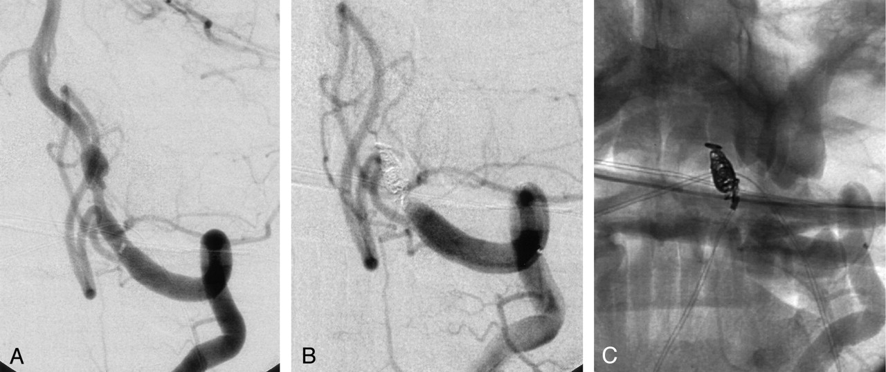
A 53-year-old male patient presented with acute subarachnoid hemorrhage. A cerebral angiogram demonstrated a dissection along the intracranial portion of the left vertebral artery. The dissection began approximately 10 mm distal to the left posterior inferior cerebellar artery (PICA) (Fig 1A). Occlusion of the dissected segment of the vertebral artery was considered the ideal treatment option. Furthermore, we hoped to avoid manipulation of the dissected portion of artery yet maintain patency of the PICA. To achieve these goals, short-segment occlusion of the left vertebral artery, immediately proximal to the most proximal aspect of the dissection, was undertaken. Using a 6Fr Envoy guiding catheter (Cordis Neurovascular, Miami Lakes, FL) and Excelsior microcatheter (Target, Boston Scientific, Fremont, CA) we placed a 4 mm T18 Micrus Spherical coil (Micrus Corp, Sunnyvale, CA) just proximal to the dissection. Subsequently, we placed two separate 2mm × 5 cm HydroCoil 14 devices within the initial platinum coil. Vessel occlusion was noted less than 5 minutes following deployment of the first HydroCoil (Fig 1B, C). The PICA was preserved.

Fig 1.
  • Download figure
  • Open in new tab
  • Download powerpoint
Fig 1.

A, Oblique digital subtraction angiogram of the left vertebral artery demonstrates a fusiform, dissecting aneurysm. The aneurysm is located approximately 10 mm distal to the origin of the posterior inferior cerebellar artery.

B, Oblique digital subtraction angiogram following coil occlusion of the left vertebral artery. The coils were placed in the segment of artery between the posterior inferior cerebellar artery origin and the proximal aspect of the dissection. The posterior inferior cerebellar artery was preserved.

C, Nonsubtracted image of the coil mass from the digital subtraction angiogram (B).

Discussion

This case series demonstrates that the HES can be used to achieve occlusion, even in short segments of vessel. In the case of vertebral PAO, device placement within vessel segments as short as 6 mm could achieve occlusion. Typically, a single platinum coil and two HES devices were needed for vessel occlusion. It remains possible that a single HES device could have achieved occlusion, but we chose to rapidly place at least two HES devices before performing control angiography to maximize attenuation of HES packing.

Preclinical experimentation with the use of HES in PAO has demonstrated durable occlusion of embolized arteries over 90 days. Significant thrombic organization proximal to the embolization site was observed (unpublished data).

Previous case series have detailed the use of detachable balloons and platinum coils, both fibered and nonfibered, for PAO procedures (1–4). Detachable silicone balloons have recently been withdrawn from the market. Nonfibered platinum coils may fail to achieve short-segment occlusion, given their nonthrombogenic nature. Fibered coils may offer improved ability to achieve vessel occlusion relative to nonfibered platinum coils. The HES expands to fill a larger volume of the vessel lumen, after it enters the artery, analogous to a balloon.

References

  1. ↵
    Debrun G, Fox A, Drake C, Peerless S, Girvin J, Ferguson G. Giant unclippable aneurysms: treatment with detachable balloons. AJNR Am J Neuroradiol 1981;2:167–173
    Abstract/FREE Full Text
  2. ↵
    Fox AJ, Vinuela F, Pelz DM, et al. Use of detachable balloons for proximal artery occlusion in the treatment of unclippable cerebral aneurysms. J Neurosurg 1987;66:40–46
    CrossRefPubMed
  3. ↵
    Kurata A, Ohmomo T, Miyasaka Y, Fujii K, Kan S, Kitahara T. Coil embolization for the treatment of ruptured dissecting vertebral aneurysms. AJNR Am J Neuroradiol 2001;22:11–18
    Abstract/FREE Full Text
  4. ↵
    Tsukahara T, Wada H, Satake K, Yaoita H, Takahashi A. Proximal balloon occlusion for dissecting vertebral aneurysms accompanied by subarachnoid hemorrhage. Neurosurgery 1995;36:914–919; discussion 919–920
    PubMed
  5. ↵
    Kallmes DF, Fujiwara NH. New expandable hydrogel-platinum coil hybrid device for aneurysm embolization. AJNR Am J Neuroradiol 2002;23:1580–1588
    Abstract/FREE Full Text
  6. ↵
    Cloft HJ, Kallmes DF. Aneurysm packing with HydroCoil Embolic System versus platinum coils: initial clinical experience. AJNR Am J Neuroradiol 2004;25:60–62
    Abstract/FREE Full Text
  • Received February 2, 2004.
  • Accepted after revision March 7, 2004.
  • Copyright © American Society of Neuroradiology
View Abstract
PreviousNext
Back to top

In this issue

American Journal of Neuroradiology: 25 (8)
American Journal of Neuroradiology
Vol. 25, Issue 8
1 Sep 2004
  • Table of Contents
  • Index by author
Advertisement
Print
Download PDF
Email Article

Thank you for your interest in spreading the word on American Journal of Neuroradiology.

NOTE: We only request your email address so that the person you are recommending the page to knows that you wanted them to see it, and that it is not junk mail. We do not capture any email address.

Enter multiple addresses on separate lines or separate them with commas.
The Use of Hydrocoil for Parent Artery Occlusion
(Your Name) has sent you a message from American Journal of Neuroradiology
(Your Name) thought you would like to see the American Journal of Neuroradiology web site.
CAPTCHA
This question is for testing whether or not you are a human visitor and to prevent automated spam submissions.
Cite this article
David F. Kallmes, Harry J. Cloft
The Use of Hydrocoil for Parent Artery Occlusion
American Journal of Neuroradiology Sep 2004, 25 (8) 1409-1410;

Citation Manager Formats

  • BibTeX
  • Bookends
  • EasyBib
  • EndNote (tagged)
  • EndNote 8 (xml)
  • Medlars
  • Mendeley
  • Papers
  • RefWorks Tagged
  • Ref Manager
  • RIS
  • Zotero
0 Responses
Respond to this article
Share
Bookmark this article
The Use of Hydrocoil for Parent Artery Occlusion
David F. Kallmes, Harry J. Cloft
American Journal of Neuroradiology Sep 2004, 25 (8) 1409-1410;
del.icio.us logo Twitter logo Facebook logo Mendeley logo
  • Tweet Widget
  • Facebook Like
  • Google Plus One

Jump to section

  • Article
    • Abstract
    • Description of Technique
    • Representative case
    • Discussion
    • References
  • Figures & Data
  • Info & Metrics
  • Responses
  • References
  • PDF

Related Articles

  • No related articles found.
  • PubMed
  • Google Scholar

Cited By...

  • Vessel occlusion using a single long oversized coil in vertebral artery dissection: a technical note
  • Treatment of a traumatic carotid-cavernous fistula by the sole use of a flow diverting stent
  • Crossref
  • Google Scholar

This article has not yet been cited by articles in journals that are participating in Crossref Cited-by Linking.

More in this TOC Section

  • SAVE vs. Solumbra Techniques for Thrombectomy
  • Delayed Reperfusion Post-Thrombectomy&Thrombolysis
  • Contrast-Induced Encephalopathy after NeuroIR
Show more Interventional

Similar Articles

Advertisement

Indexed Content

  • Current Issue
  • Accepted Manuscripts
  • Article Preview
  • Past Issues
  • Editorials
  • Editors Choice
  • Fellow Journal Club
  • Letters to the Editor

Cases

  • Case Collection
  • Archive - Case of the Week
  • Archive - Case of the Month
  • Archive - Classic Case

Special Collections

  • Special Collections

Resources

  • News and Updates
  • Turn around Times
  • Submit a Manuscript
  • Author Policies
  • Manuscript Submission Guidelines
  • Evidence-Based Medicine Level Guide
  • Publishing Checklists
  • Graphical Abstract Preparation
  • Imaging Protocol Submission
  • Submit a Case
  • Become a Reviewer/Academy of Reviewers
  • Get Peer Review Credit from Publons

Multimedia

  • AJNR Podcast
  • AJNR SCANtastic
  • Video Articles

About Us

  • About AJNR
  • Editorial Board
  • Not an AJNR Subscriber? Join Now
  • Alerts
  • Feedback
  • Advertise with us
  • Librarian Resources
  • Permissions
  • Terms and Conditions

American Society of Neuroradiology

  • Not an ASNR Member? Join Now

© 2025 by the American Society of Neuroradiology All rights, including for text and data mining, AI training, and similar technologies, are reserved.
Print ISSN: 0195-6108 Online ISSN: 1936-959X

Powered by HighWire