Advertisement

Main menu

  • Home
  • Content
    • Current Issue
    • Accepted Manuscripts
    • Article Preview
    • Past Issue Archive
    • AJNR Case Collection
    • Case of the Week Archive
    • Classic Case Archive
    • Case of the Month Archive
  • Special Collections
    • Spinal CSF Leak Articles (Jan 2020-June 2024)
    • 2024 AJNR Journal Awards
    • Most Impactful AJNR Articles
  • Multimedia
    • AJNR Podcast
    • AJNR Scantastics
    • Video Articles
  • For Authors
    • Submit a Manuscript
    • Author Policies
    • Fast publishing of Accepted Manuscripts
    • Graphical Abstract Preparation
    • Manuscript Submission Guidelines
    • Imaging Protocol Submission
    • Submit a Case for the Case Collection
  • About Us
    • About AJNR
    • Editorial Board
  • More
    • Become a Reviewer/Academy of Reviewers
    • Subscribers
    • Permissions
    • Alerts
    • Feedback
    • Advertisers
    • ASNR Home
  • Other Publications
    • ajnr

User menu

  • Alerts
  • Log in

Search

  • Advanced search
American Journal of Neuroradiology

American Journal of Neuroradiology

ASHNR American Society of Functional Neuroradiology ASHNR American Society of Pediatric Neuroradiology ASSR
  • Alerts
  • Log in

Advanced Search

  • Home
  • Content
    • Current Issue
    • Accepted Manuscripts
    • Article Preview
    • Past Issue Archive
    • AJNR Case Collection
    • Case of the Week Archive
    • Classic Case Archive
    • Case of the Month Archive
  • Special Collections
    • Spinal CSF Leak Articles (Jan 2020-June 2024)
    • 2024 AJNR Journal Awards
    • Most Impactful AJNR Articles
  • Multimedia
    • AJNR Podcast
    • AJNR Scantastics
    • Video Articles
  • For Authors
    • Submit a Manuscript
    • Author Policies
    • Fast publishing of Accepted Manuscripts
    • Graphical Abstract Preparation
    • Manuscript Submission Guidelines
    • Imaging Protocol Submission
    • Submit a Case for the Case Collection
  • About Us
    • About AJNR
    • Editorial Board
  • More
    • Become a Reviewer/Academy of Reviewers
    • Subscribers
    • Permissions
    • Alerts
    • Feedback
    • Advertisers
    • ASNR Home
  • Follow AJNR on Twitter
  • Visit AJNR on Facebook
  • Follow AJNR on Instagram
  • Join AJNR on LinkedIn
  • RSS Feeds

Welcome to the new AJNR, Updated Hall of Fame, and more. Read the full announcements.


AJNR is seeking candidates for the position of Associate Section Editor, AJNR Case Collection. Read the full announcement.


OtherHEAD AND NECK

Clinical Applications of Tumor Volume Measurements for Predicting Outcome in Patients with Squamous Cell Carcinoma of the Upper Aerodigestive Tract

Suresh K. Mukherji, Ilona M. Schmalfuss, Jonas Castelijns and Anthony A. Mancuso
American Journal of Neuroradiology September 2004, 25 (8) 1425-1432;
Suresh K. Mukherji
  • Find this author on Google Scholar
  • Find this author on PubMed
  • Search for this author on this site
Ilona M. Schmalfuss
  • Find this author on Google Scholar
  • Find this author on PubMed
  • Search for this author on this site
Jonas Castelijns
  • Find this author on Google Scholar
  • Find this author on PubMed
  • Search for this author on this site
Anthony A. Mancuso
  • Find this author on Google Scholar
  • Find this author on PubMed
  • Search for this author on this site
  • Article
  • Figures & Data
  • Info & Metrics
  • Responses
  • References
  • PDF
Loading

Article Figures & Data

Figures

  • Fig 1.
    • Download figure
    • Open in new tab
    • Download powerpoint
    Fig 1.

    Technique of volume contouring.

    A, Axial enhanced CT scan obtained at the level of the cricoarytenoid joint shows a true vocal cord carcinoma on the left. A white contour has been drawn around the tumor.

    B, Axial CT scan obtained at the aryepiglottic fold shows transglottic spread of the tumor (white contour). This finding precludes voice-conservation laryngectomy (hemilaryngectomy).

    C, Tumor (white contour) also extends inferior to the level of the subglottis, preventing partial laryngectomy (supracricoid laryngectomy). The only surgical alternative was total laryngectomy. Total tumor volume was 3.2 mL. The patient was treated with combined chemotherapy and RT.

  • Fig 2.
    • Download figure
    • Open in new tab
    • Download powerpoint
    Fig 2.

    Patient with an 8.4-mL supraglottic carcinoma. Pre-RT enhanced CT scan obtained at the lower supraglottic level shows a mass (arrow) infiltrating the paraglottic space on the left. Despite the large tumor volume, the patient elected definitive RT, which failed. The patient then underwent successful salvage resection of the supraglottic larynx (supraglottic laryngectomy).
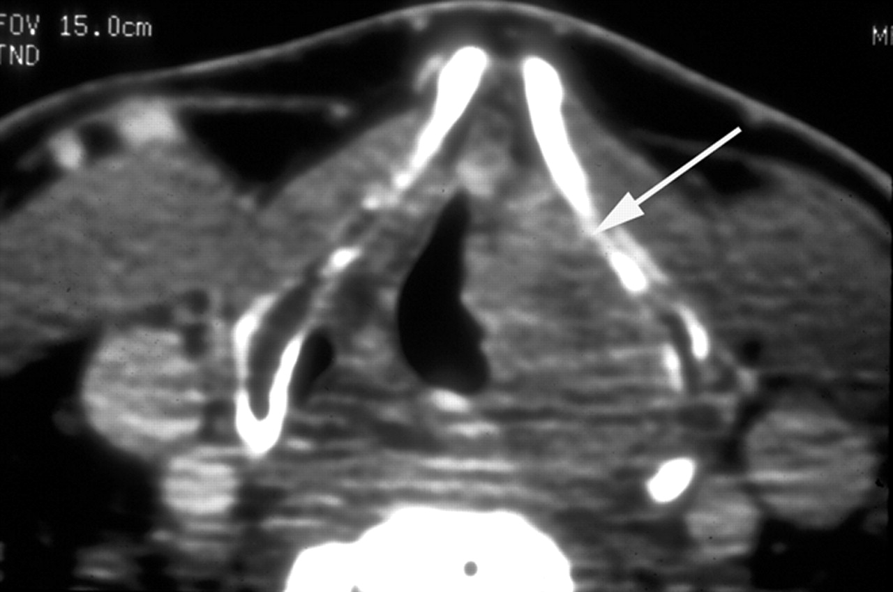
  • Fig 3.
    • Download figure
    • Open in new tab
    • Download powerpoint
    Fig 3.

    Pre-RT enhanced CT scans at the level of the true vocal cords in a patient with a T3 glottic carcinoma and sclerosis of the arytenoid and cricoid cartilages on the left. Calculated tumor volume was 4.3 mL. The patient elected curative RT alone, though the likelihood of control was <20% based on the CT risk profile. Because of the high risk of recurrence, close post-treatment surveillance with CT was chosen. Treatment failed, and total laryngectomy was performed.

    A, Soft tissue window shows cancer of the true vocal cords on the left (arrow) without involvement of the anterior commissure (arrowhead).

    B, Bone window shows sclerosis of the left lingual cortex of the left lamina of the thyroid cartilage (arrowhead) and the left arytenoid cartilage (arrow).
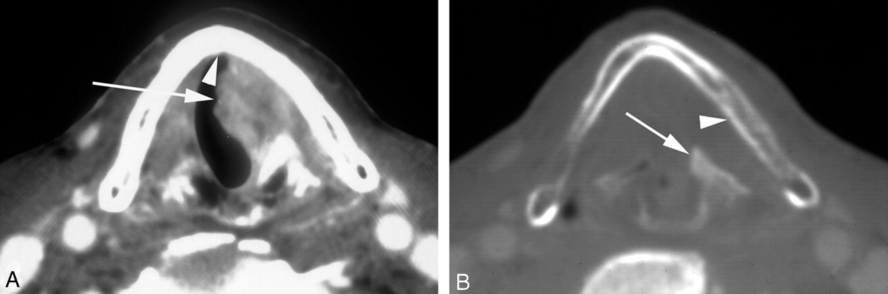
  • Fig 4.
    • Download figure
    • Open in new tab
    • Download powerpoint
    Fig 4.

    Patient with a 4.5-mL cancer of the right pyriform sinus with minimal involvement of its apex. The patient was cured with RT alone.

    A, Pretreatment enhanced CT scan through the lower third of the sinus shows a large mass (arrow) predominantly along the anterior and lateral walls of the sinus on the right.

    B, Axial image at the level of the true vocal cords shows minimal involvement of the apex of the sinus on the involved side. Compare the mildly enhancing fullness of the apex on the right with normally enhancing mucosa on the left (arrows). Note subtle obliteration of the submucosal fat plane on the right compared with the left (arrowheads).

PreviousNext
Back to top

In this issue

American Journal of Neuroradiology: 25 (8)
American Journal of Neuroradiology
Vol. 25, Issue 8
1 Sep 2004
  • Table of Contents
  • Index by author
Advertisement
Print
Download PDF
Email Article

Thank you for your interest in spreading the word on American Journal of Neuroradiology.

NOTE: We only request your email address so that the person you are recommending the page to knows that you wanted them to see it, and that it is not junk mail. We do not capture any email address.

Enter multiple addresses on separate lines or separate them with commas.
Clinical Applications of Tumor Volume Measurements for Predicting Outcome in Patients with Squamous Cell Carcinoma of the Upper Aerodigestive Tract
(Your Name) has sent you a message from American Journal of Neuroradiology
(Your Name) thought you would like to see the American Journal of Neuroradiology web site.
CAPTCHA
This question is for testing whether or not you are a human visitor and to prevent automated spam submissions.
Cite this article
Suresh K. Mukherji, Ilona M. Schmalfuss, Jonas Castelijns, Anthony A. Mancuso
Clinical Applications of Tumor Volume Measurements for Predicting Outcome in Patients with Squamous Cell Carcinoma of the Upper Aerodigestive Tract
American Journal of Neuroradiology Sep 2004, 25 (8) 1425-1432;

Citation Manager Formats

  • BibTeX
  • Bookends
  • EasyBib
  • EndNote (tagged)
  • EndNote 8 (xml)
  • Medlars
  • Mendeley
  • Papers
  • RefWorks Tagged
  • Ref Manager
  • RIS
  • Zotero
0 Responses
Respond to this article
Share
Bookmark this article
Clinical Applications of Tumor Volume Measurements for Predicting Outcome in Patients with Squamous Cell Carcinoma of the Upper Aerodigestive Tract
Suresh K. Mukherji, Ilona M. Schmalfuss, Jonas Castelijns, Anthony A. Mancuso
American Journal of Neuroradiology Sep 2004, 25 (8) 1425-1432;
del.icio.us logo Twitter logo Facebook logo Mendeley logo
  • Tweet Widget
  • Facebook Like
  • Google Plus One

Jump to section

  • Article
    • Abstract
    • Technique
    • Primary Sites
    • Lymph Nodes
    • Discussion
    • References
  • Figures & Data
  • Info & Metrics
  • Responses
  • References
  • PDF

Related Articles

  • No related articles found.
  • PubMed
  • Google Scholar

Cited By...

  • Does primary tumour volumetry performed early in the course of definitive concomitant chemoradiotherapy for head and neck squamous cell carcinoma improve prediction of primary site outcome?
  • Crossref
  • Google Scholar

This article has not yet been cited by articles in journals that are participating in Crossref Cited-by Linking.

More in this TOC Section

  • Correlation of Apparent Diffusion Coefficient at 3T with Prognostic Parameters of Retinoblastoma
  • Parathyroid Lesions: Characterization with Dual-Phase Arterial and Venous Enhanced CT of the Neck
  • Efficacy of Diffusion-Weighted Imaging for the Differentiation between Lymphomas and Carcinomas of the Nasopharynx and Oropharynx: Correlations of Apparent Diffusion Coefficients and Histologic Features
Show more Head and Neck

Similar Articles

Advertisement

Indexed Content

  • Current Issue
  • Accepted Manuscripts
  • Article Preview
  • Past Issues
  • Editorials
  • Editors Choice
  • Fellow Journal Club
  • Letters to the Editor

Cases

  • Case Collection
  • Archive - Case of the Week
  • Archive - Case of the Month
  • Archive - Classic Case

Special Collections

  • Special Collections

Resources

  • News and Updates
  • Turn around Times
  • Submit a Manuscript
  • Author Policies
  • Manuscript Submission Guidelines
  • Evidence-Based Medicine Level Guide
  • Publishing Checklists
  • Graphical Abstract Preparation
  • Imaging Protocol Submission
  • Submit a Case
  • Become a Reviewer/Academy of Reviewers
  • Get Peer Review Credit from Publons

Multimedia

  • AJNR Podcast
  • AJNR SCANtastic
  • Video Articles

About Us

  • About AJNR
  • Editorial Board
  • Not an AJNR Subscriber? Join Now
  • Alerts
  • Feedback
  • Advertise with us
  • Librarian Resources
  • Permissions
  • Terms and Conditions

American Society of Neuroradiology

  • Not an ASNR Member? Join Now

© 2025 by the American Society of Neuroradiology All rights, including for text and data mining, AI training, and similar technologies, are reserved.
Print ISSN: 0195-6108 Online ISSN: 1936-959X

Powered by HighWire