
Article Figures & Data
Figures
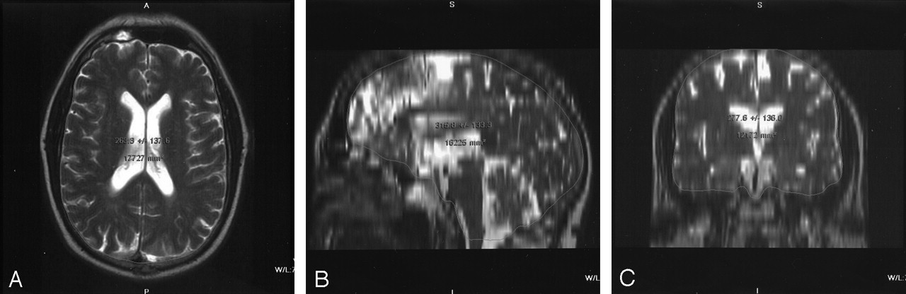
- Fig 1.
Measurement of intracranial volume. Hyperintense CSF is outlined on T2-weighted images, and volume is calculated. Gaps have been interpolated to provide smooth surface contours.
A, Axial image.
B, Reformatted sagittal image.
C, Reformatted coronal image.
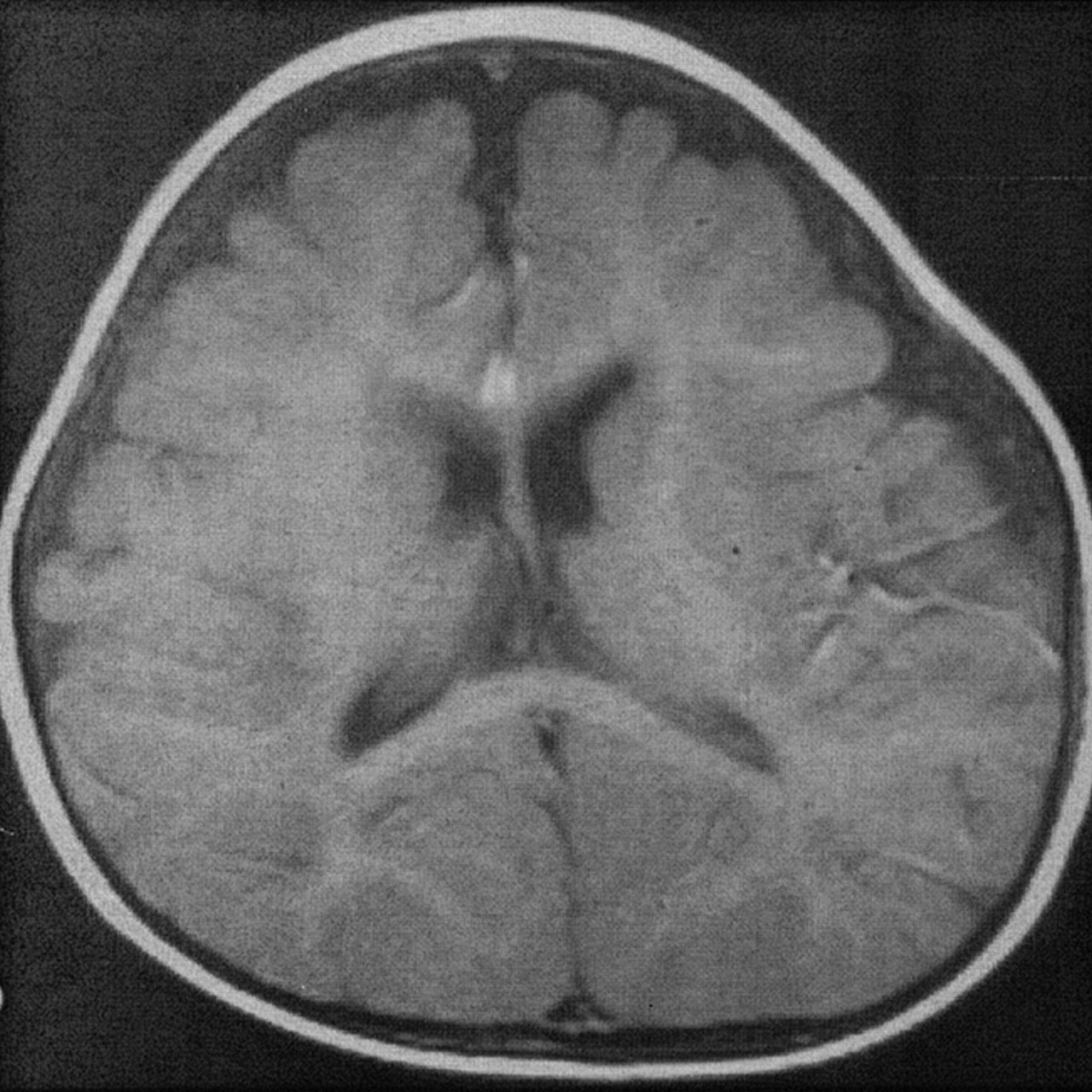
- Fig 2.
Benign external hydrocephalus in 7-month-old infant results in enlargement of the frontal subarachnoid spaces with minimal ventricular enlargement.
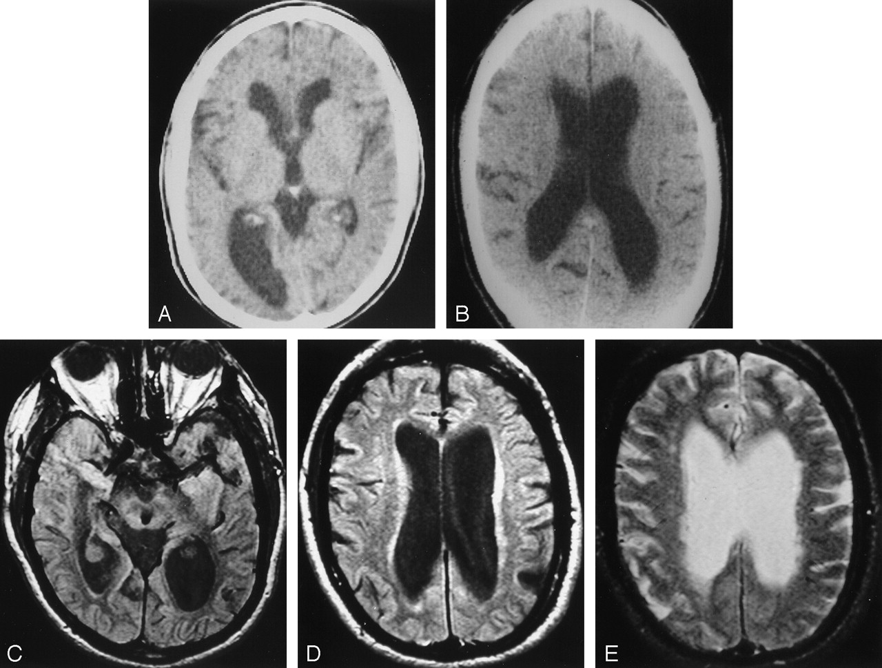
- Fig 3.
Chronically enlarged ventricles in a patient developing NPH.
A, At age 67 years, this patient had clearly enlarged ventricles. CT was performed to evaluate suspected giant cell arteritis. The patient was walking 20 miles a day and underwent successful shunt placement for NPH 19 years later.
B, At age 70, the patient still had no symptoms of NPH.
C–E, At age 76, the ventricles are markedly enlarged. Proton density–weighted image (C) shows a prominent CSF flow void in the aqueduct. The patient will not develop symptoms of NPH for another 10 years (when a pacemaker precluded MR imaging).


In this issue


Jump to section
Related Articles
Cited By...
- Molecular Signatures of Normal Pressure Hydrocephalus: A Large-scale Proteomic Analysis of Cerebrospinal Fluid
- Aqueductal CSF Stroke Volume Is Increased in Patients with Idiopathic Normal Pressure Hydrocephalus and Decreases after Shunt Surgery
- Lumbar Puncture Test in Normal Pressure Hydrocephalus: Does the Volume of CSF Removed Affect the Response to Tap?
- CSF Flow in the Brain in the Context of Normal Pressure Hydrocephalus
- Evidence that congenital hydrocephalus is a precursor to idiopathic normal pressure hydrocephalus in only a subset of patients
- Idiopathic normal pressure hydrocephalus