
Article Figures & Data
Figures
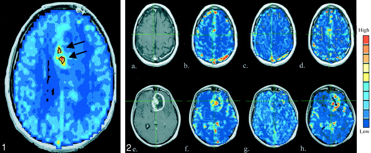
- Fig 1.
Sample SE rCBV map shows hot-spot ROIs (black outlines and arrows), which represent the highest approximate 10–20% of microvascular rCBVs. These were used to mask the image maps and perform hot-spot analysis.
- Fig 2.
T1-weighted contrast-enhanced images (a and e) GE rCBV maps (b and f), SE rCBV maps (c and g), and ratio maps (d and h) obtained in a 53-year-old man with an anaplastic oligodendroglioma (a–d) and a 55-year-old man with a glioblastoma multiforme (e–h).
- Fig 3.
Whole-tumor ROI analysis for gliomas.
A, GE rCBV, which is sensitive to total blood volume, is significantly correlated with tumor grade (n = 72).
B, Conversely, SE rCBV, which is sensitive to microvascular blood volume, is not significantly correlated with tumor grade (n = 67).
C, ΔR2*/ΔR2 ratio, an index of mVD, is significantly correlated with tumor grade (n = 67).
- Fig 4.
Microvascular hot-spot ROI analysis for gliomas.
A, GE rCBV results maintain a significant correlation (n = 67).
B, Contrary to whole-tumor results, SE rCBV shows a significant correlation with tumor grade (n = 67).
C, While a significant correlation with tumor grade is retained, the correlation is weaker than that of whole-tumor ROIs (n = 67).
- Fig 5.
SE rCBV data as a function of GE rCBV data. The K nearest-neighbor analysis (K = 5) resulted in correct classification of five (69%) of 16 grade I–II tumors (circles) and 49 (95%) of 51 grade III–IV tumors (squares). Asterisk indicates misclassified data points; the arrows indicate two low-grade tumors where diagnosis was based on biopsy.
- Fig 6.
MRI-derived (a) post-contrast (b) GE rCBV, (c) SE rCBV and (d) ratio maps obtained in a 66-year-old female patient prior to CT-guided biopsy. The biopsy pathologic results suggest low-grade astrocytoma. This diagnosis is not consistent with either the rCBV results or the clinical course of this patient.


Tables
Patient histology and imaging results
Patient/Age (y)/Sex Pathologic Diagnosis 1/80/M Ependymoma grade II 2/40/F Anaplastic astrocytoma grade III* 3/50/M Glioblastoma multiforme 4/32/F Anaplastic astrocytoma grade III 5/52/F Recurrent glioblastoma multiforme 6/73/M Glioblastoma multiforme 7/45/F Anaplastic oligodendroglioma grade IV 8/37/F Anaplastic astrocytoma grade III 9/66/M Mixed glioblastoma multiforme and low-grade astrocytoma 10/69/M Anaplastic astrocytoma grade III 11/47/M Anaplastic astrocytoma grade III 12/48/F Astrocytoma II, with markers of glioblastoma multiforme 13/48/M Recurrent glioblastoma multiforme 14/68/M Glioblastoma multiforme 15/50/F Recurrent glioblastoma multiforme 16/50/M Low-grade glioma grade II* 17/77/M Anaplastic astrocytoma grade III 18/41/M Glioblastoma multiforme 19/59/M Oligodendroglioma 20/23/F Recurrent central neurocytoma 21/50/M Recurrent anaplastic astrocytoma grade III 22/42/M Recurrent glioblastoma multiforme 23/74/M Glioblastoma multiforme 24/39/M Oligodendroglioma grade II 25/49/F Glioblastoma multiforme 26/50/M Astrocytoma grade II* 27/71/F Glioblastoma multiforme 28/30/M Glioblastoma multiforme 29/41/M Malignant oligodendroglioma grade III 30/53/M Astrocytoma grade II 31/41/M Recurrent glioblastoma multiforme 32/65/F Glioblastoma multiforme 33/42/M Glioblastoma multiforme 34/66/M Glioblastoma multiforme 35/41/M Anaplastic astrocytoma grade III 36/56/M Recurrent glioblastoma multiforme 37/77/M Glioblastoma multiforme 38/66/F Astrocytoma grade II* 39/30/M Mixed glioma: astrocytoma/oligodendroglioma 40/55/M Anaplastic oligodendroglioma grade III 41/54/F Recurrent glioblastoma multiforme 42/45/M Recurrent glioblastoma multiforme 43/19/M Astrocytoma grade II 44/55/M Glioblastoma multiforme 45/40/M Recurrent glioblastoma multiforme 46/78/M Glioblastoma multiforme 47/54/F Glioblastoma multiforme 48/46/F Mixed anaplastic astrocytoma 49/43/M Recurrent glioblastoma multiforme 50/45/F Recurrent malignant mixed glioma 51/32/M Recurrent glioblastoma multiforme 52/42/M Recurrent glioblastoma multiforme 53/40/M Mixed gliomas: mostly oligodendroglioma, some astrocytoma 54/25/F Astrocytoma grade II 55/33/F Glioblastoma multiforme 56/41/M Glioma grade II 57/34/F Oligodendroglioma grade II 58/52/M Glioblastoma multiforme 59/68/M Anaplastic astrocytoma 60/44/F Anaplastic astrocytoma 61/56/F Anaplastic astrocytoma 62/42/M Anaplastic astrocytoma grade III 63/64/F Glioblastoma multiforme 64/28/M Anaplastic astrocytoma 65/23/M Giant cell astrocytoma grade I 66/40/F Anaplastic astrocytoma 67/36/M Anaplastic astrocytoma 68/53/M Recurrent anaplastic oligodendroglioma 69/62/M Glioblastoma multiforme 70/68/F Glioblastoma multiforme 71/52/F Glioblastoma multiforme 72/52/F Glioblastoma multiforme 73/54/F Glioblastoma multiforme * Pathologic diagnosis was based on tissue biopsy.
In this issue


Jump to section
Related Articles
Cited By...
- Moving Toward a Consensus DSC-MRI Protocol: Validation of a Low-Flip Angle Single-Dose Option as a Reference Standard for Brain Tumors
- Optimization of Acquisition and Analysis Methods for Clinical Dynamic Susceptibility Contrast MRI Using a Population-Based Digital Reference Object
- Multisite Concordance of DSC-MRI Analysis for Brain Tumors: Results of a National Cancer Institute Quantitative Imaging Network Collaborative Project
- Impact of Software Modeling on the Accuracy of Perfusion MRI in Glioma
- ASFNR Recommendations for Clinical Performance of MR Dynamic Susceptibility Contrast Perfusion Imaging of the Brain
- Differentiation between Oligodendroglioma Genotypes Using Dynamic Susceptibility Contrast Perfusion-Weighted Imaging and Proton MR Spectroscopy
- The Effect of Pulse Sequence Parameters and Contrast Agent Dose on Percentage Signal Recovery in DSC-MRI: Implications for Clinical Applications
- Semi-automated and automated glioma grading using dynamic susceptibility-weighted contrast-enhanced perfusion MRI relative cerebral blood volume measurements
- The Role of Preload and Leakage Correction in Gadolinium-Based Cerebral Blood Volume Estimation Determined by Comparison with MION as a Criterion Standard
- Correlations between Perfusion MR Imaging Cerebral Blood Volume, Microvessel Quantification, and Clinical Outcome Using Stereotactic Analysis in Recurrent High-Grade Glioma
- Recent advances in imaging epilepsy
- Acquisition of MR perfusion images and contrast-enhanced MR angiography in acute ischaemic stroke patients: which procedure should be done first?
- Magnetic Resonance As a Cancer Imaging Biomarker