
Article Figures & Data
Figures
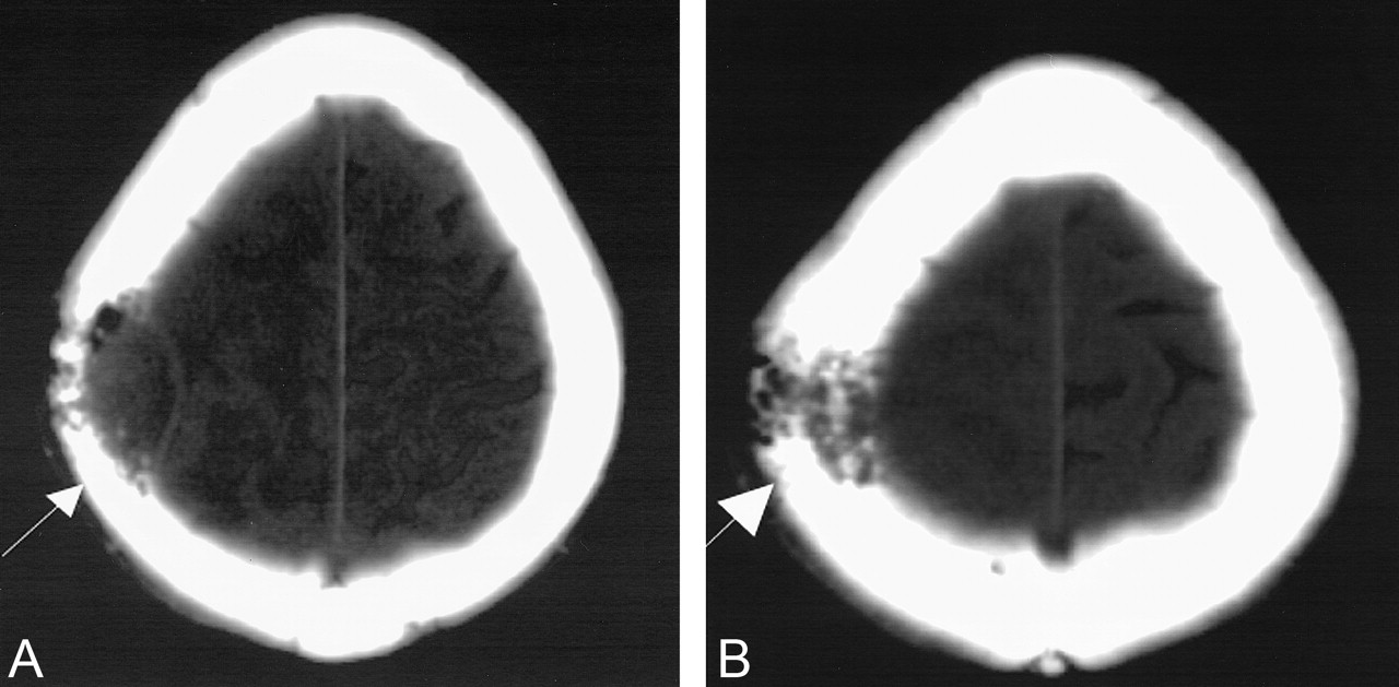
- Fig 1.
CT scans.
A and B, Nonenhanced scans obtained at slightly different heights show a rounded heterogeneous mass in the right frontoparietal region (arrow).
C, Higher section shows inward dural displacement (arrow) and overlying calvarial destruction, suggesting extra-axial extension of the mass.
- Fig 2.
Bone window settings from the same nonenhanced CT study as in Figure 1 shows calvarial destruction (arrow).
- Fig 3.
MR images demonstrate intra- and extra-axial locations of the mass eroding through the calvaria (arrow). Note inward displacement of the hypointense dura.
A, T2-weighted image.
B, T1-weighted image.
C, Gadolinium-enhanced T1-weighted image.
- Fig 4.
Sequential T2-weighted images demonstrate no definite gross violation of the dura.
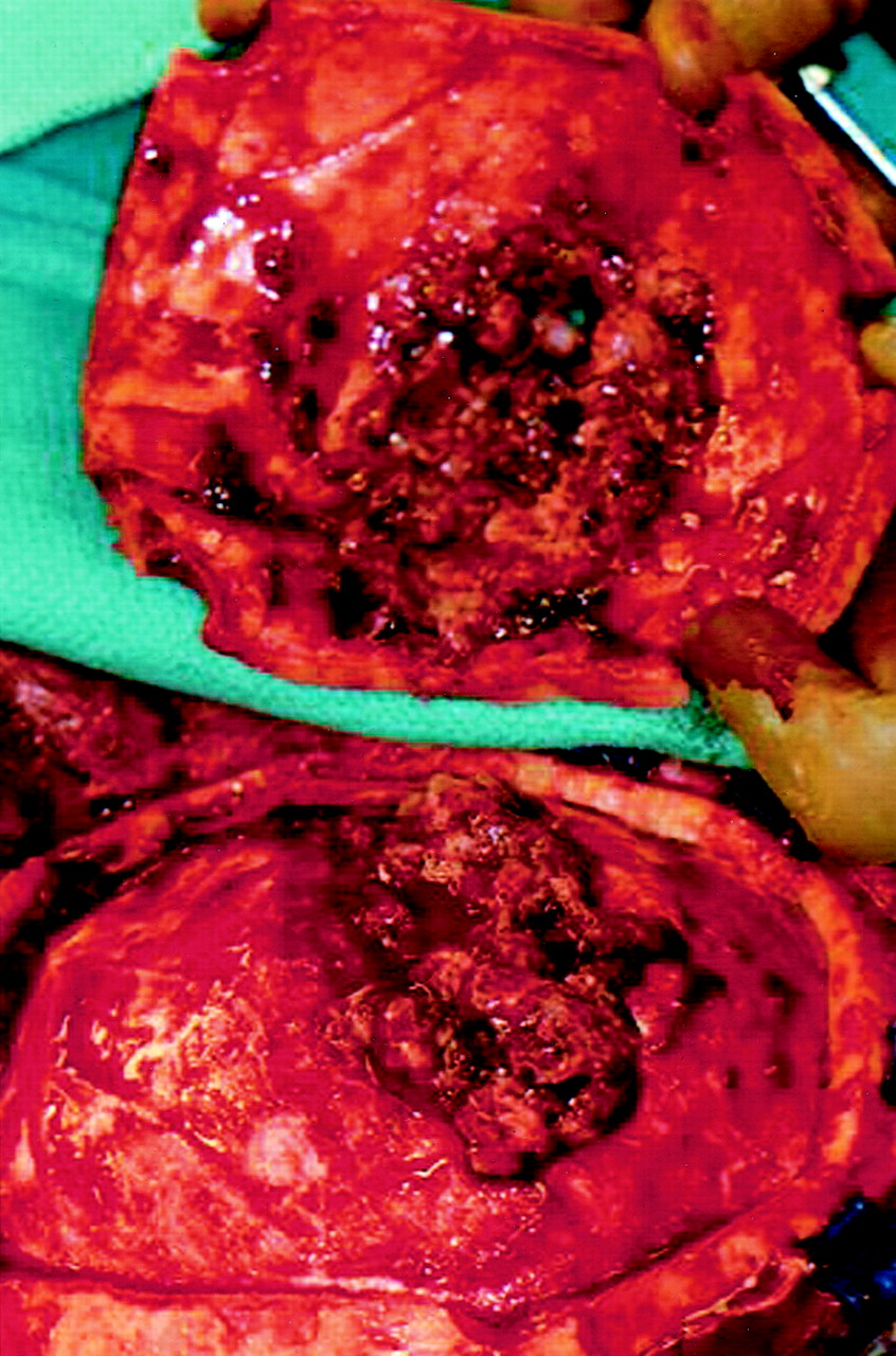
- Fig 5.
Tumor eroding through the bone before the bone flap was created.
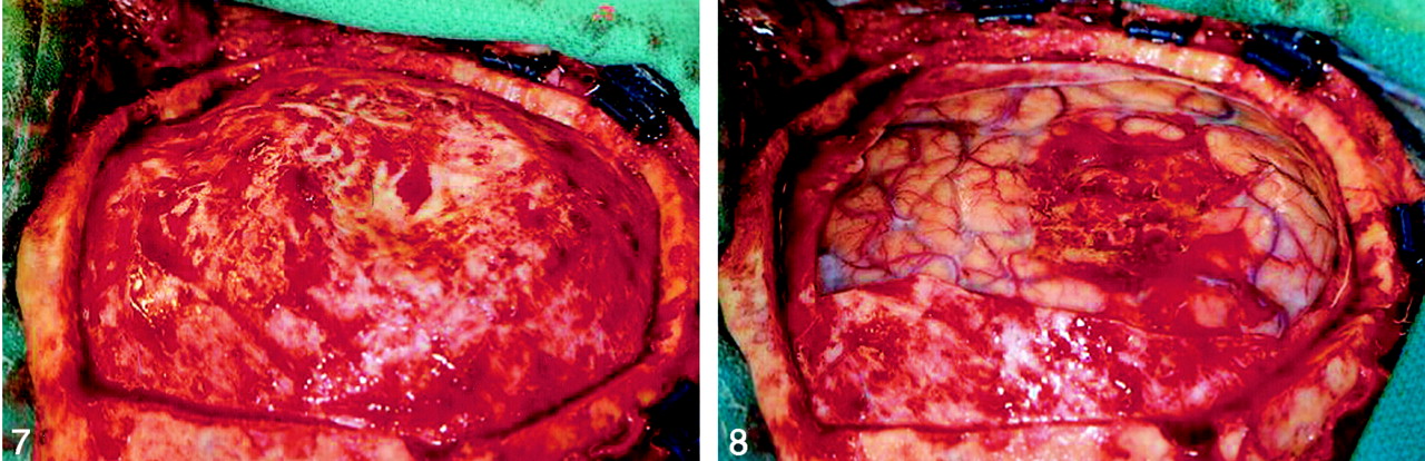
- Fig 6.
Epidural tumor leaving the dura macroscopically intact without substantial dural adherence.
- Fig 7.
Macroscopically intact dura after the epidural portion of tumor was removed.
- Fig 8.
Intracerebral component is visible and surrounded by normal cortex.
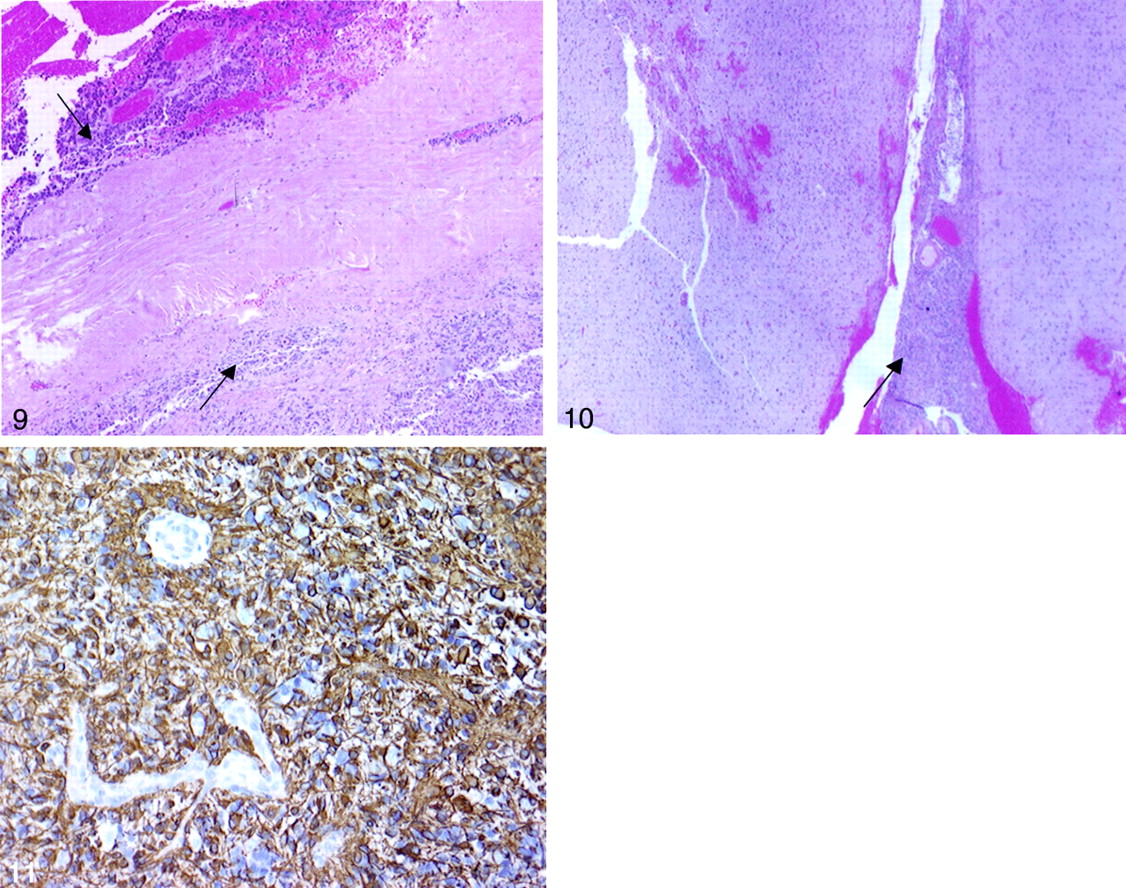
- Fig 9.
Right parietal dura with tumor (arrows) on both sides of the dura (original magnification ×40).
- Fig 10.
Right parietal intracerebral tumor (arrow). Image shows the GBM arising in the brain and growing outward as a mass outside the brain that penetrated the dura and the skull (original magnification ×20).
- Fig 11.
Right parietal tumor shows positivity for immunoperoxidase staining against GFAP (original magnification ×40).


