
Article Figures & Data
Figures
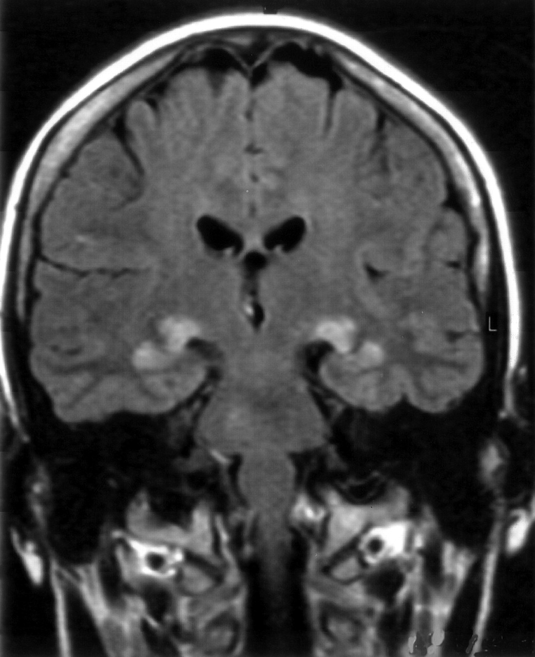
- Fig 1.
Coronal FLAIR image obtained 1 day after the acute onset of anaphylactic shock symptoms, showing bilateral hyperintense areas symmetrically located within the lateral geniculate bodies and extending laterally within the white matter of the parahippocampal regions.
- Fig 2.
Coronal Gd-enhanced T1WI obtained at day 6. Enhancement within both geniculate bodies extending to the superior parahippocampal regions is clearly visible.
- Fig 3.
Control MR images obtained 16 days after onset of symptoms.
A, Coronal T1WI, in which the geniculate bodies appear hyperintense.
B, Coronal FLAIR image, in which lesions are still visible but the extent and intensity of the abnormal high signal intensity are decreased.
C, Coronal Gd-enhanced T1WI, in which mild contrast uptake is still evident.
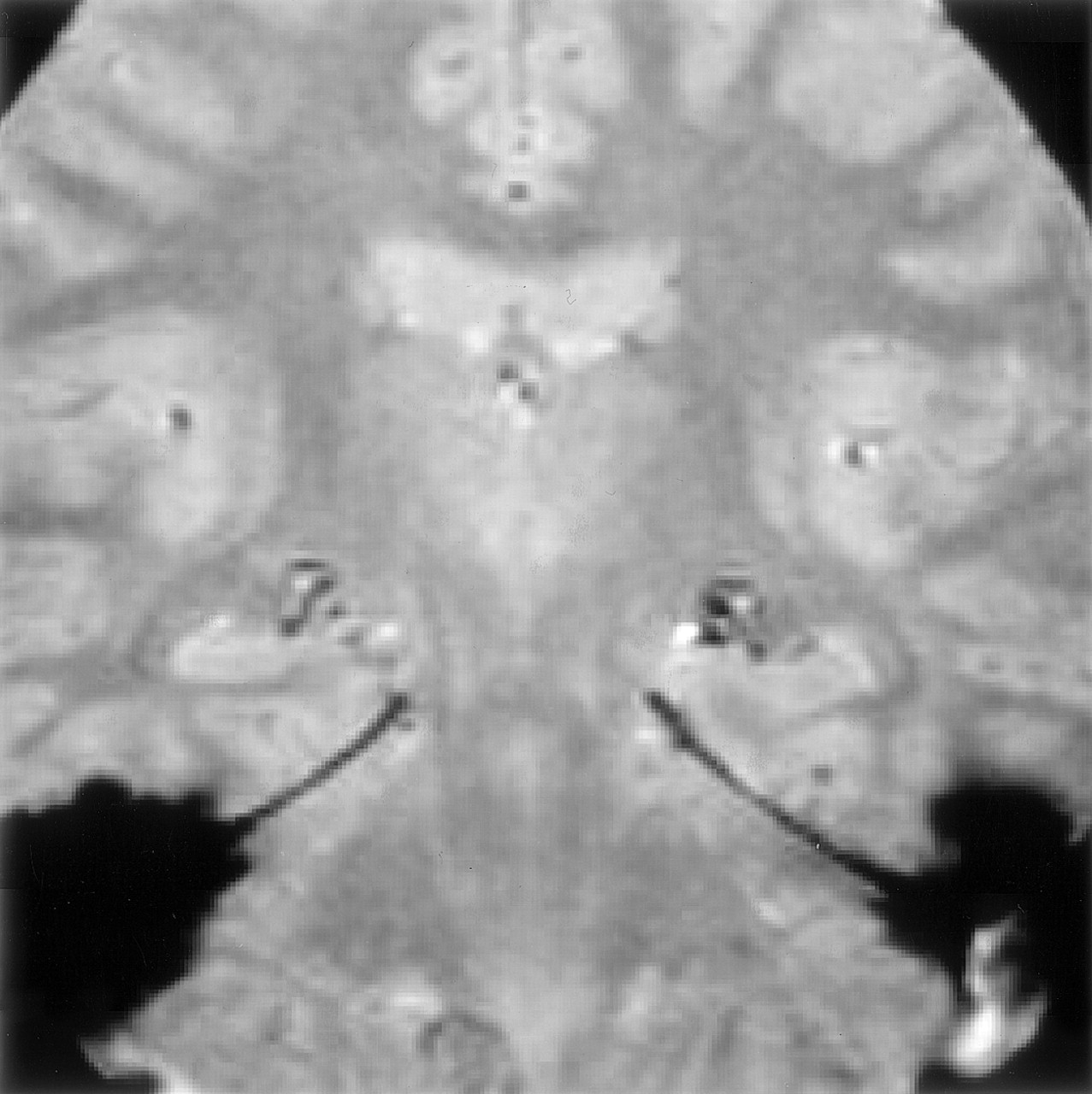
- Fig 4.
Coronal gradient echo T2WI obtained at day 16, demonstrating small hypointense areas in both geniculate bodies corresponding to hemosiderin deposits.
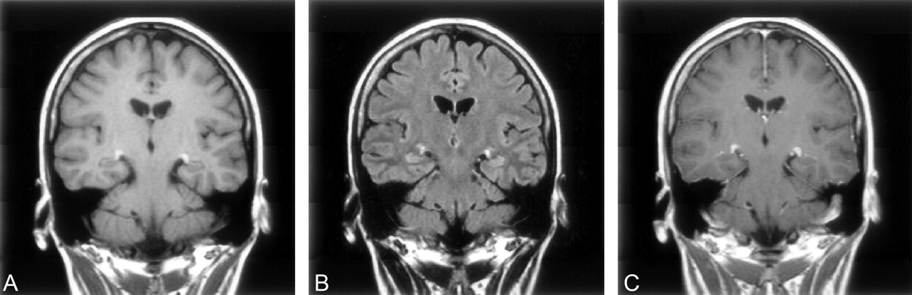
- Fig 5.
Follow-up MR images obtained at day 51.
A, Coronal T1WI, in which lesions are very discreetly hyperintense.
B, Normal FLAIR image.
C, Gd-enhanced T1WI, in which the lesions are still visible but are reduced in size and contrast uptake is mild.


In this issue


Jump to section
Related Articles
Cited By...
- Loss of Vision Because of Bilateral Lateral Geniculate Body Infarction After Traumatic Brain Injury
- Eyes and stroke: the visual aspects of cerebrovascular disease
- Reversible severe bilateral visual loss in an unusual case of bilateral lateral geniculate myelinolysis during acute pancreatitis
- Atrophy of the lateral geniculate nucleus in human glaucoma detected by magnetic resonance imaging