
Article Figures & Data
Figures
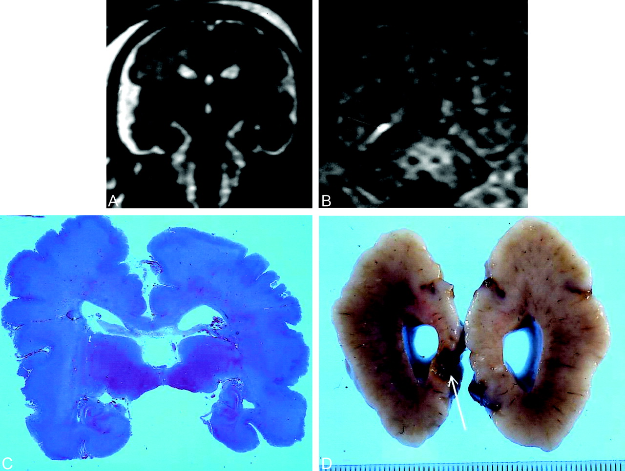
- Fig 1.
Transitory ventriculomegaly at 21 weeks’ gestation. At 29 weeks, US revealed linear hyperechogenicities around the frontal horns. MR imaging was performed at 30 weeks.
A, T2-weighted coronal section at the level of the temporal lobes shows bilateral opercular dysplasia, gyration delay (no frontal or temporal sulci), and a polymicrogyric cortical pattern.
B, T1-weighted coronal section at the level of the atria. Hyperintensity along the inferior aspect of the right atrium (arrow) was overlooked on T2-weighted images. The pregnancy was terminated at 30 weeks’ gestation. NFP findings confirmed gyration and parenchymal lesions.
C, Coronal histologic section at the level of the temporal lobes show diffuse polymicrogyria with opercular dysplasia.
D, Macroscopic coronal view shows right occipital cavitation (arrow). Calcifications were present on histologic examination.
- Fig 2.
At 32 weeks’ gestation, US showed decreased cerebellar transverse diameter (<10th percentile) without supratentorial abnormality. MR imaging was performed at 32.5 weeks. Infratentorial biometry was evaluated with MR study at about 28 weeks.
A, T2-weighted coronal section at the level of the thalami shows marked bilateral hyperintensity beneath the thalami just above the hippocampal fissure (arrows). Because of biometric and signal intensity abnormalities, termination of pregnancy was proposed and performed at 34 weeks. NFP confirmed the biometric abnormalities and showed a diffuse gliosis with neuronal necrosis lesions involving the cerebral peduncles, thalami, and hippocampal cortex.
B, Coronal histologic section shows bilateral cavitation (arrows) above both hippocampal fissures.
- Fig 3.
MR imaging performed at 34 weeks’ gestation because of mild (12-mm) dilatation of the left lateral ventricle at US. T2-weighted axial section at the level of the vertex shows diffuse bilateral frontal hyperintensity (hypointensity on T1-weighted imaging, not shown). Images also showed left lateral ventriculomegaly (12.5 mm) and a left subependymal pseudocyst. An ischemic process was suggested and because of the diffuse frontal abnormality, a termination of pregnancy was performed at 36 weeks’ gestation. NFP revealed a diffuse softening of the white matter with periventricular gliosis.
- Fig 4.
Huge subdural hematoma (after a fall down stairs a week earlier) in a fetus of 34 weeks’ gestation. US showed the bilateral hematoma with a fluid-fluid level, an enlarged right choroid plexus, and a hyperechogenicity in the posterior part of the right hemisphere.
A and B, Axial T1-weighted (A) and T2-weighted (B) sections at the level of the lateral ventricles. The hematoma is hyperintense in A and has intermediate signal intensity in B. Right hemisphere and frontal part of the left hemisphere are hypointense in A and hyperintense in B in relation to gliosis. Cortical hyperintensity in the right hemisphere (arrows) and posterior part of the left hemisphere corresponded to laminar necrosis during NFP study (not shown).
C, T2*-weighted coronal section shows marked hypointensity of the right choroid plexus (arrow) in relation to hemorrhage.


Tables
Etiology No. of Fetuses Vascular: preeclampsia, intrauterine growth restriction with abnormalities on uterine/cerebral Doppler study 5 Infection: toxoplasmosis, cytomegaloviral infection 3 Metabolic: diabetes with severe maternal ketoacidosis 1 Space-occupying intracranial lesion: brain tumor, huge bilateral subdural hematoma 2 No. of Lesions Definition and Extent Diffuse Focal Unique (n = 13) 11 2 Multiple (n = 13) 3 10 Sequence Hypointensity Hyperintensity T1 weighted 17 8 T2 weighted 5 16 T2* weighted 2 0


