
Article Figures & Data
Figures
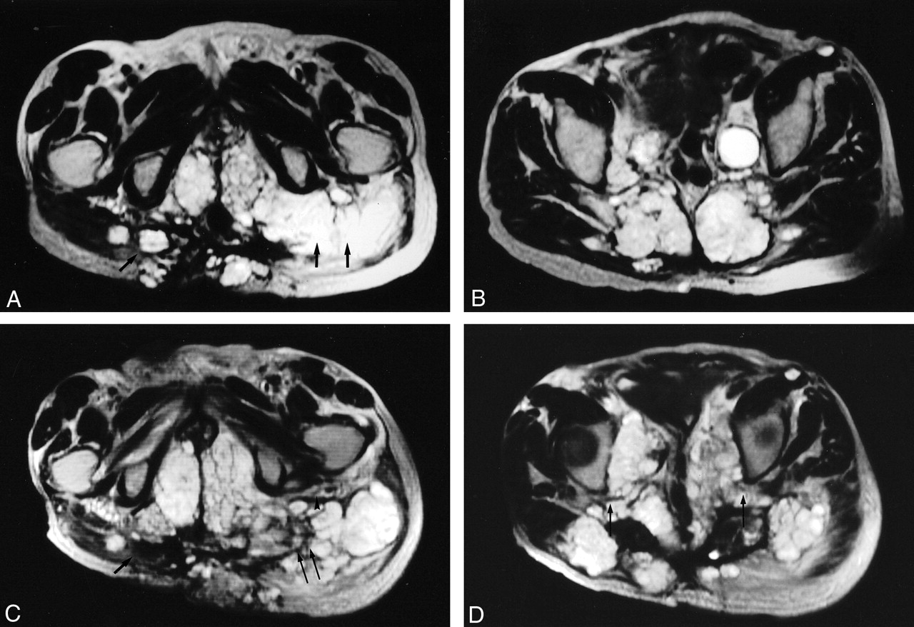
- Fig 1.
A 72-year-old male patient with recurrent sacral chordoma and regional invasion. All the images are obtained in the transverse plane T2-weighted Turbo spin-echo. Images A and B were obtained in December 2002; C and D depict the same regions as those of A and B (obtained August 2003).
A, the single arrow points to invasion of the right gluteus muscle with extension into the subcutaneous tissues and the skin. The double arrow points to the involvement of the left gluteus muscle with encasement of the tract of the left sciatic nerve, which is not clearly defined and probably encased. Note the presence of invasion of the perineum bilaterally (B) shows regional invasion of the posterior pelvic wall with invasion of the sacrum, the piriformis muscle, and bilateral extension into the gluteus muscles. In addition, a metastatic deposit is seen anterior to the left sartorius muscle. Large metastatic iliac nodes are also noted.
C, obtained in August 2003 (8 months later) at approximately the same level as A, shows the first site treated devoid of tumor tissue (single arrow). The component at the skin remains intact. The third site treated (double arrows) shows debulking of the tumor. Note that the left sciatic nerve tract and the fat planes surrounding it are better visualized, because of the retraction of the tumor secondary to ablation (arrowheads). Also note the increase in the extent of the involvement of the perineum with significant compression of the structures within it. The nontreated mass in the left gluteus muscle, laterally, increased in size.
D, obtained in August 2003 at approximately the same level as B, shows the sacral masses, which were treated with significant loss of substance and volume. Note the reexpansion of the spaces at both sciatic notches with resultant decompression of both sciatic nerves tracts (single arrow). Also note the remarkable increase in the involvement of the pelvic cavity with compression of the pelvic structures along the midline. There are also now more tumoral masses in the nontreated areas at the gluteus muscles bilaterally with atrophy of the left gluteus muscle.

In this issue


Jump to section
Related Articles
Cited By...
- No citing articles found.