
Article Figures & Data
Figures
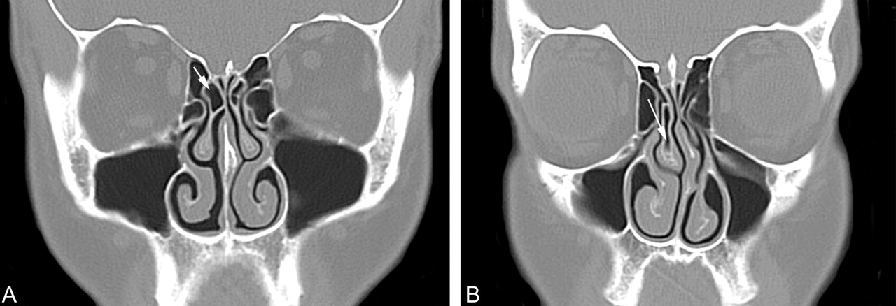
- Fig 1.
Coronal CT scans showing the definition of a concha bullosa.
A, Coronal CT scan of the paranasal sinuses shows pneumatization (arrow) of just under 50% of the vertical height of the right middle turbinate. This was not considered to be a concha bullosa in this study. There is no deviation of the nasal septum. There is inflammatory mucosal thickening obstructing the left infundibulum.
B, Coronal CT scan of the paranasal sinuses shows pneumatization (arrow) of more than 50% of the vertical height of the right middle turbinate. This pneumatization extends into the caudal bulbous portion of the turbinate. This was considered to be a small concha bullosa in this study. Also note that the nasal septum is moderately deviated convexity to the left and there is preservation of the air channel between the concha and the nasal septum. There is some mucosal thickening in both maxillary sinuses.
- Fig 2.
Coronal CT scan of the paranasal sinuses shows a moderate-sized left concha bullosa with moderate deviation of the nasal septum convexity to the right. Note that there is preservation of the air channel between the concha and the nasal septum. There is mucosal disease in both ethmoid sinuses.
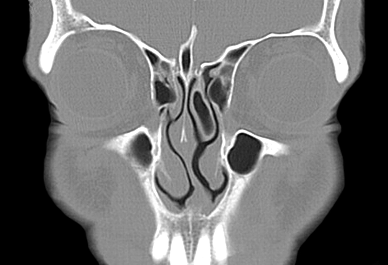
- Fig 3.
Coronal CT scan of the paranasal sinuses shows a large left concha bullosa with severe deviation of the nasal septum convexity to the right. Note that there is preservation of the air channel between the concha and the nasal septum. There is mucosal disease in both maxillary sinuses.
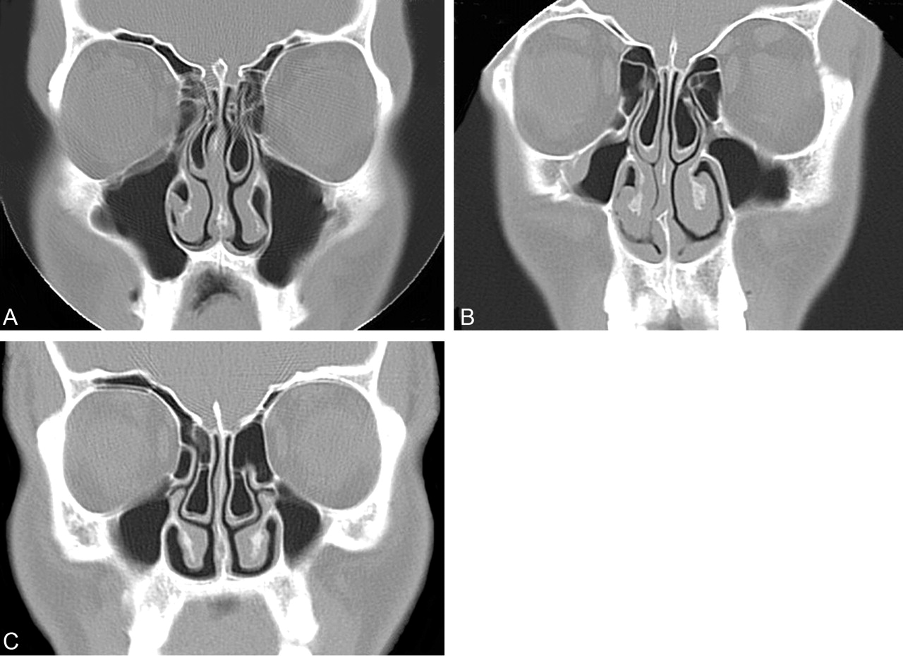
- Fig 4.
Coronal CT scans showing variation in concha size wth preservation of nasal air channels.
A, Coronal CT scan of the paranasal sinuses shows moderate-sized concha bullosa bilaterally, with the one on the left side being slightly larger, or dominant. There is mild deviation of the nasal septum convexity to the right. Note that there is preservation of the air channel between the dominant concha and the nasal septum. There is, however, some loss of the air channel between the nasal septum and the right concha. There is mucosal disease in both ethmoid and maxillary sinuses.
B, Coronal CT scan of the paranasal sinuses shows moderate-sized concha bullosa bilaterally, with the one on the left side being slightly larger, or dominant. There is mild deviation of the nasal septum convexity to the right. Note that there is preservation of the air channels between each concha and the nasal septum. There is mucosal disease in both maxillary sinuses.
C, Coronal CT scan of the paranasal sinuses shows a moderate-sized bilateral concha bullosa with mild deviation of the nasal septum convexity to the right. Neither concha was considered to be dominant. Note that there is preservation of the air channels between each concha and the nasal septum. There is mucosal disease in both maxillary sinuses.


Tables
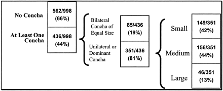
- TABLE 1:
Summary of incidence, unilateral or bilateral occurrence, and size of concha bullosa

- TABLE 2:
Incidence of nasal septal deviation in patients with and without unilateral or dominant, if bilateral concha bullosa
No Septal Deviation Septal Deviation No unilateral or dominant concha 275 /647 (43%) 357 /647 (55%) Unilateral or dominant concha present 75 /351 (21%) 276 /351 (79%) - TABLE 3:
Incidence of nasal septal deviation broken down by convexity compared to the presence and location of unilateral or dominant, if bilateral concha bullosa
No Septal Deviation Rightward Septal Deviation Leftward Septal Deviation No concha or equal bilateral concha 275 /632 (44%) 190 /632 (30%) 167 /632 (26%) Right unilateral or dominant concha 37 /185 (20%) 21 /185 (11%) 127 /185 (69%) Left unilateral or dominant concha 38 /166 (23%) 117 /166 (70%) 11 /166 (7%) - TABLE 4:
Distribution of nasal septal deviation and concha bullosa in the patient population studied
Sinus Disease Present No septal deviation 253 /350 (72%) Septal deviation 504 /648 (78%) No concha 439 /562 (78%) Concha 318 /436 (73%)


