
Article Figures & Data
Figures
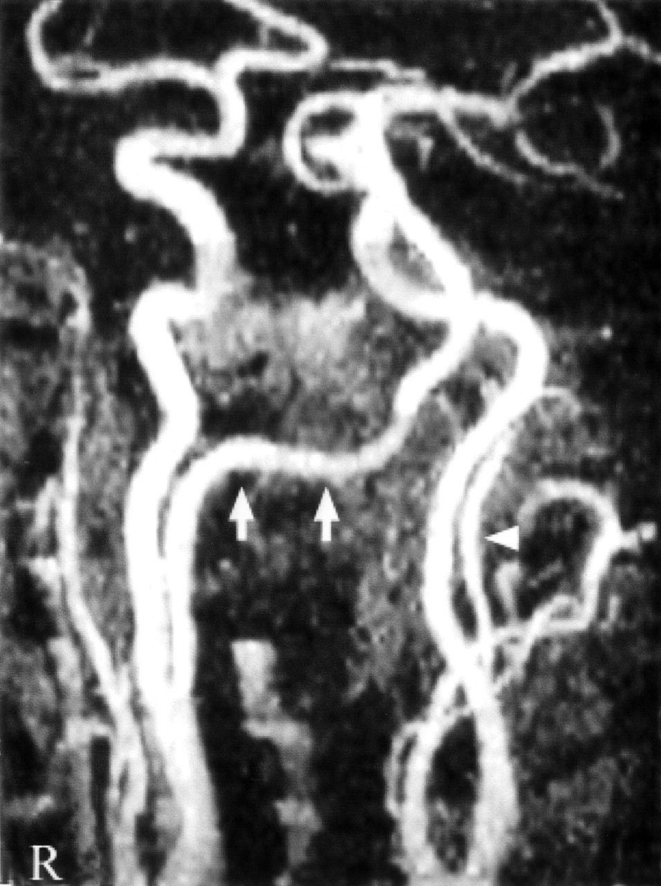
- Fig 1.
MR angiogram (2D time of flight). Right proatlantal artery originating from internal carotid artery can be seen in its full course (arrows), but only the proximal portion of left proatlantal artery can be seen (arrowhead).
- Fig 2.
Aortic arch angiogram, showing the absence of both vertebral arteries.
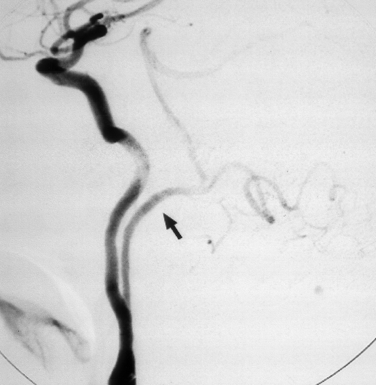
- Fig 3.
Right common carotid artery digital subtraction arteriogram, lateral projection. Proatlantal artery (arrow) originates from the internal carotid artery. The vessel extends to the posterior aspect of atlas with a horizontal sweep characteristic of a type 1 proatlantal artery (arrowhead) before turning upward to join the horizontal segment of the vertebral artery.
- Fig 4.
Left common carotid artery digital subtraction arteriogram, lateral projection. Proatlantal artery (arrow) courses dorsally above C1 before joining the vertebral artery.
- Fig 5.
A, At the 4–5-mm embryonic stage bilateral longitudinal neural arteries (arrows)—one set of longitudinal neural arteries, dorsal aorta, and cervical intersegmental arteries is shown—are supplied by trigeminal artery (TA), otic artery (OA), hypoglossal artery (HA), proatlantal intersegmental artery (PA), and cervical intersegmental arteries (CIA1–6)
B, At the 7–12-mm embryo vertebral artery (VA) develops through the transverse anastomoses between adjacent cervical intersegmental arteries and distal part of the proatlantal artery becomes the horizontal portion of the vertebral artery (arrowheads) while proximal part regresses completely. Failure of this regression results as persistent proatlantal artery (dashed lines). Also note that at this stage of embryo TA, OA, and HA has disappeared after development of posterior communicating artery (PCA). AA, fourth aortic arch; DAo, dorsal aorta; ECA, external carotid artery; ICA, internal carotid artery; VAo, ventral aorta.
- Fig 6.
A, Persistent proatlantal artery type I (PPA 1) arises from the caudal part of the internal carotid artery and courses along the anterior aspect of the vertebral bodies to the level of the occipitoatlantal space before coursing dorsally.
B, Persistent proatlantal artery type II (PPA 2) arises from the external carotid artery; it crosses the C1 or C2 vertebra obliquely. Both PPA-1 and PPA-2 enter the skull via the foramen magnum.


