
Article Figures & Data
Figures
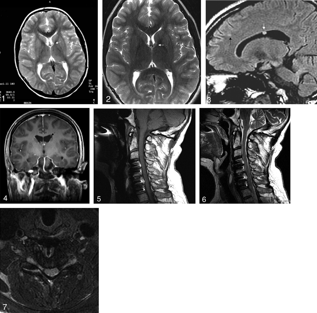
- Fig 1.
5 mm Axial T2 TSE obtained on 1.5T in a patient with tuberous scvlerosis showing equivocal lesion (arrow tip) adjacent to left foramen of Monro.
- Fig 2.
4 mm Axial T2 FSE, 512 × 384, obtained on 3T 12 weeks later in same patient as Figure 1 with arrow tip on more obvious lesin adjacent to left foramen of Monro. Note increased flow artifacts in phase direction which are exacerbated by 3T. These will be significantly diminished with multidirectional flow comp or with a 3D T2FSE acquisition.
- Fig 3.
1 mm Direct Sagittal FLAIR FSE, 256 × 256, demonstrating the only lesions (3mm subependymal nodule [white arrow] and a 2mm subcortical tuber [black arrow]) in another patient with tuberous sclerosis.
- Fig 4.
1 mm Reformatted Coronal T1 FSPGR from same patient as Figure 3 obtained in the axial plane with 1mm isotropic voxels, 256 × 256, revealing the subependymal nodule (black arrow) in the superolateral aspect of the left lateral ventricle.
- Fig 5.
2 mm Sagittal T1 FSE obtained on 3T showing adequate cord visualization (in patient with a previous anterior fusion) by varying 5 parameters (increasing bandwidth and echo train length [ETL], decreasing slice thickness and TE, and orienting frequency encoding gradient parallel to long axis of metal).
- Fig 6.
2 mm Sagittal T2 FSE at 3T from same patient as Figure 5 demonstrating good visualization of the spinal cord by using the aforementionted techniques.
- Fig 7.
3 mm T2 Axial T2 FSE with fat sat through the C6-7 foramina, demonstrates adequate visualization of cord and nerve roots (except proximal left C7). If this acquisition had been obtained with 2 mm slice thickness as well as maximum bandwidth and ETL and the lowest TE, the left C7 root may have been seen in its entirety.


In this issue


Jump to section
Related Articles
Cited By...
- No citing articles found.