
Article Figures & Data
Figures
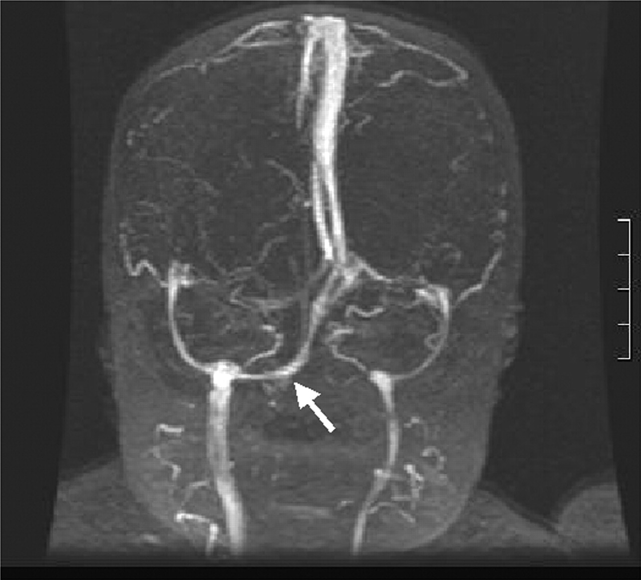
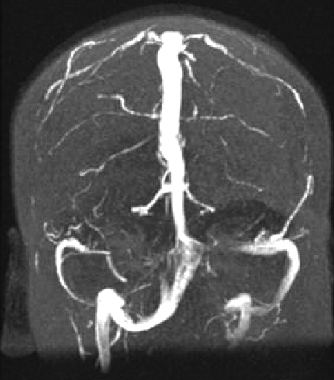
- Fig 1.
Coronal MIP from coronal MRV shows symmetric hypoplasia of the transverse sinuses associated with a persistent occipital sinus (arrow).
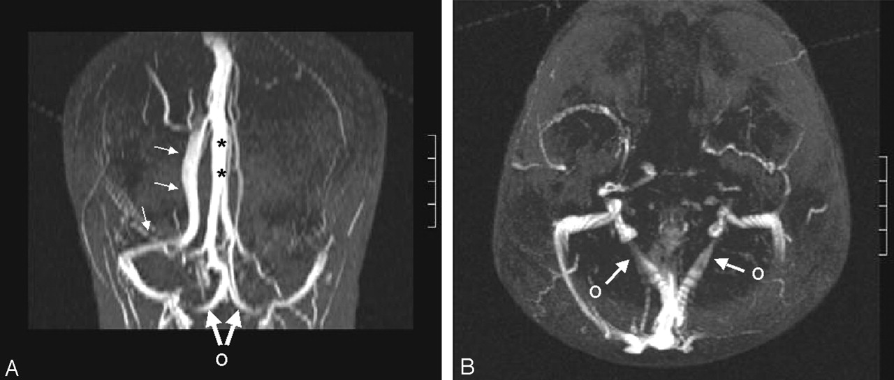
- Fig 2.
Absence of the left transverse sinus and a persistent occipital sinus.
A, Coronal MIP from axial MRV shows that the superior sagittal sinus continues as the right transverse sinus (small arrows). Straight sinus (asterisks) courses upward to the high-riding torcula, which drains downward into the occipital sinus. Large arrows and O indicate bifurcation of the occipital sinus at the foramen magnum.
B, Axial MIP shows the occipital sinus bifurcating (arrows, O) and draining into the IJVs.
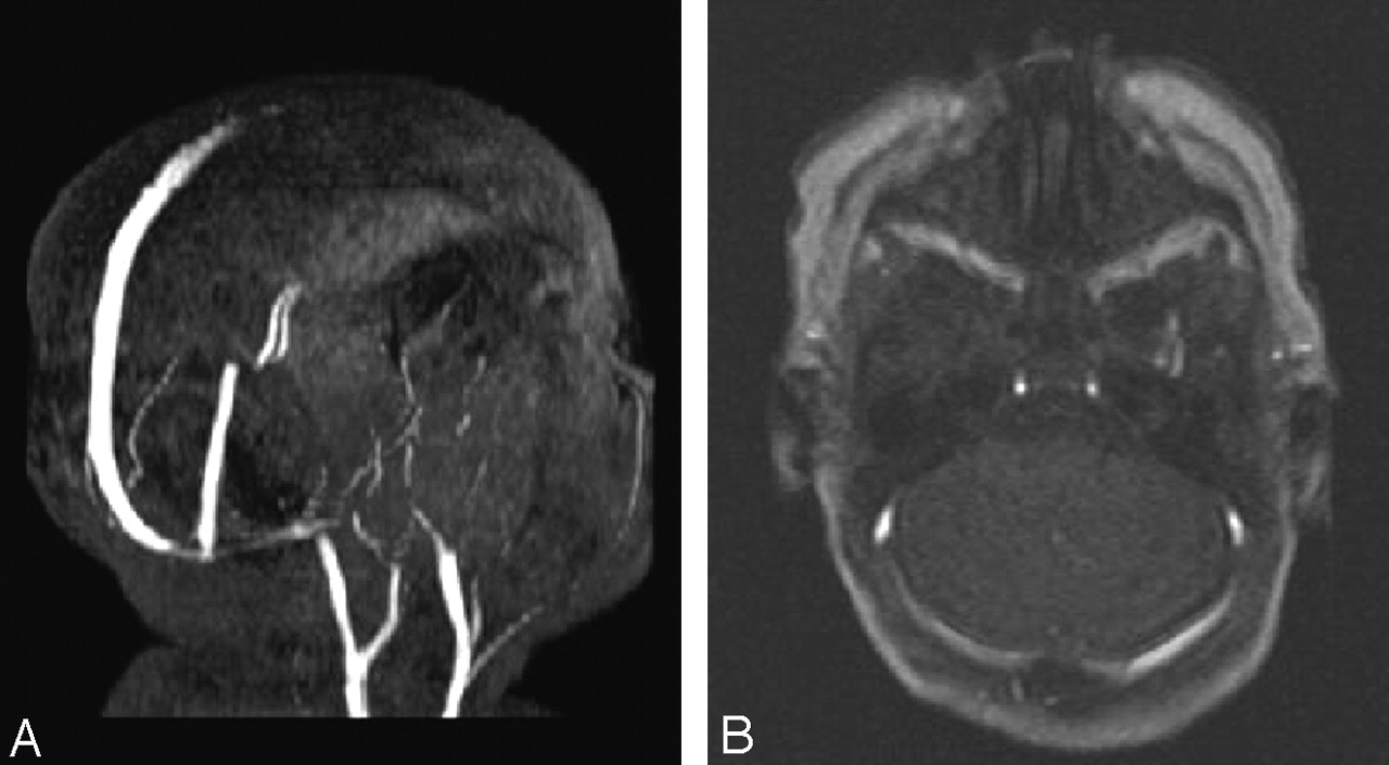
- Fig 3.
Saturation effects due to in-plane flow in the transverse sinuses.
A, Oblique sagittal MIP from axial MRV in shows diminished signal intensity from the transverse and sigmoid sinuses.
B, Axial image shows patent transverse sinuses.
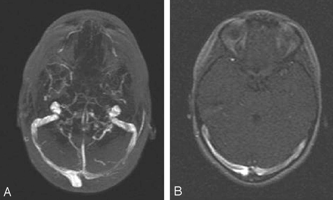
- Fig 4.
Artifactual signal intensity loss due to the MIP algorithm.
A, Axial MIP from the axial images shows diminished signal intensity from the medial two-thirds of the left transverse sinus.
B, Axial image shows nearly equal signal intensity in both transverse sinuses.
- Fig 5.
Asymmetric transverse sinus hypoplasia associated with a large persistent occipital sinus. Coronal MIP from coronal MRV.
- Fig 6.
Comparison of coronal MIPs from 2D TOF MRV in the axial (A) and coronal planes (B).
A, Focal area of diminished signal intensity in the left transverse sinus (arrow). Internal jugular veins are well depicted.
B, Diminished signal intensity in the medial thirds of both transverse sinuses (arrows), while the lateral aspect of the left transverse sinus is well depicted. Loss of signal intensity in the IJVs is due to in-plane flow. Lack of a caudal presaturation pulse accounts for visualization of arterial structures.
- Fig 7.
Narrowing at the junction of the sigmoid sinus and the IJV seen in >60% of patients older than 24 months.
A, Parasagittal subvolume MIP from coronal MRV shows marked luminal narrowing at this junction (long arrow). Note the prominent posterior condylar vein (short arrow).
B, Comparative digital subtraction angiogram, venous phase. Long arrow indicates the narrowed junction; short arrow, a prominent posterior condylar vein.
C, Narrowed junction not associated with a prominent posterior condylar vein in another patient. Arrow indicates loss of signal intensity in the posterior aspect of the superior sagittal sinus due to in-plane flow; coronal MRV was performed.


Tables
Variations in posterior fossa venous drainage with age
Group Right Left Codominant Atretic Transverse Sinus Occipital Sinus I (n = 38) 14 (37) 8 (21) 16 (42) 2 (5) 5 (13) II (n = 17) 6 (35) 5 (30) 6 (35) 3 (18) 2 (12) III (n = 53) 26 (50) 9 (16) 18 (34) 7 (13) 1 (2) Total (n = 108) 46 (44) 22 (20) 40 (37) 6 (5) 8 (7) Note.—Data in parentheses are percentages.


