
Article Figures & Data
Figures
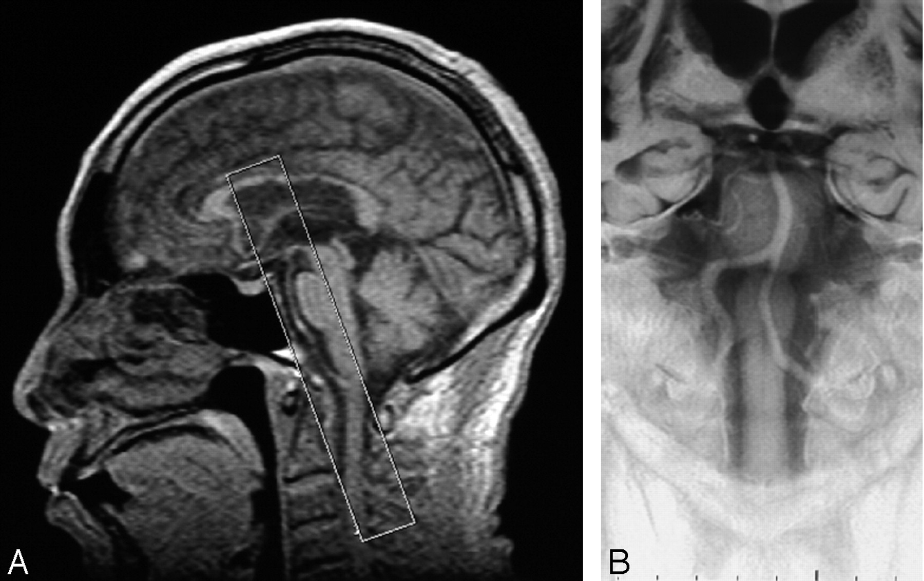
- Fig 1.
A midsagittal scout view (A) shows the location of a BPAS scan. White box indicates a thick coronal section posteriorly parallel to the clivus. BPAS-MR imaging (B) demonstrates an overview of the vertebrobasilar artery within the cistern.
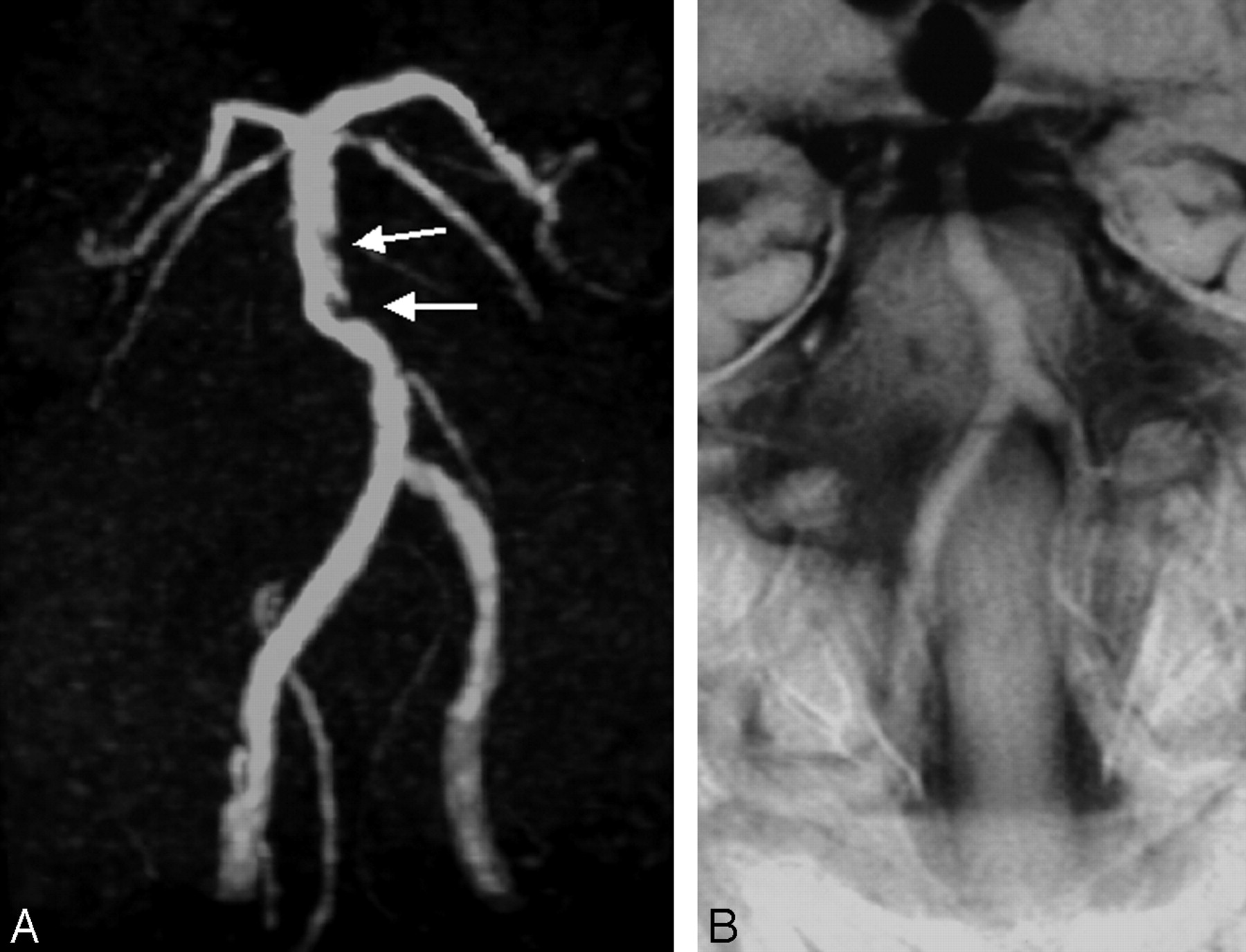
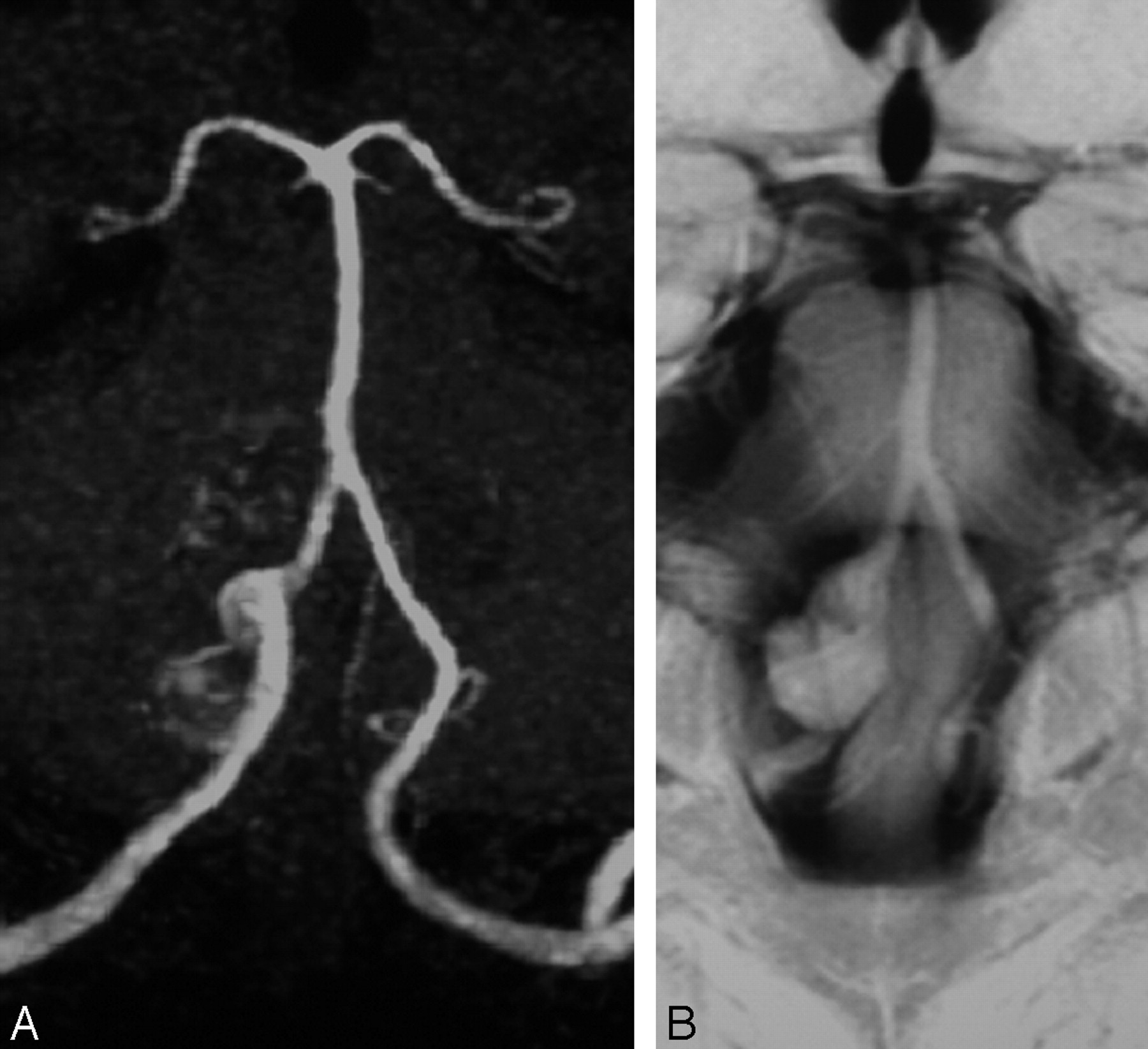
- Fig 2.
A typical case of “atherosclerosis.” MRA (A) reveals irregular defects within the basilar trunk (arrows). Its outer contour on BPAS-MR imaging (B) is relatively smooth, not stenotic.
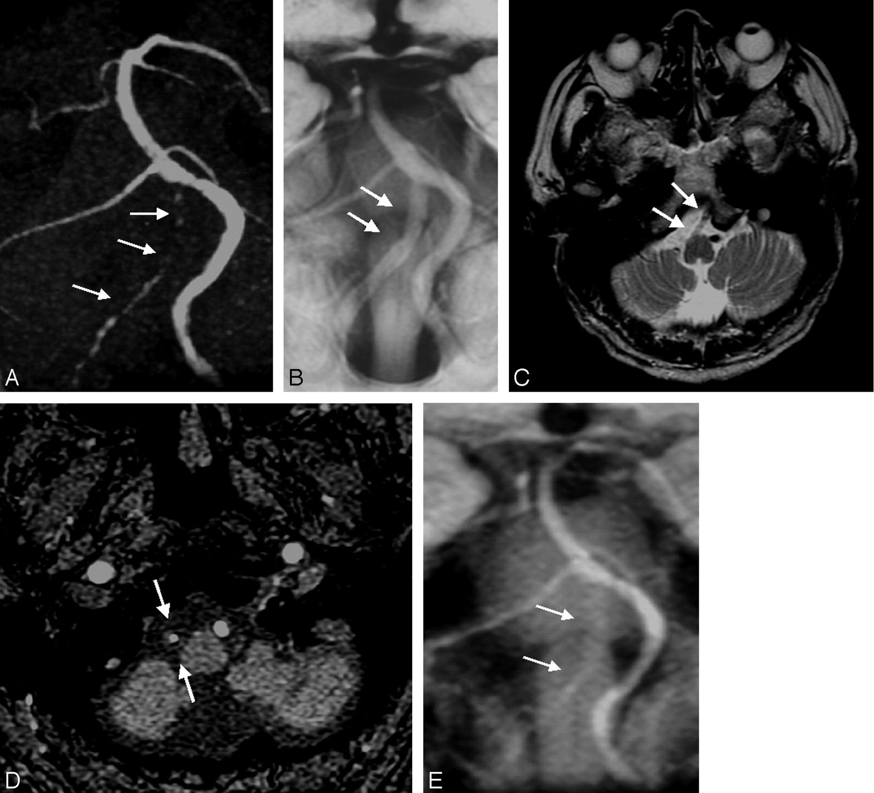
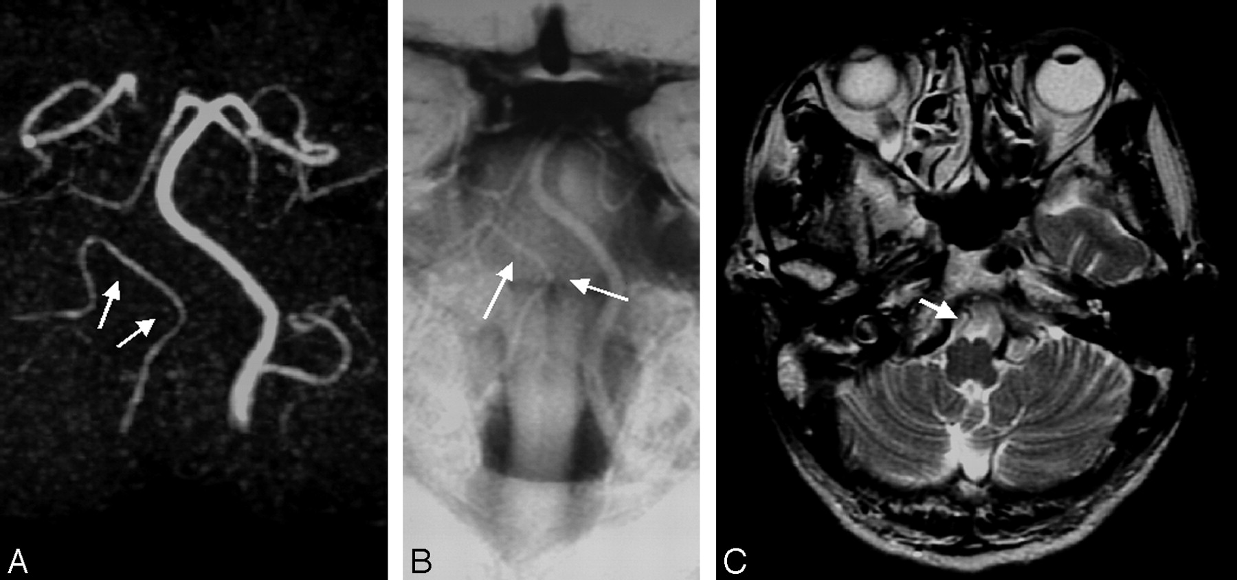
- Fig 3.
A typical case of unilateral VA “occlusive condition.” Although the right VA is very narrow and not visualized clearly on MRA (A, arrows), its sufficient outer caliber is confirmed on BPAS-MR imaging (B, arrows). T2-weighted image (C) shows absence of normal “flow void” in the right VA (arrows) that suggests the arterial occlusion. A source image of 3D TOF MRA (D) and a reformatted thick coronal image (E) hardly show the outer diameter of the occluded right VA (D and E, arrows)
- Fig 4.
A case of unilateral hypoplastic VA, classified as “hypoplastic VA.” The right VA is not seen clearly on MRA (A, arrows). A small and hypoplastic right VA is confirmed on BPAS-MR imaging (B, arrows). T2-weighted image (C) shows no vascular structure suggesting the right VA.
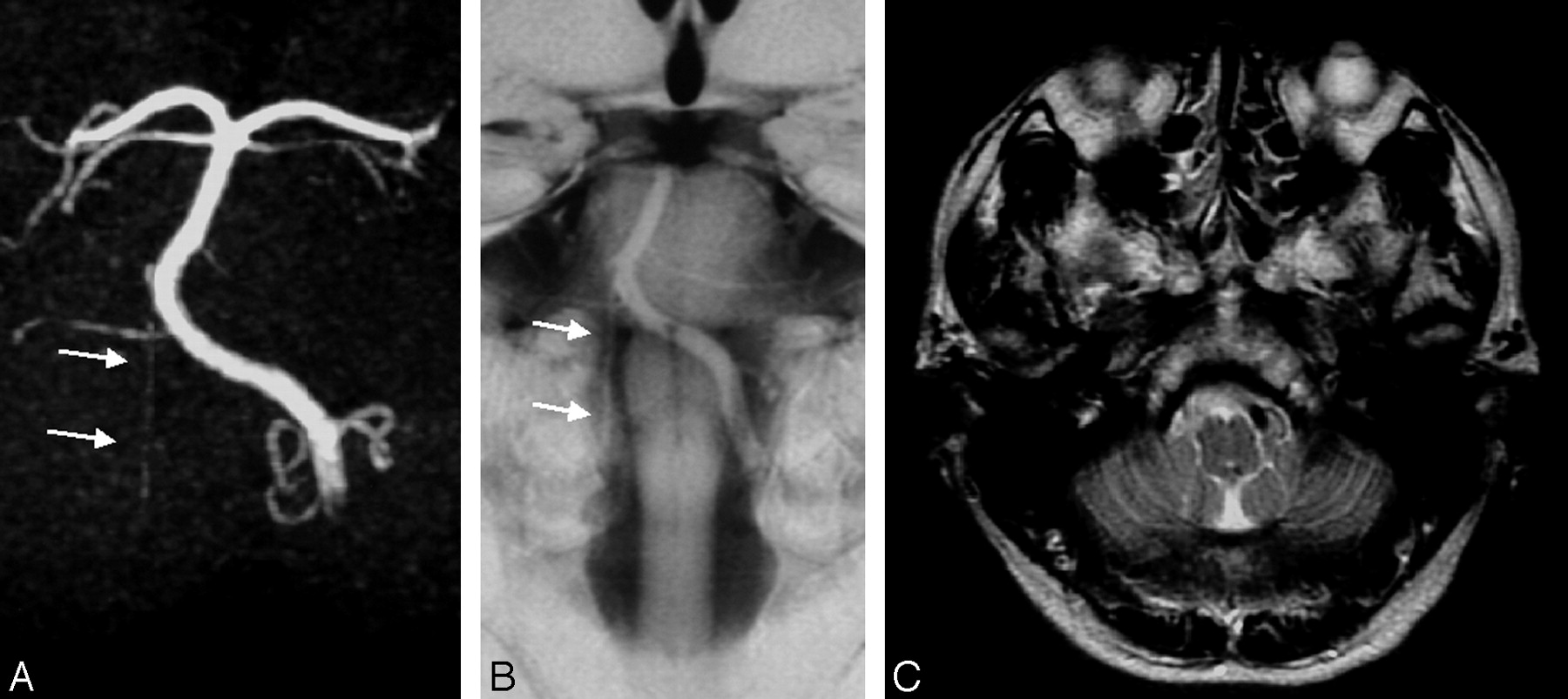
- Fig 5.
Another case of “hypoplastic VA.” The right VA terminates in the posterior inferior cerebellar artery (PICA) on both MRA (A, arrows) and BPAS-MR imaging (B, arrows). Note the absence of the distal right VA on BPAS-MR imaging (B). On T2-weighted axial image (C), it is actually difficult to distinguish from the right PICA from the hypoplastic small VA.
- Fig 6.
A case of right VA aneurysm. The right VA aneurysm on MRA (A) is not clear. Its shape is not equal to what is evident on BPAS-MR imaging (B), perhaps due to partial thrombosis and/or turbulent flow within the aneurysm.


Tables
- TABLE 1:
Vertebrobasilar abnormalities classified by the combination of MR angiography and BPAS-MRI findings
Classified Conditions MR Angiography Finding BPAS-MR Imaging Finding Atherosclerosis Irregular or stenotic Almost normal Occlusive condition Not visible or hardly visible Almost normal Hypoplastic vertebral artery Not visible or hardly visible Narrow or not visible Aneurysm Aneurysmal dilation (unnecessary) Aneurysmal dilation Note.—BPAS indicates basiparallel anatomical scanning.
Atherosclerosis (S) 28 (7.3%) Occlusive condition (O) 18 (4.7%) Hypoplastic vertebral artery (H) 23 (6.0%) Aneurysm (A) 6 (1.6%) S + O 10 (2.6%) S + H 3 (0.8%) S + O + A 2 (0.5%) Total 90 (23.4%)
In this issue


Jump to section
Related Articles
Cited By...
- No citing articles found.