
Article Figures & Data
Figures
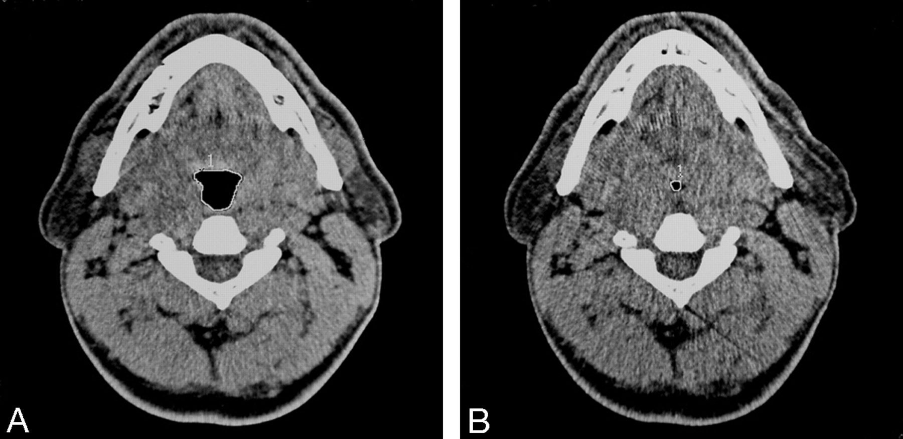
- Fig 1.
A, Cross-sectional image of a patient at the level of uvula in tidal breathing. B, The significant narrowing at the same level in forced expiration is seen. The region of interest (white line) was used to assess total cross-sectional areas in each image.
- Fig 2.
Lateral scout view showing skeletal and soft tissue profile landmarks and the levels at which CT sections were obtained. S, sella; N, nasion; A, subspinale; B, supramental; H, hyoid; MP, mandibular plane; U, tip of the uvula; PNS, posterior nasal spine; SP-max, maximum thickness of the soft palate; level 1, level of tip of the uvula; level 2, level of hypopharynx.
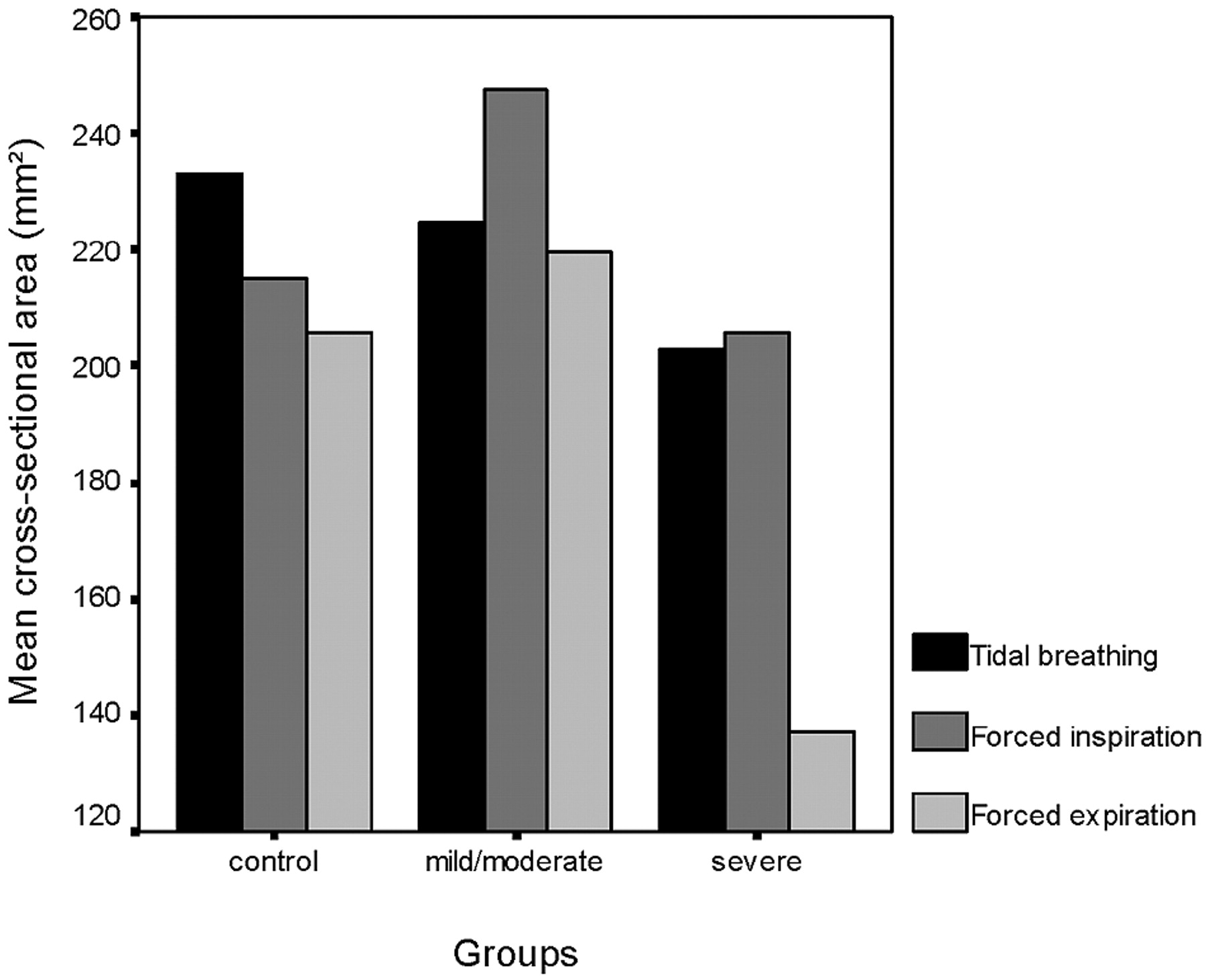
- Fig 3.
Mean cross-sectional areas at the level of uvula in tidal breathing, at the end of both forced inspiration and expiration in each group.
- Fig 4.
Mean cross-sectional areas at the level of hypopharynx in tidal breathing, at the end of both forced inspiration and expiration in each group.


Tables
Parameters Control Group (n = 24) (mean ± SD) Mild/Moderate OSAS (n = 27) (mean ± SD) Severe OSAS (n = 20) (mean ± SD) P* Value Age (y) 46.0 ± 14.3 49.0 ± 6.0 49.0 ± 10.0 .533 BMI (kg/m2) 34.1 ± 7.0 33.9 ± 5.7 31.7 ± 5.1 .357 Neck circumference (cm) 37.4 ± 4.4 38.3 ± 4.2 40.6 ± 3.8 .043 AHI 2.1 ± 1.4 13.9 ± 6.7 50.8 ± 15.6 .000 Uvula tidal breathing area (mm2) 233.2 ± 89.9 224.7 ± 68.6 202.8 ± 76.7 .418 Uvula inspiration area (mm2) 215.2 ± 94.3 247.4 ± 107.6 205.7 ± 146.7 .425 Uvula expiration area (mm2) 205.6 ± 106.3 219.6 ± 82.7 137.2 ± 73.6 .007 Hypopharynx tidal breathing area (mm2) 349.2 ± 117.8 257.1 ± 88.7 366.2 ± 137.2 .453 Hypopharynx inspiration area (mm2) 333.8 ± 118.4 338.6 ± 131.8 345.6 ± 146.8 .698 Hypopharynx expiration area (mm2) 272.8 ± 107.5 323.6 ± 150.0 307.0 ± 140.2 .344 MP-H (mm) 15.0 ± 5.7 15.2 ± 6.4 20.0 ± 8.1 .029 PNS-U (mm) 35.0 ± 3.6 34.9 ± 4.1 37.5 ± 6.1 .120 SP-max (mm) 10.0 ± 2.0 9.6 ± 2.2 11.6 ± 2.2 .009 SNA (angle) 81.5 ± 4.9 81.3 ± 6.2 84.0 ± 5.0 .211 SNB (angle) 79.3 ± 4.0 79.1 ± 5.1 81.1 ± 5.3 .347 ANB (angle) 2.1 ± 3.6 1.9 ± 3.4 2.9 ± 3.4 .628 Note.—OSAS indicates obstructive sleep apnea syndrome; AHI, apnea-hypopnea index; MP-H, distance from mandibulas plane to the hyoid bone; PNS-U, distance from the posterior nasal spine to the tip of the uvula; SP-max, maximum distance of the soft palate perpendicular to PNS-U; SNA, SNB, ANB, angles where S is the midpoint of the sella, N is the nasion, A is the deepest point on the premaxillary outer contour, and B is the deepest point on the outer mandibular contour.
* Compared by using the one-way ANOVA. Significant values are shown in boldface.
- TABLE 2:
Significant correlations among AHI, neck circumference, cephalometric parameters, and upper airway size in patients with OSAS
Parameters NC (cm) PNS-U (mm) SP-max (mm) MP-H (mm) EU NH IH AHI .411 .363 .339 .420 −.446 .376 NC 1000 .383 PNS-U .383 1000 .422 .545 SP-max .422 1000 −.365 MP-H .545 1000 .306 SNB (angle) .380 IU .325 Note.—Values are correlation coefficients. Significance is defined as P < .05. AHI indicates apnea-hypopnea index; OSAS, obstructive sleep apnea syndrome; NC, neck circumference; PNS-U, distance from the posterior nasal spine to the tip of the uvula; SP-max, maximum distance of the soft palate perpendicular to PNS-U; MP-H, distance from mandibular plane to the hyoid bone; EU, cross-sectional area at level of uvula in expiration; NH, cross-sectional area at level of hypopharynx in tidal breathing; IH, cross-sectional area at level of hypopharynx in inspiration; SNB, angle where S is the midpoint of the sella, N is the nasion, and B is the deepest point on the outer mandibular contour; IU, cross-sectional area at level of uvula in inspiration.


