Advertisement

Main menu

  • Home
  • Content
    • Current Issue
    • Accepted Manuscripts
    • Article Preview
    • Past Issue Archive
    • AJNR Case Collection
    • Case of the Week Archive
    • Classic Case Archive
    • Case of the Month Archive
  • Special Collections
    • Spinal CSF Leak Articles (Jan 2020-June 2024)
    • 2024 AJNR Journal Awards
    • Most Impactful AJNR Articles
  • Multimedia
    • AJNR Podcast
    • AJNR Scantastics
    • Video Articles
  • For Authors
    • Submit a Manuscript
    • Author Policies
    • Fast publishing of Accepted Manuscripts
    • Graphical Abstract Preparation
    • Manuscript Submission Guidelines
    • Imaging Protocol Submission
    • Submit a Case for the Case Collection
  • About Us
    • About AJNR
    • Editorial Board
  • More
    • Become a Reviewer/Academy of Reviewers
    • Subscribers
    • Permissions
    • Alerts
    • Feedback
    • Advertisers
    • ASNR Home
  • Other Publications
    • ajnr

User menu

  • Alerts
  • Log in

Search

  • Advanced search
American Journal of Neuroradiology

American Journal of Neuroradiology

ASHNR American Society of Functional Neuroradiology ASHNR American Society of Pediatric Neuroradiology ASSR
  • Alerts
  • Log in

Advanced Search

  • Home
  • Content
    • Current Issue
    • Accepted Manuscripts
    • Article Preview
    • Past Issue Archive
    • AJNR Case Collection
    • Case of the Week Archive
    • Classic Case Archive
    • Case of the Month Archive
  • Special Collections
    • Spinal CSF Leak Articles (Jan 2020-June 2024)
    • 2024 AJNR Journal Awards
    • Most Impactful AJNR Articles
  • Multimedia
    • AJNR Podcast
    • AJNR Scantastics
    • Video Articles
  • For Authors
    • Submit a Manuscript
    • Author Policies
    • Fast publishing of Accepted Manuscripts
    • Graphical Abstract Preparation
    • Manuscript Submission Guidelines
    • Imaging Protocol Submission
    • Submit a Case for the Case Collection
  • About Us
    • About AJNR
    • Editorial Board
  • More
    • Become a Reviewer/Academy of Reviewers
    • Subscribers
    • Permissions
    • Alerts
    • Feedback
    • Advertisers
    • ASNR Home
  • Follow AJNR on Twitter
  • Visit AJNR on Facebook
  • Follow AJNR on Instagram
  • Join AJNR on LinkedIn
  • RSS Feeds

Welcome to the new AJNR, Updated Hall of Fame, and more. Read the full announcements.


AJNR is seeking candidates for the position of Associate Section Editor, AJNR Case Collection. Read the full announcement.


OtherSPINE

Inflammatory Pseudotumor in the Epidural Space of the Thoracic Spine: A Case Report and Literature Review of MR Imaging Findings

Ho Jun Seol, Sam Soo Kim, Ji Eun Kim, Sang Hyung Lee and Jun Yeon Won
American Journal of Neuroradiology November 2005, 26 (10) 2667-2670;
Ho Jun Seol
  • Find this author on Google Scholar
  • Find this author on PubMed
  • Search for this author on this site
Sam Soo Kim
  • Find this author on Google Scholar
  • Find this author on PubMed
  • Search for this author on this site
Ji Eun Kim
  • Find this author on Google Scholar
  • Find this author on PubMed
  • Search for this author on this site
Sang Hyung Lee
  • Find this author on Google Scholar
  • Find this author on PubMed
  • Search for this author on this site
Jun Yeon Won
  • Find this author on Google Scholar
  • Find this author on PubMed
  • Search for this author on this site
  • Article
  • Figures & Data
  • Info & Metrics
  • Responses
  • References
  • PDF
Loading

Article Figures & Data

Figures

  • Tables
  • Fig 1.
    • Download figure
    • Open in new tab
    • Download powerpoint
    Fig 1.

    Thoracic spine CT scan shows an epidural mass compressing the spinal cord without bony destruction.
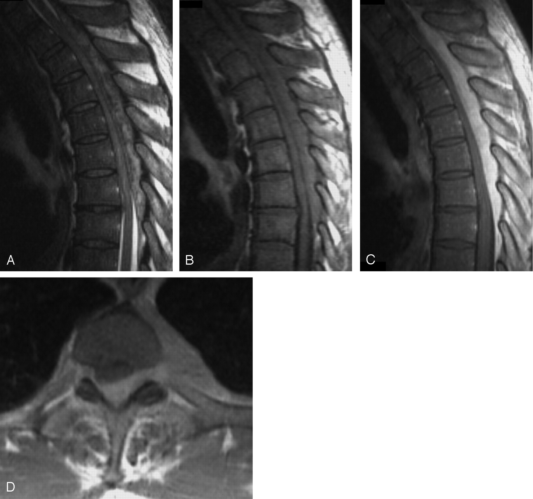
  • Fig 2.
    • Download figure
    • Open in new tab
    • Download powerpoint
    Fig 2.

    MR imaging demonstrates the expansile epidural mass from T1 to T7, showing heterogeneous iso- and hyperintensity on the sagittal T2-weighted image (A) and homogeneous isointensity on the T1-weighted image (B). The postcontrast sagittal (C) and axial (D) images reveal a homogeneous enhancing lesion with cord compression. There is no abnormality in the adjacent bone.
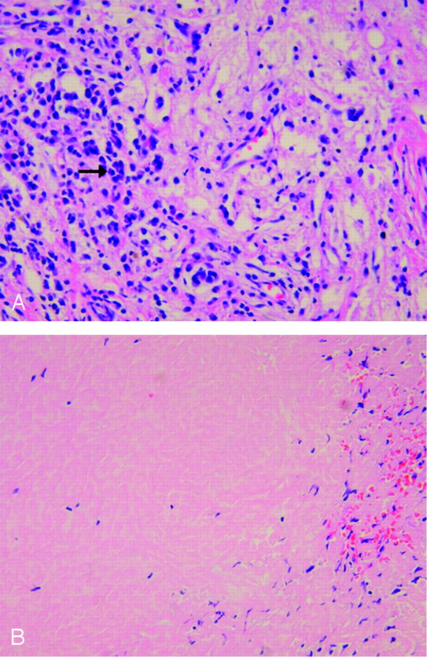
  • Fig 3.
    • Download figure
    • Open in new tab
    • Download powerpoint
    Fig 3.

    Microscopically, there is a polymorphic infiltrate composed of plump myofibroblasts (arrows) and lymphoplasma cells in loose fibromyxoid stroma, suggesting inflammatory myofibroblastic tumor (hematoxylin and eosin, original magnification ×100).

  • Fig 4.
    • Download figure
    • Open in new tab
    • Download powerpoint
    Fig 4.

    A, Abundant inflammatory cells such as plasma cells (arrow) in rich vascular stroma are seen in some areas (hematoxylin and eosin, original magnification ×200.)

    B, Paucicellular area shows platelike collagen resembling scar tissue (hematoxylin and eosin, original magnification ×200).

Tables

  • Figures
  • Characteristics of cases of inflammatory pseudotumors reported in the literature that originated in the spinal canal

    SourceAge (y)/SexLocationRelation with MeningesBony DestructionSignal Intensity on MR Images Compared with Spinal Cord
    T1-weightedT2-weightedContrast-enhanced
    Roberts et al, 1997 (1)58/FT9–T11EpiduralYesIsoHypoNR
    Aizawa et al, 2002 (3)46/MC3–C7IntramedullaryNoIsoHypoWell
    Jeon et al, 2005 (4)60/FLEtramedullary intraduralNRNRNRWell
    Hsieh and Lin, 1995 (5)37/MT5, T12–L1Extramedullary intraduralNoLowNRNR
    Eimoto et al, 1978 (6)37/MC4–C5Extramedullary intraduralNoNRNRNR
    Gilliard et al, 2000 (7)45/MC3–T2EpiduralYesIsoNRWell
    Hsiang et al, 1994 (8)57/MT12–T13Intra- and extraduralYesLowNRWell
    Roberts et al, 2001 (9)39/FT5–T6EpiduralNoIsoHypoNR
    Lee et al, 1998 (10)3/FC2–T10IntramedullaryNoNRNRWell
    Kilinc et al, 2002 (11)34/MT9–T12IntramedullaryNoLowHyperWell
    Despeyroux-Ewers et al, 2003 (12)22/FT9Extramedullary intraduralNoIsoHypoWell
    • Note.—Iso indicates isointensity; Hypo, hypointensity; NR, not reported.

PreviousNext
Back to top

In this issue

American Journal of Neuroradiology: 26 (10)
American Journal of Neuroradiology
Vol. 26, Issue 10
1 Nov 2005
  • Table of Contents
  • Index by author
Advertisement
Print
Download PDF
Email Article

Thank you for your interest in spreading the word on American Journal of Neuroradiology.

NOTE: We only request your email address so that the person you are recommending the page to knows that you wanted them to see it, and that it is not junk mail. We do not capture any email address.

Enter multiple addresses on separate lines or separate them with commas.
Inflammatory Pseudotumor in the Epidural Space of the Thoracic Spine: A Case Report and Literature Review of MR Imaging Findings
(Your Name) has sent you a message from American Journal of Neuroradiology
(Your Name) thought you would like to see the American Journal of Neuroradiology web site.
CAPTCHA
This question is for testing whether or not you are a human visitor and to prevent automated spam submissions.
Cite this article
Ho Jun Seol, Sam Soo Kim, Ji Eun Kim, Sang Hyung Lee, Jun Yeon Won
Inflammatory Pseudotumor in the Epidural Space of the Thoracic Spine: A Case Report and Literature Review of MR Imaging Findings
American Journal of Neuroradiology Nov 2005, 26 (10) 2667-2670;

Citation Manager Formats

  • BibTeX
  • Bookends
  • EasyBib
  • EndNote (tagged)
  • EndNote 8 (xml)
  • Medlars
  • Mendeley
  • Papers
  • RefWorks Tagged
  • Ref Manager
  • RIS
  • Zotero
0 Responses
Respond to this article
Share
Bookmark this article
Inflammatory Pseudotumor in the Epidural Space of the Thoracic Spine: A Case Report and Literature Review of MR Imaging Findings
Ho Jun Seol, Sam Soo Kim, Ji Eun Kim, Sang Hyung Lee, Jun Yeon Won
American Journal of Neuroradiology Nov 2005, 26 (10) 2667-2670;
del.icio.us logo Twitter logo Facebook logo Mendeley logo
  • Tweet Widget
  • Facebook Like
  • Google Plus One

Jump to section

  • Article
    • Abstract
    • Case Report
    • Discussion
    • Footnotes
    • References
  • Figures & Data
  • Info & Metrics
  • Responses
  • References
  • PDF

Related Articles

  • No related articles found.
  • PubMed
  • Google Scholar

Cited By...

  • Inflammatory pseudotumour arising in the epidural space of a dog
  • Intraosseous Inflammatory Myofibroblastic Tumor of the Twelfth Thoracic Vertebra: Report of a Rare Case with Histological Diagnosis and Surgical Treatment
  • Imaging Features of Meningeal Inflammatory Myofibroblastic Tumor
  • Crossref
  • Google Scholar

This article has not yet been cited by articles in journals that are participating in Crossref Cited-by Linking.

More in this TOC Section

  • Bern Score Validity for SIH
  • MP2RAGE 7T in MS Lesions of the Cervical Spine
  • Deep Learning for STIR Spine MRI Quality
Show more Spine

Similar Articles

Advertisement

Indexed Content

  • Current Issue
  • Accepted Manuscripts
  • Article Preview
  • Past Issues
  • Editorials
  • Editors Choice
  • Fellow Journal Club
  • Letters to the Editor

Cases

  • Case Collection
  • Archive - Case of the Week
  • Archive - Case of the Month
  • Archive - Classic Case

Special Collections

  • Special Collections

Resources

  • News and Updates
  • Turn around Times
  • Submit a Manuscript
  • Author Policies
  • Manuscript Submission Guidelines
  • Evidence-Based Medicine Level Guide
  • Publishing Checklists
  • Graphical Abstract Preparation
  • Imaging Protocol Submission
  • Submit a Case
  • Become a Reviewer/Academy of Reviewers
  • Get Peer Review Credit from Publons

Multimedia

  • AJNR Podcast
  • AJNR SCANtastic
  • Video Articles

About Us

  • About AJNR
  • Editorial Board
  • Not an AJNR Subscriber? Join Now
  • Alerts
  • Feedback
  • Advertise with us
  • Librarian Resources
  • Permissions
  • Terms and Conditions

American Society of Neuroradiology

  • Not an ASNR Member? Join Now

© 2025 by the American Society of Neuroradiology All rights, including for text and data mining, AI training, and similar technologies, are reserved.
Print ISSN: 0195-6108 Online ISSN: 1936-959X

Powered by HighWire