
Article Figures & Data
Figures
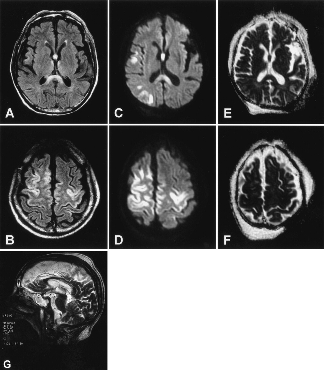
- Fig 1.
MR images in patient 1. Axial FLAIR images on admission show hyperintensity in the corpus callosum (A) and the cerebral cortex (B). Diffusion-weighted images (C and D) also show hyperintensity in these regions with relatively decreased ADC values (see Fig 3). The decreased ADC values, however, are inconspicuous on ADC mapping (E and F). Follow-up T2-weighted sagittal image obtained 10 days after the initial study shows callosal lesions mainly involving the central part of the splenium and body (G).
- Fig 2.
MR images in patient 2. Axial FLAIR images on admission show hyperintensity in the corpus callosum (A) and the cerebral cortex (B). Diffusion-weighted images (C and D) also show hyperintensity in these regions with relatively decreased ADC values (see Fig 3). The decreased ADC values, however, are inconspicuous on ADC mapping (E and F). Follow-up T2-weighted sagittal image obtained 10 days after the initial study shows callosal lesions mainly involving the central part of the splenium and body (G).
- Fig 3.
Comparison of signal intensities on diffusion-weighted images with ADC values on ADC maps. Each coordinate of regions of interest in the ADC maps is the same as that in diffusion-weighted images. ADC values are reduced in regions with high signal intensity on diffusion-weighted images (regions 3 and 4 in each patient). Asterisks denote regions in affected cortex identified by high signal intensity on diffusion-weighted images.


