
Article Figures & Data
Figures
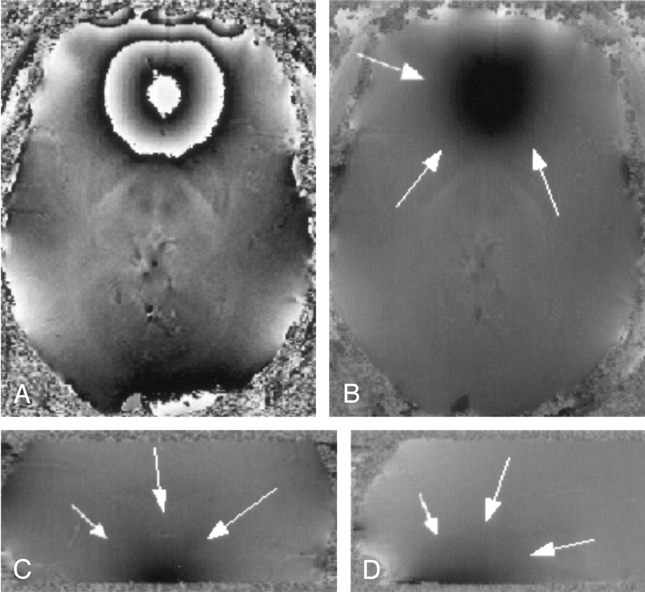
- Fig 1.
Wrapped (A) and unwrapped phase images (B–D). The sphenoid sinuses cause strong field inhomogeneities that extend far into the interior of the brain (white arrows). Parameters: TR = 67 ms, TE = 40 ms, α = 25°, FOV = 256 × 256 × 64, imaging matrix = 512 × 512 × 128.
- Fig 2.
Minimum intensity projections over 4 sections of the magnitude data (A), the corresponding high-pass filtered phase images (B), and a reproduction from an anatomic atlas for comparison (C). In the phase image (B) the capsula interna, capsula externa, putamen, nucleus caudatus, corpus callosum and the claustrum are better visualized than in the magnitude image (A). Also improved is the contrast between gray and white matter. The ventricles are not easily visible in the phase images due to the fact that the magnetic susceptibility of CSF and brain tissue is very similar. TR = 67, TE = 40, α = 25°, FOV = 256 × 256 × 192 mm3, matrix = 512 × 256 × 96, w = 21. (Image C is reproduced with permission from Putz/Pabst: Sobotta, Atlas der Anatomie des Menschen, 21. Auflage 2000, Elsevier GmbH, Urban & Fischer Verlag München, Jena.)
- Fig 3.
Minimum intensity projections over 4 sections of magnitude image (A), the corresponding high-pass filtered phase images (B), and a reproduction from an anatomic atlas (C). In the phase image (B) the caudate nucleus, the putamen, the globus pallidus, and the thalamus with parts of the thalamostriate veins, amongst other features, can be identified. TR = 67 ms, TE = 40 ms, α = 25°, FOV = 256 × 256 × 96 mm3, matrix = 512 × 256 × 96, w = 21. (Image C is reproduced with permission from Putz/Pabst: Sobotta, Atlas der Anatomie des Menschen, 21. Auflage 2000, Elsevier GmbH, Urban & Fischer Verlag München-Jena.)
- Fig 4.
Magnitude image (A), a mIP over 5 sections from the phase images (B) and a reproduction from an anatomic atlas. The red nuclei, the substantia nigra, the crus cerebri and venous vessels are displayed well in the phase image. The medullary lamellae in the red nuclei can be clearly identified. TR = 67 ms, TE = 40 ms, α = 25°, FOV = 256 × 256 × 96 mm3, matrix = 512 × 256 × 96, w = 21. (Image C is reproduced with permission from Putz/Pabst: Sobotta, Atlas der Anatomie des Menschen, 21. Auflage 2000, Elsevier GmbH, Urban & Fischer Verlag München-Jena.)
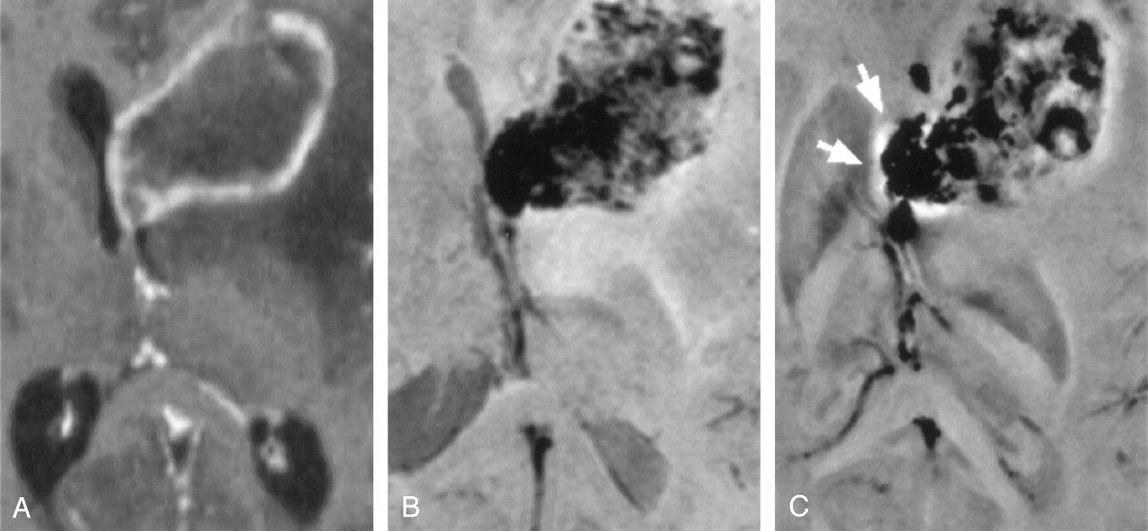
- Fig 5.
Contrast-enhanced T1-weighted MP-RAGE scan (A) (TR = 15 ms, TE = 5 ms, FOV = 224 × 256 mm2, section thickness = 1.0 mm), minimum intensity projections over four sections of magnitude (B), and phase (C) of the susceptibility weighted 3D scan of a 29-year-old female patient with a glioblastoma multiforme (TR = 67 ms, TE = 40 ms, α = 25°, FOV = 169 × 256 × 64 mm3, matrix = 160 × 160 × 64, w = 23).


In this issue


Jump to section
Related Articles
Cited By...
- Advanced computational and statistical multiparametric analysis of Susceptibility-Weighted Imaging to characterize gliomas and brain metastases
- Gadolinium-Enhanced Susceptibility-Weighted Imaging in Multiple Sclerosis: Optimizing the Recognition of Active Plaques for Different MR Imaging Sequences
- Lesion Heterogeneity on High-Field Susceptibility MRI Is Associated with Multiple Sclerosis Severity
- Magnetic resonance frequency shifts during acute MS lesion formation
- Biophysical mechanisms of MRI signal frequency contrast in multiple sclerosis
- Combination of high-resolution susceptibility-weighted imaging and the apparent diffusion coefficient: added value to brain tumour imaging and clinical feasibility of non-contrast MRI at 3 T
- Localization of the Subthalamic Nucleus: Optimization with Susceptibility-Weighted Phase MR Imaging
- Added Value and Diagnostic Performance of Intratumoral Susceptibility Signals in the Differential Diagnosis of Solitary Enhancing Brain Lesions: Preliminary Study
- Biophysical mechanisms of phase contrast in gradient echo MRI
- Semiquantitative Assessment of Intratumoral Susceptibility Signals Using Non-Contrast-Enhanced High-Field High-Resolution Susceptibility-Weighted Imaging in Patients with Gliomas: Comparison with MR Perfusion Imaging
- Usefulness of Susceptibility-Weighted Imaging for Voxel Placement in MR Spectroscopy
- Characterizing the Mesencephalon Using Susceptibility-Weighted Imaging
- Susceptibility-Weighted Imaging: Technical Aspects and Clinical Applications, Part 2
- Susceptibility-Weighted Imaging: Technical Aspects and Clinical Applications, Part 1
- High-field MRI of brain cortical substructure based on signal phase