Advertisement

Main menu

  • Home
  • Content
    • Current Issue
    • Accepted Manuscripts
    • Article Preview
    • Past Issue Archive
    • Video Articles
    • AJNR Case Collection
    • Case of the Week Archive
    • Case of the Month Archive
    • Classic Case Archive
  • Special Collections
    • AJNR Awards
    • Low-Field MRI
    • Alzheimer Disease
    • ASNR Foundation Special Collection
    • Photon-Counting CT
    • View All
  • Multimedia
    • AJNR Podcasts
    • AJNR SCANtastic
    • Trainee Corner
    • MRI Safety Corner
    • Imaging Protocols
  • For Authors
    • Submit a Manuscript
    • Submit a Video Article
    • Submit an eLetter to the Editor/Response
    • Manuscript Submission Guidelines
    • Statistical Tips
    • Fast Publishing of Accepted Manuscripts
    • Graphical Abstract Preparation
    • Imaging Protocol Submission
    • Author Policies
  • About Us
    • About AJNR
    • Editorial Board
    • Editorial Board Alumni
  • More
    • Become a Reviewer/Academy of Reviewers
    • Subscribers
    • Permissions
    • Alerts
    • Feedback
    • Advertisers
    • ASNR Home

User menu

  • Alerts
  • Log in

Search

  • Advanced search
American Journal of Neuroradiology

American Journal of Neuroradiology

ASHNR American Society of Functional Neuroradiology ASHNR American Society of Pediatric Neuroradiology ASSR
  • Alerts
  • Log in

Advanced Search

  • Home
  • Content
    • Current Issue
    • Accepted Manuscripts
    • Article Preview
    • Past Issue Archive
    • Video Articles
    • AJNR Case Collection
    • Case of the Week Archive
    • Case of the Month Archive
    • Classic Case Archive
  • Special Collections
    • AJNR Awards
    • Low-Field MRI
    • Alzheimer Disease
    • ASNR Foundation Special Collection
    • Photon-Counting CT
    • View All
  • Multimedia
    • AJNR Podcasts
    • AJNR SCANtastic
    • Trainee Corner
    • MRI Safety Corner
    • Imaging Protocols
  • For Authors
    • Submit a Manuscript
    • Submit a Video Article
    • Submit an eLetter to the Editor/Response
    • Manuscript Submission Guidelines
    • Statistical Tips
    • Fast Publishing of Accepted Manuscripts
    • Graphical Abstract Preparation
    • Imaging Protocol Submission
    • Author Policies
  • About Us
    • About AJNR
    • Editorial Board
    • Editorial Board Alumni
  • More
    • Become a Reviewer/Academy of Reviewers
    • Subscribers
    • Permissions
    • Alerts
    • Feedback
    • Advertisers
    • ASNR Home
  • Follow AJNR on Twitter
  • Visit AJNR on Facebook
  • Follow AJNR on Instagram
  • Join AJNR on LinkedIn
  • RSS Feeds

AJNR Awards, New Junior Editors, and more. Read the latest AJNR updates

Research ArticleBrain

Diffusion Tensor Imaging in Cases with Visual Field Defect after Anterior Temporal Lobectomy

Toshiaki Taoka, Masahiko Sakamoto, Satoru Iwasaki, Hiroyuki Nakagawa, Akio Fukusumi, Shinji Hirohashi, Keiko Taoka, Kimihiko Kichikawa, Tohru Hoshida and Toshisuke Sakaki
American Journal of Neuroradiology April 2005, 26 (4) 797-803;
Toshiaki Taoka
  • Find this author on Google Scholar
  • Find this author on PubMed
  • Search for this author on this site
Masahiko Sakamoto
  • Find this author on Google Scholar
  • Find this author on PubMed
  • Search for this author on this site
Satoru Iwasaki
  • Find this author on Google Scholar
  • Find this author on PubMed
  • Search for this author on this site
Hiroyuki Nakagawa
  • Find this author on Google Scholar
  • Find this author on PubMed
  • Search for this author on this site
Akio Fukusumi
  • Find this author on Google Scholar
  • Find this author on PubMed
  • Search for this author on this site
Shinji Hirohashi
  • Find this author on Google Scholar
  • Find this author on PubMed
  • Search for this author on this site
Keiko Taoka
  • Find this author on Google Scholar
  • Find this author on PubMed
  • Search for this author on this site
Kimihiko Kichikawa
  • Find this author on Google Scholar
  • Find this author on PubMed
  • Search for this author on this site
Tohru Hoshida
  • Find this author on Google Scholar
  • Find this author on PubMed
  • Search for this author on this site
Toshisuke Sakaki
  • Find this author on Google Scholar
  • Find this author on PubMed
  • Search for this author on this site
  • Article
  • Figures & Data
  • Info & Metrics
  • Responses
  • References
  • PDF
Loading

Article Figures & Data

Figures

  • Tables
  • Fig 1.
    • Download figure
    • Open in new tab
    • Download powerpoint
    Fig 1.

    Schematic drawing of the classification by visual field defect. Anterior fibers of Meyer’s loop correspond to the medial sector and posterior fibers correspond to lateral sectors of the upper quadrant visual field of the contralateral side.

    Group A, incomplete defect in the medial sector of the upper quadrant visual field.

    Group B, complete defect in the medial sector and incomplete defect within the lateral sector.

    Group C, complete defect in both the medial and lateral sectors.

  • Fig 2.
    • Download figure
    • Open in new tab
    • Download powerpoint
    Fig 2.

    Case 1, a 23-year-old man.

    A and B, T2-weighted images. The patient had a left temporal lobectomy (A). No abnormal signal intensity is seen within the sagittal stratum, which includes optic radiation (B). C, Visual field: Partial visual field defect in the medial sector is seen (group A). D, Color-displayed tensor image: Sagittal strata including the optic radiation are recognized as a green area indicating an anterior to posterior directed tract. Regions of interest for measurement are shown as dotted lines. E, FA image 255 . FA of the left optic radiation shows a slightly lower value as compared with the opposite side (right, 0.499; left, 0.425). F, ADC image 255 . There is no apparent difference between the optic radiations of both sides (right, 5.90 × 10 −4 mm2/s; left, 5.70 × 10⁻⁴ mm2/s).
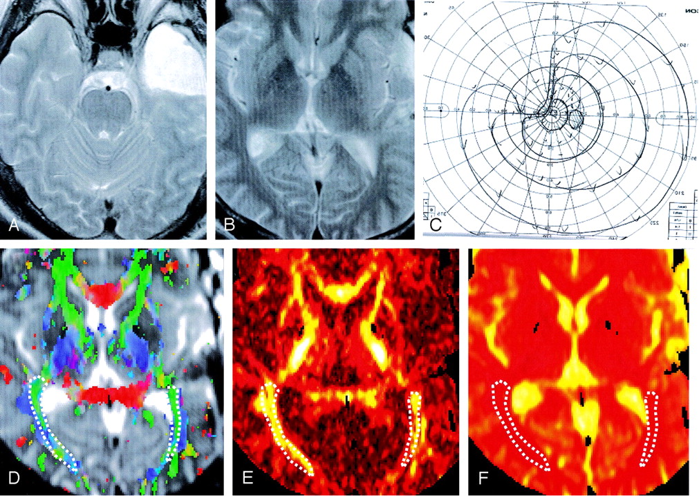
  • Fig 3.
    • Download figure
    • Open in new tab
    • Download powerpoint
    Fig 3.

    Case 9, a 44-year-old man.

    A and B, T2-weighted image. The patient had a left temporal lobectomy (A). No abnormal signal intensity can be seen along the sagittal stratum (B). C, Visual field. The medial sector of the upper quadrant visual field is completely impaired, and partial impairment in the lateral sector area can be seen. D, Color-displayed tensor image. Sagittal strata are recognized as a green area, which indicates an anterior to posterior directed tract. Regions of interest for measurement of ADC and FA values are shown as dotted lines. E, FA image. The FA value of the left optic radiation shows a lower value as compared with the opposite side (right, 0.526; left, 0.488). F, ADC image. There is no apparent difference between the optic radiations of both sides (right, 6.20 × 10⁻⁴ mm2/s; left, 6.10 × 10⁻⁴ mm2/s).

  • Fig 4.
    • Download figure
    • Open in new tab
    • Download powerpoint
    Fig 4.

    Case 8, a 49-year-old man.

    A and B, T2-weighted image. The patient had left anterior temporal lobectomy (A), and there is a high signal intensity area in the anterior part of left sagittal stratum (B, arrow). C, Visual field. The upper quadrant visual field is completely impaired. D, Color-displayed tensor image. Sagittal strata including the optic radiation were recognized as a green area even in operated side. Regions of interest for measurement are shown as dotted lines. E, FA image. The FA value of the left sagittal strata shows a noticeably lower value as compared with the opposite side (right, 0.506; left, 0. 394). F, ADC image. No apparent difference between the optic radiations of both sides is observed (right, 6.10 × 10⁻⁴ mm2/s; left, 7.00 × 10⁻⁴ mm2/s).
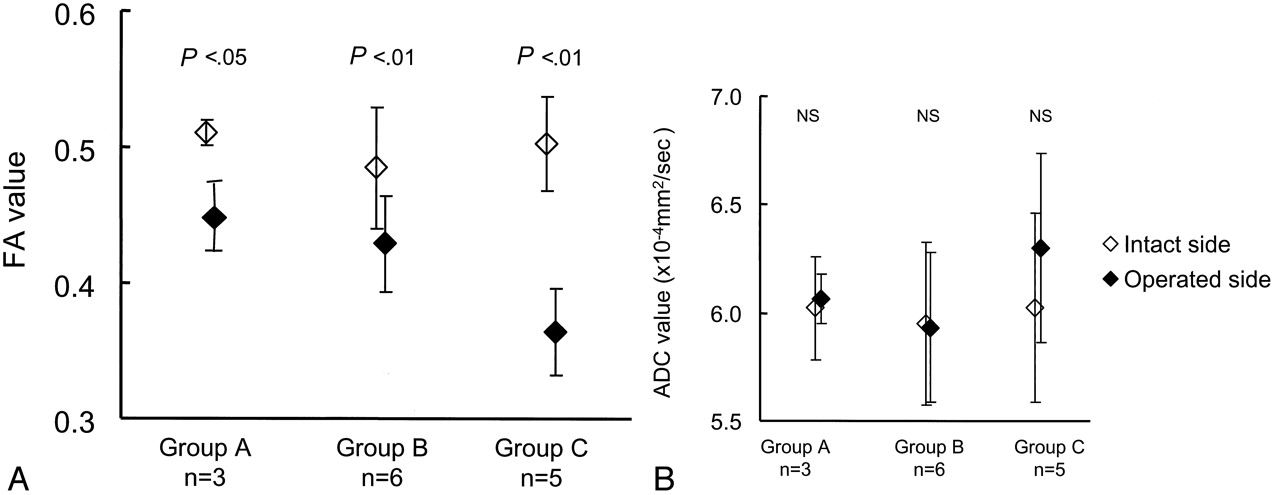
  • Fig 5.
    • Download figure
    • Open in new tab
    • Download powerpoint
    Fig 5.

    A, FA values for the lobectomy and intact side in groups A, B, and C. The FA value of the lobectomy side shows a lower value compared with the intact side in each group. There are statistically significant differences between the operated and intact side in group A (P < .05), group B (P < .01) and C (P < .01). B, ADC values for the lobectomy and intact side in groups A, B, and C. There is no statistical difference between the ADC value of the operative side and intact side when evaluated overall.
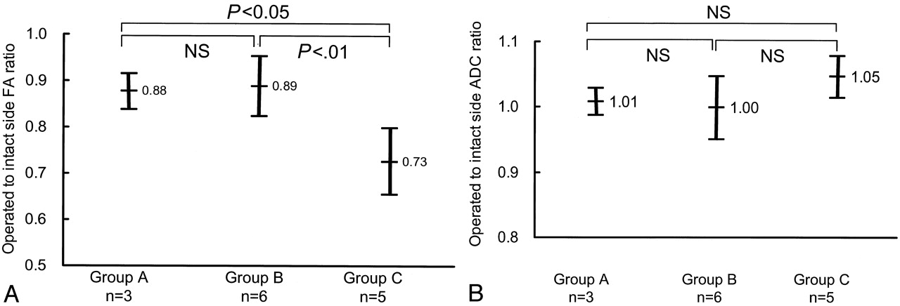
  • Fig 6.
    • Download figure
    • Open in new tab
    • Download powerpoint
    Fig 6.

    A, Mean operated-to-intact side FA ratio. The mean operated-to-intact side FA ratio in the sagittal strata is shown. The more severe groups show a smaller FA in the operated side. Groups A and C, and groups B and C showed statistically significant differences. B, Mean operated-to-intact side ADC ratio. The mean operated-to-intact side ADC ratio in the sagittal strata is shown. There is no statistically significant difference between the three groups when evaluated overall.

  • Fig 7.
    • Download figure
    • Open in new tab
    • Download powerpoint
    Fig 7.

    A, The mean operative to intact side FA ratio as classified by the severity of visual defect and duration after surgery. The mean operated-to-intact side FA ratio as classified by visual field and duration after surgery is shown. Even in early cases after surgery, the FA value is found to decrease, and for the groups examined long after surgery, there is also an obvious decrease. There were no cases that were examined within 4–10 weeks in groups A or B. B, Mean operated-to-intact side ADC ratio as classified by the severity of visual defect and duration after surgery. Although a statistically significant difference is not detected when evaluated overall, there is a trend for the ADC value to decrease in the early period after surgery and increase during the late period after surgery. In the groups with time periods of >10 weeks after surgery, the increase of ADC is larger in group C. There were no cases that were examined within 4–10 weeks for groups A or B.

Tables

  • Figures
    • View popup
    TABLE 1:

    Cases consists of three groups and their duration after surgery

    GroupCase No.AgeSexDuration after surgery
    A123M2 weeks
    225M2 weeks
    355F9 years
    B429M2 weeks
    530F3 weeks
    642M3 weeks
    729F3 weeks
    812M3 months
    944M1 year
    C1022F3 weeks
    1149M5 weeks
    1231M2 months
    1330F3 months
    1426F1 year
    • View popup
    TABLE 2:

    Number of cases that show a high signal along the optic radiation after temporal lobectomy. (There were no cases that were examined within 4–10 weeks for groups A and B.)

    Duration after surgeryGroup AGroup BGroup C
    Less than 4 weeks0/20/40/1
    4 to 10 weeks––1/2
    More than 10 weeks0/11/22/2
PreviousNext
Back to top

In this issue

American Journal of Neuroradiology: 26 (4)
American Journal of Neuroradiology
Vol. 26, Issue 4
1 Apr 2005
  • Table of Contents
  • Index by author
Advertisement
Print
Download PDF
Email Article

Thank you for your interest in spreading the word on American Journal of Neuroradiology.

NOTE: We only request your email address so that the person you are recommending the page to knows that you wanted them to see it, and that it is not junk mail. We do not capture any email address.

Enter multiple addresses on separate lines or separate them with commas.
Diffusion Tensor Imaging in Cases with Visual Field Defect after Anterior Temporal Lobectomy
(Your Name) has sent you a message from American Journal of Neuroradiology
(Your Name) thought you would like to see the American Journal of Neuroradiology web site.
CAPTCHA
This question is for testing whether or not you are a human visitor and to prevent automated spam submissions.
Cite this article
Toshiaki Taoka, Masahiko Sakamoto, Satoru Iwasaki, Hiroyuki Nakagawa, Akio Fukusumi, Shinji Hirohashi, Keiko Taoka, Kimihiko Kichikawa, Tohru Hoshida, Toshisuke Sakaki
Diffusion Tensor Imaging in Cases with Visual Field Defect after Anterior Temporal Lobectomy
American Journal of Neuroradiology Apr 2005, 26 (4) 797-803;

Citation Manager Formats

  • BibTeX
  • Bookends
  • EasyBib
  • EndNote (tagged)
  • EndNote 8 (xml)
  • Medlars
  • Mendeley
  • Papers
  • RefWorks Tagged
  • Ref Manager
  • RIS
  • Zotero
0 Responses
Respond to this article
Share
Bookmark this article
Diffusion Tensor Imaging in Cases with Visual Field Defect after Anterior Temporal Lobectomy
Toshiaki Taoka, Masahiko Sakamoto, Satoru Iwasaki, Hiroyuki Nakagawa, Akio Fukusumi, Shinji Hirohashi, Keiko Taoka, Kimihiko Kichikawa, Tohru Hoshida, Toshisuke Sakaki
American Journal of Neuroradiology Apr 2005, 26 (4) 797-803;
del.icio.us logo Twitter logo Facebook logo Mendeley logo
  • Tweet Widget
  • Facebook Like
  • Google Plus One

Jump to section

  • Article
    • Abstract
    • Methods
    • Results
    • Discussion
    • Conclusion
    • References
  • Figures & Data
  • Info & Metrics
  • Responses
  • References
  • PDF

Related Articles

  • No related articles found.
  • PubMed
  • Google Scholar

Cited By...

  • Changes in fiber tract integrity and visual fields after anterior temporal lobectomy
  • Quantitative Fiber Tracking Analysis of the Optic Radiation Correlated with Visual Performance in Premature Newborns
  • Identifying the human optic radiation using diffusion imaging and fiber tractography
  • Crossref
  • Google Scholar

This article has not yet been cited by articles in journals that are participating in Crossref Cited-by Linking.

More in this TOC Section

  • Statin Therapy Does Not Affect the Radiographic and Clinical Profile of Patients with TIA and Minor Stroke
  • Usefulness of Quantitative Susceptibility Mapping for the Diagnosis of Parkinson Disease
  • White Matter Alterations in the Brains of Patients with Active, Remitted, and Cured Cushing Syndrome: A DTI Study
Show more Brain

Similar Articles

Advertisement

Indexed Content

  • Current Issue
  • Accepted Manuscripts
  • Article Preview
  • Past Issues
  • Editorials
  • Editor's Choice
  • Fellows' Journal Club
  • Letters to the Editor
  • Video Articles

Cases

  • Case Collection
  • Archive - Case of the Week
  • Archive - Case of the Month
  • Archive - Classic Case

More from AJNR

  • Trainee Corner
  • Imaging Protocols
  • MRI Safety Corner
  • Book Reviews

Multimedia

  • AJNR Podcasts
  • AJNR Scantastics

Resources

  • Turnaround Time
  • Submit a Manuscript
  • Submit a Video Article
  • Submit an eLetter to the Editor/Response
  • Manuscript Submission Guidelines
  • Statistical Tips
  • Fast Publishing of Accepted Manuscripts
  • Graphical Abstract Preparation
  • Imaging Protocol Submission
  • Evidence-Based Medicine Level Guide
  • Publishing Checklists
  • Author Policies
  • Become a Reviewer/Academy of Reviewers
  • News and Updates

About Us

  • About AJNR
  • Editorial Board
  • Editorial Board Alumni
  • Alerts
  • Permissions
  • Not an AJNR Subscriber? Join Now
  • Advertise with Us
  • Librarian Resources
  • Feedback
  • Terms and Conditions
  • AJNR Editorial Board Alumni

American Society of Neuroradiology

  • Not an ASNR Member? Join Now

© 2025 by the American Society of Neuroradiology All rights, including for text and data mining, AI training, and similar technologies, are reserved.
Print ISSN: 0195-6108 Online ISSN: 1936-959X

Powered by HighWire