Advertisement

Main menu

  • Home
  • Content
    • Current Issue
    • Accepted Manuscripts
    • Article Preview
    • Past Issue Archive
    • AJNR Case Collection
    • Case of the Week Archive
    • Classic Case Archive
    • Case of the Month Archive
  • Special Collections
    • Spinal CSF Leak Articles (Jan 2020-June 2024)
    • 2024 AJNR Journal Awards
    • Most Impactful AJNR Articles
  • Multimedia
    • AJNR Podcast
    • AJNR Scantastics
    • Video Articles
  • For Authors
    • Submit a Manuscript
    • Author Policies
    • Fast publishing of Accepted Manuscripts
    • Graphical Abstract Preparation
    • Manuscript Submission Guidelines
    • Imaging Protocol Submission
    • Submit a Case for the Case Collection
  • About Us
    • About AJNR
    • Editorial Board
  • More
    • Become a Reviewer/Academy of Reviewers
    • Subscribers
    • Permissions
    • Alerts
    • Feedback
    • Advertisers
    • ASNR Home
  • Other Publications
    • ajnr

User menu

  • Alerts
  • Log in

Search

  • Advanced search
American Journal of Neuroradiology

American Journal of Neuroradiology

ASHNR American Society of Functional Neuroradiology ASHNR American Society of Pediatric Neuroradiology ASSR
  • Alerts
  • Log in

Advanced Search

  • Home
  • Content
    • Current Issue
    • Accepted Manuscripts
    • Article Preview
    • Past Issue Archive
    • AJNR Case Collection
    • Case of the Week Archive
    • Classic Case Archive
    • Case of the Month Archive
  • Special Collections
    • Spinal CSF Leak Articles (Jan 2020-June 2024)
    • 2024 AJNR Journal Awards
    • Most Impactful AJNR Articles
  • Multimedia
    • AJNR Podcast
    • AJNR Scantastics
    • Video Articles
  • For Authors
    • Submit a Manuscript
    • Author Policies
    • Fast publishing of Accepted Manuscripts
    • Graphical Abstract Preparation
    • Manuscript Submission Guidelines
    • Imaging Protocol Submission
    • Submit a Case for the Case Collection
  • About Us
    • About AJNR
    • Editorial Board
  • More
    • Become a Reviewer/Academy of Reviewers
    • Subscribers
    • Permissions
    • Alerts
    • Feedback
    • Advertisers
    • ASNR Home
  • Follow AJNR on Twitter
  • Visit AJNR on Facebook
  • Follow AJNR on Instagram
  • Join AJNR on LinkedIn
  • RSS Feeds

Welcome to the new AJNR, Updated Hall of Fame, and more. Read the full announcements.


AJNR is seeking candidates for the position of Associate Section Editor, AJNR Case Collection. Read the full announcement.


OtherSPINE

Cervical Diskography Performed with a “Prong Deflector” for Improved Access to the Cervical Disk Spaces

Walter S. Bartynski, Stephen Z. Grahovac and William E. Rothfus
American Journal of Neuroradiology May 2005, 26 (5) 1004-1007;
Walter S. Bartynski
  • Find this author on Google Scholar
  • Find this author on PubMed
  • Search for this author on this site
Stephen Z. Grahovac
  • Find this author on Google Scholar
  • Find this author on PubMed
  • Search for this author on this site
William E. Rothfus
  • Find this author on Google Scholar
  • Find this author on PubMed
  • Search for this author on this site
  • Article
  • Figures & Data
  • Info & Metrics
  • Responses
  • References
  • PDF
Loading

Article Figures & Data

Figures

  • Tables
  • Fig 1.
    • Download figure
    • Open in new tab
    • Download powerpoint
    Fig 1.

    The prong deflector tool has an aluminum handle for operator grasp and control attached to a plastic covered steel two tine active end that is shaped to resemble the operator’s fingertips with slight tine curvature and a 3–3.5-cm gap.

  • Fig 2.
    • Download figure
    • Open in new tab
    • Download powerpoint
    Fig 2.

    Example of thyroid cartilage displacement visualized at fluoroscopy during cervical diskography in a 49-year-old man with severe neck pain radiating to his left arm and shoulder.

    A, The prong deflector (arrow) lies adjacent to the right lateral margin of the thyroid cartilage (arrowheads). The cartilage is difficult to visualize as it overlies the disk spaces, uncinate processes and Lushka’s joints on the patients right.

    B, With displacement of the thyroid cartilage toward the midline, the lateral margin of the cartilage is now visible (arrowheads) and can be seen, no longer overlying the lateral aspect of the disk space and no longer overlying the intended target.

  • Fig 3.
    • Download figure
    • Open in new tab
    • Download powerpoint
    Fig 3.

    Operator insertion of the 25-gauge needle by using the prong deflector for neck tissue control.

    A, The prong deflector maintains control of the neck structures while the needle is inserted (arrow). Fluoroscopic inspection of the needle position and trajectory can be obtained without direct operator exposure of the central beam.

    B, Example of fluoroscopic visualization of needle insertion during cervical diskography with assistance of the prong deflector tool in a 44-year-old man with persistent neck pain radiating to his posterior left shoulder. The prong deflector is being used during needle insertion of the C5–6 disk space with thyroid cartilage displaced medial to the prong deflector tines (white arrow) and clear access to the disk space established (black arrowhead).

  • Fig 4.
    • Download figure
    • Open in new tab
    • Download powerpoint
    Fig 4.

    Postdiskogram CT image in a 46-year-old woman with unrelenting neck pain radiating down both arms that demonstrates close proximity of the carotid artery and neurovascular sheath to thyroid cartilage (arrow) as is typically seen in most patients.
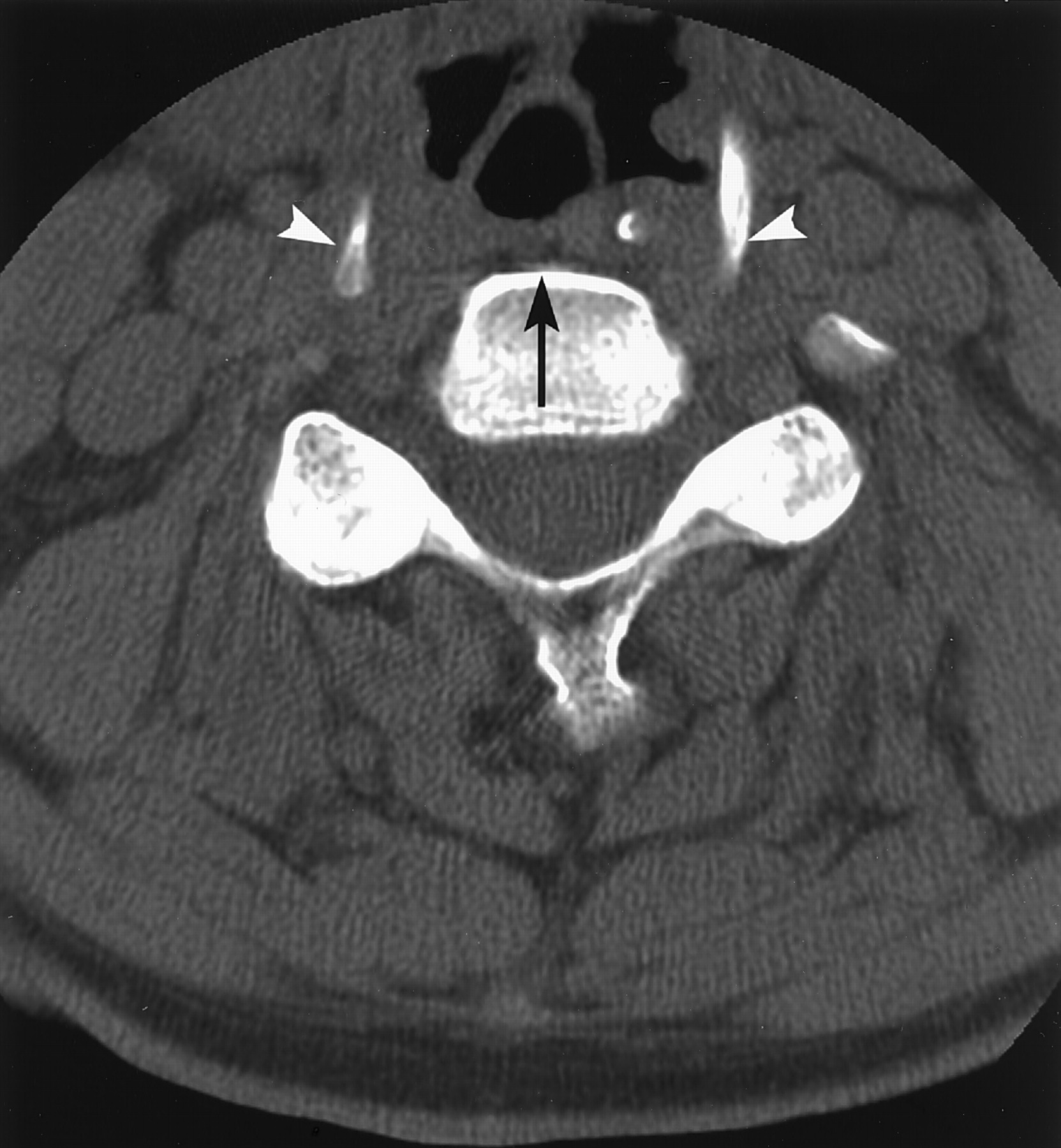
  • Fig 5.
    • Download figure
    • Open in new tab
    • Download powerpoint
    Fig 5.

    Postdiskogram CT images in the 48-year-old man with chronic neck and right arm pain where difficulty was encountered in access to the C4–5 disk space despite attempted displacement of the larynx with the prong deflector tool. The hyoid bone is wide and the anterior aspect of the C4 vertebral body (black arrow) projects deeply into the gap between the hyoid cornua (arrowheads), preventing displacement of the hyoid and thyroid cartilages across the midline. Without hyoid and thyroid cartilage displacement, clear and safe access to the disk space could not be obtained.

Tables

  • Figures
    • View popup
    TABLE 1:

    Number of levels studied by cervical discography

    Procedure GroupC23 LevelC34 LevelC45 LevelC56 LevelC67 Level
    Prong Deflector1913118
    Traditional139109
    Total212222117
    • View popup
    TABLE 2:

    Thyroid cartilage overlap of disc spaces in the “prong deflector” group: observations at discography

    Fluoroscopy observation of disc space access: Pre Cartilage Deflection → Post Cartilage DeflectionC34 LevelC45 LevelC56 LevelC67 LevelTotal
    Partial covered → uncovered334111 (29%)
    Covered → uncovered21210327 (71%)
    • View popup
    TABLE 3:

    Procedure sedation, disc access and fluoroscopic time

    Procedure GroupSedation dose/procedureSedation dose/levelProcedure time/level (min)Fluoroscopy time/level (min)
    Prong Deflector2.1.7610.71.4 ± .35
    Traditional1.8.8411.92.1 ± 1.4
    Average2.8011.241.68
PreviousNext
Back to top

In this issue

American Journal of Neuroradiology: 26 (5)
American Journal of Neuroradiology
Vol. 26, Issue 5
1 May 2005
  • Table of Contents
  • Index by author
Advertisement
Print
Download PDF
Email Article

Thank you for your interest in spreading the word on American Journal of Neuroradiology.

NOTE: We only request your email address so that the person you are recommending the page to knows that you wanted them to see it, and that it is not junk mail. We do not capture any email address.

Enter multiple addresses on separate lines or separate them with commas.
Cervical Diskography Performed with a “Prong Deflector” for Improved Access to the Cervical Disk Spaces
(Your Name) has sent you a message from American Journal of Neuroradiology
(Your Name) thought you would like to see the American Journal of Neuroradiology web site.
CAPTCHA
This question is for testing whether or not you are a human visitor and to prevent automated spam submissions.
Cite this article
Walter S. Bartynski, Stephen Z. Grahovac, William E. Rothfus
Cervical Diskography Performed with a “Prong Deflector” for Improved Access to the Cervical Disk Spaces
American Journal of Neuroradiology May 2005, 26 (5) 1004-1007;

Citation Manager Formats

  • BibTeX
  • Bookends
  • EasyBib
  • EndNote (tagged)
  • EndNote 8 (xml)
  • Medlars
  • Mendeley
  • Papers
  • RefWorks Tagged
  • Ref Manager
  • RIS
  • Zotero
0 Responses
Respond to this article
Share
Bookmark this article
Cervical Diskography Performed with a “Prong Deflector” for Improved Access to the Cervical Disk Spaces
Walter S. Bartynski, Stephen Z. Grahovac, William E. Rothfus
American Journal of Neuroradiology May 2005, 26 (5) 1004-1007;
del.icio.us logo Twitter logo Facebook logo Mendeley logo
  • Tweet Widget
  • Facebook Like
  • Google Plus One

Jump to section

  • Article
    • Abstract
    • Description of Technique
    • The Prong Deflector Tool
    • Cervical Diskogram Technique with the Prong Deflector Tool
    • Parameters Evaluated
    • Observations
    • Discussion
    • Conclusion
    • References
  • Figures & Data
  • Info & Metrics
  • Responses
  • References
  • PDF

Related Articles

  • No related articles found.
  • PubMed
  • Google Scholar

Cited By...

  • No citing articles found.
  • Crossref
  • Google Scholar

This article has not yet been cited by articles in journals that are participating in Crossref Cited-by Linking.

More in this TOC Section

  • Bern Score Validity for SIH
  • MP2RAGE 7T in MS Lesions of the Cervical Spine
  • Deep Learning for STIR Spine MRI Quality
Show more Spine

Similar Articles

Advertisement

Indexed Content

  • Current Issue
  • Accepted Manuscripts
  • Article Preview
  • Past Issues
  • Editorials
  • Editors Choice
  • Fellow Journal Club
  • Letters to the Editor

Cases

  • Case Collection
  • Archive - Case of the Week
  • Archive - Case of the Month
  • Archive - Classic Case

Special Collections

  • Special Collections

Resources

  • News and Updates
  • Turn around Times
  • Submit a Manuscript
  • Author Policies
  • Manuscript Submission Guidelines
  • Evidence-Based Medicine Level Guide
  • Publishing Checklists
  • Graphical Abstract Preparation
  • Imaging Protocol Submission
  • Submit a Case
  • Become a Reviewer/Academy of Reviewers
  • Get Peer Review Credit from Publons

Multimedia

  • AJNR Podcast
  • AJNR SCANtastic
  • Video Articles

About Us

  • About AJNR
  • Editorial Board
  • Not an AJNR Subscriber? Join Now
  • Alerts
  • Feedback
  • Advertise with us
  • Librarian Resources
  • Permissions
  • Terms and Conditions

American Society of Neuroradiology

  • Not an ASNR Member? Join Now

© 2025 by the American Society of Neuroradiology All rights, including for text and data mining, AI training, and similar technologies, are reserved.
Print ISSN: 0195-6108 Online ISSN: 1936-959X

Powered by HighWire