
Article Figures & Data
Figures
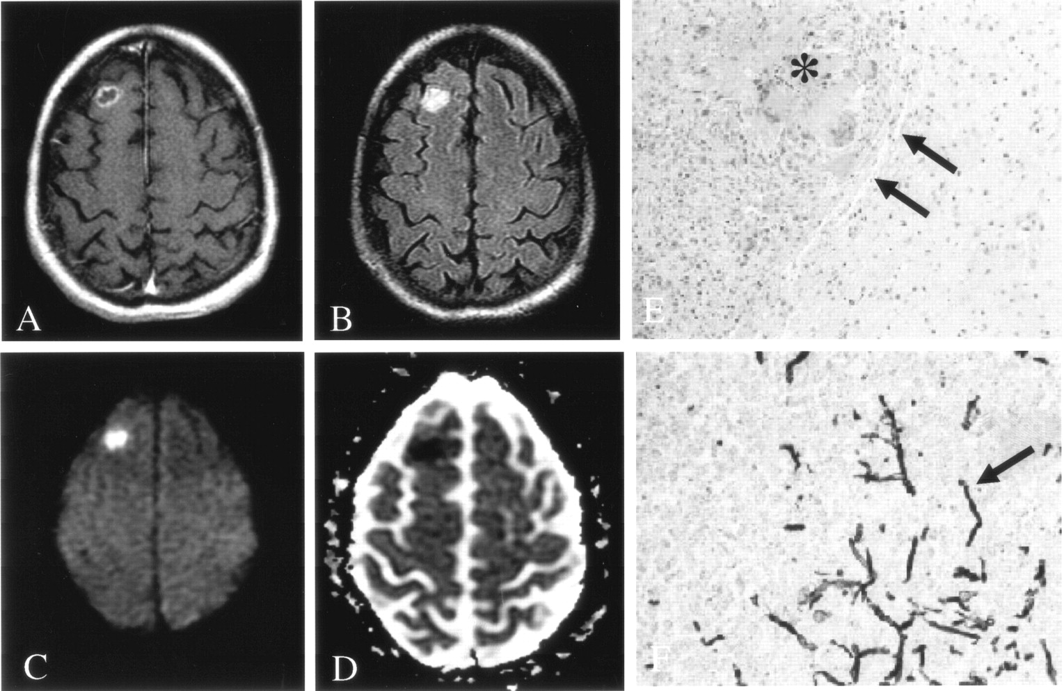
- Fig 1.
Patient 1. Fungal cerebritis due to Rhizopus infection of the left basal ganglia and corona radiata.
A, On Gd-enhanced T1-weighted imaging, the lesion is hypointense with minimal peripheral enhancement.
B, On FLAIR imaging, the lesion has heterogeneous signal intensity with moderate surrounding edema.
C and D, DWI (C) and ADC (D) images show predominantly decreased diffusion (short arrow in D) with a smaller region of elevated diffusion (long arrow in D).
E, Hematoxylin-eosin stain (40×) shows perivascular ring hemorrhages and necrosis. Moderate amount of acute and chronic inflammation was also found (data not shown).
F, Methenamine silver stain (450×) shows fungal organisms predominantly in the lumen of blood vessels (arrows).
- Fig 2.
Patient 7. Fungal abscess due to Scedosporium infection.
A, On Gd-enhanced T1-weighted imaging, the lesion is ring enhancing.
B, On FLAIR imaging, the lesion is isointense to brain parenchyma, with moderate surrounding edema.
C and D, DWI (C) and ADC (D) images show homogeneously decreased diffusion in the center of the lesion, similar to that seen with pyogenic abscess.
E and F, Hematoxylin-eosin (E) (450×) and methenamine silver stain (F) (250×) stains show fungal organisms (arrow) in necrotic tissue and chronic inflammation.
- Fig 3.
Patient 6. Fungal abscess due to Aspergillus infection.
A, On Gd-enhanced T1-weighted imaging, the lesion is ring enhancing.
B, On FLAIR imaging, the lesion is hyperintense to brain parenchyma, without surrounding edema.
C and D, DWI (C) and ADC (D) images show homogeneously decreased diffusion in the center of the lesion.
E, Hematoxylin-eosin stain (100×) shows a lesion discrete from brain with a well-defined capsule (arrows). Lesions were composed of granulomatous chronic inflammation with numerous histiocytes and giant cells engulfing fungal organisms (asterisk).
F, Methenamine silver stain (250×) shows fungal organisms (arrow) with 45° angle branching, consistent with Aspergillus infection.
- Fig 4.
Patient 3. Early fungal abscess due to Aspergillus infection.
A, On Gd-enhanced T1-weighted imaging, the lesion has a thin rim of peripheral enhancement.
B, On FLAIR imaging, the lesion has heterogeneous signal intensity with minimal surrounding edema.
C and D, DWI (C) and ADC (D) maps show peripherally decreased diffusion, with elevated diffusion in the center of the lesion.
E, Hematoxylin-eosin stain (100×) shows acute and chronic inflammation in the brain parenchyma, without a well-defined capsule.
F, Methenamine silver stain (250×) shows septate and 45°-branching hyphae in necrotic parenchyma.


Tables
Clinical presentation and pathologic and DWI findings in patients with fungal cerebral infection or pyogenic bacterial abscess
Patient Clinical Presentation Lesion Aspiration Organism MR Imaging Finding ADC (×10−3 mm2/s) ADC Ratio Lesion WM 1 Acute Not done, autopsy Rhizopus Minimal enhancement, DWI heterogeneously hyperintense 0.20 ± 0.05 0.86 ± 0.06 0.23 2 Acute Not done, autopsy Aspergillus No enhancement, DWI heterogeneously hyperintense 0.23 ± 0.04 0.85 ± 0.05 0.27 3 Subacute Not done, autopsy Aspergillus Ring-enhancement, DWI peripherally hyperintense 0.13 ± 0.07 0.69 ± 0.05 0.19 4 Subacute Mucoid fluid Aspergillus Ring-enhancement, DWI centrally hyperintense 0.59 ± 0.05 0.80 ± 0.06 0.74 5 Subacute Not done, autopsy Aspergillus Ring-enhancement, DWI centrally hyperintense 0.71 ± 0.03 0.87 ± 0.04 0.81 6 Subacute Mucoid fluid Aspergillus Ring-enhancement, DWI centrally hyperintense 0.39 ± 0.08 0.74 ± 0.05 0.53 7 Subacute Mucoid fluid Scedosporium Ring-enhancement, DWI centrally hyperintense 0.22 ± 0.05 0.58 ± 0.06 0.39 8 Subacute Mucoid fluid Aspergillus Ring-enhancement, DWI hyperintense 0.21 ± 0.08 0.66 ± 0.06 0.31 Note.—Mean lesion and white matter ADC and ADC ratio were, respectively, 0.33 ± 0.06 × 10⁻³ mm2/s, 0.76 ± 0.05 × 10⁻³ mm2/s, and 0.43 in the fungal group and 0.46 ± 0.06 × 10⁻³ mm2/s (difference not significant), 0.76 ± 0.05 × 10⁻³ mm2/s, and 0.61 (difference not significant) in the bacterial group.


